
Chinese Journal of Magnetic Resonance ›› 2023, Vol. 40 ›› Issue (3): 270-279.doi: 10.11938/cjmr20223047
• Articles • Previous Articles Next Articles
TIAN Hui1,WU Jie1,*( ),BIAN Yun2,#(),ZHANG Zhiwei1,SHAO Chengwei2
Received:2022-12-18
Published:2023-09-05
Online:2023-03-22
Contact:
*Tel: 021-55271116, E-mail: CLC Number:
TIAN Hui, WU Jie, BIAN Yun, ZHANG Zhiwei, SHAO Chengwei. Classification of Pancreatic Cystic Tumors Based on DenseNet and Transfer Learning[J]. Chinese Journal of Magnetic Resonance, 2023, 40(3): 270-279.
Add to citation manager EndNote|Reference Manager|ProCite|BibTeX|RefWorks
Table 1
DenseNet161 network structure
| Layers | DenseNet161 |
|---|---|
| Convolution | 7×7 conv |
| Pooling | 3×3 max pool |
| Dense Block 1 | $\left[ \begin{matrix} 1\times 1\ \text{conv} \\ 3\times 3\ \text{conv} \\ \end{matrix} \right]\times 6$ |
| Transition Layer 1 | 1×1 conv, 2×2 average pool |
| Dense Block 2 | $\left[ \begin{matrix} 1\times 1\ \text{conv} \\ 3\times 3\ \text{conv} \\ \end{matrix} \right]\times 12$ |
| Transition Layer 2 | 1×1 conv, 2×2 average pool |
| Dense Block 3 | $\left[ \begin{matrix} 1\times 1\ \text{conv} \\ 3\times 3\ \text{conv} \\ \end{matrix} \right]\times 36$ |
| Transition Layer 3 | 1×1 conv, 2×2 average pool |
| Dense Block 4 | $\left[ \begin{matrix} 1\times 1\ \text{conv} \\ 3\times 3\ \text{conv} \\ \end{matrix} \right]\times 24$ |
| Classification Layer | 7×7 global average pool, fully-connected, softmax |
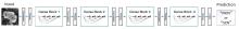
Fig. 1
MCN and SCN classification flowchart
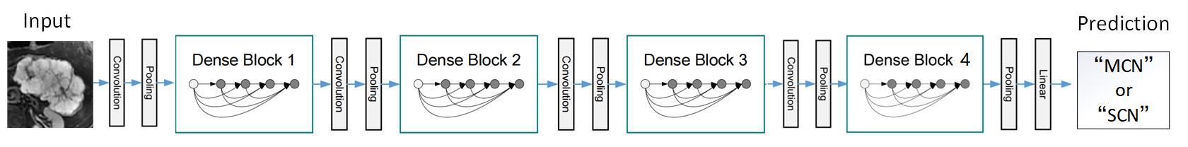
Table 2
Comparison of the effect of DenseNet161 network combined with transfer learning
| 模型 | ACC | AUC |
|---|---|---|
| DenseNet161 | 0.767 | 0.837 |
| DenseNet161+迁移学习 | 0.943 | 0.989 |
Table 3
Evaluation index values of MCN and SCN by DenseNet161 combined with transfer learning
| 类别 | ACC | Precision | Recall | F1-Score |
|---|---|---|---|---|
| SCN | 0.943 | 0.948 | 0.937 | 0.942 |
| MCN | 0.943 | 0.938 | 0.949 | 0.943 |
| 均值 | 0.943 | 0.943 | 0.943 | 0.943 |
Table 4
Experimental results of MCN and SCN recognition by different deep learning models
| 模型 | ACC | Precision | Recall | Specificity | F1-Score | AUC | 参数量 |
|---|---|---|---|---|---|---|---|
| AlexNet | 0.844 | 0.846 | 0.842 | 0.847 | 0.844 | 0.930 | 6100840 |
| MobileNet_v3 | 0.889 | 0.894 | 0.884 | 0.895 | 0.889 | 0.960 | 5483032 |
| Vgg16 | 0.898 | 0.907 | 0.886 | 0.909 | 0.896 | 0.966 | 138365992 |
| ResNet50 | 0.912 | 0.923 | 0.898 | 0.926 | 0.910 | 0.971 | 25557032 |
| 本文 | 0.943 | 0.938 | 0.949 | 0.937 | 0.943 | 0.989 | 28681000 |
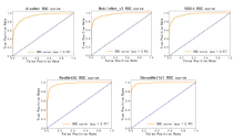
Fig. 2
ROC curve and area under curve of different models
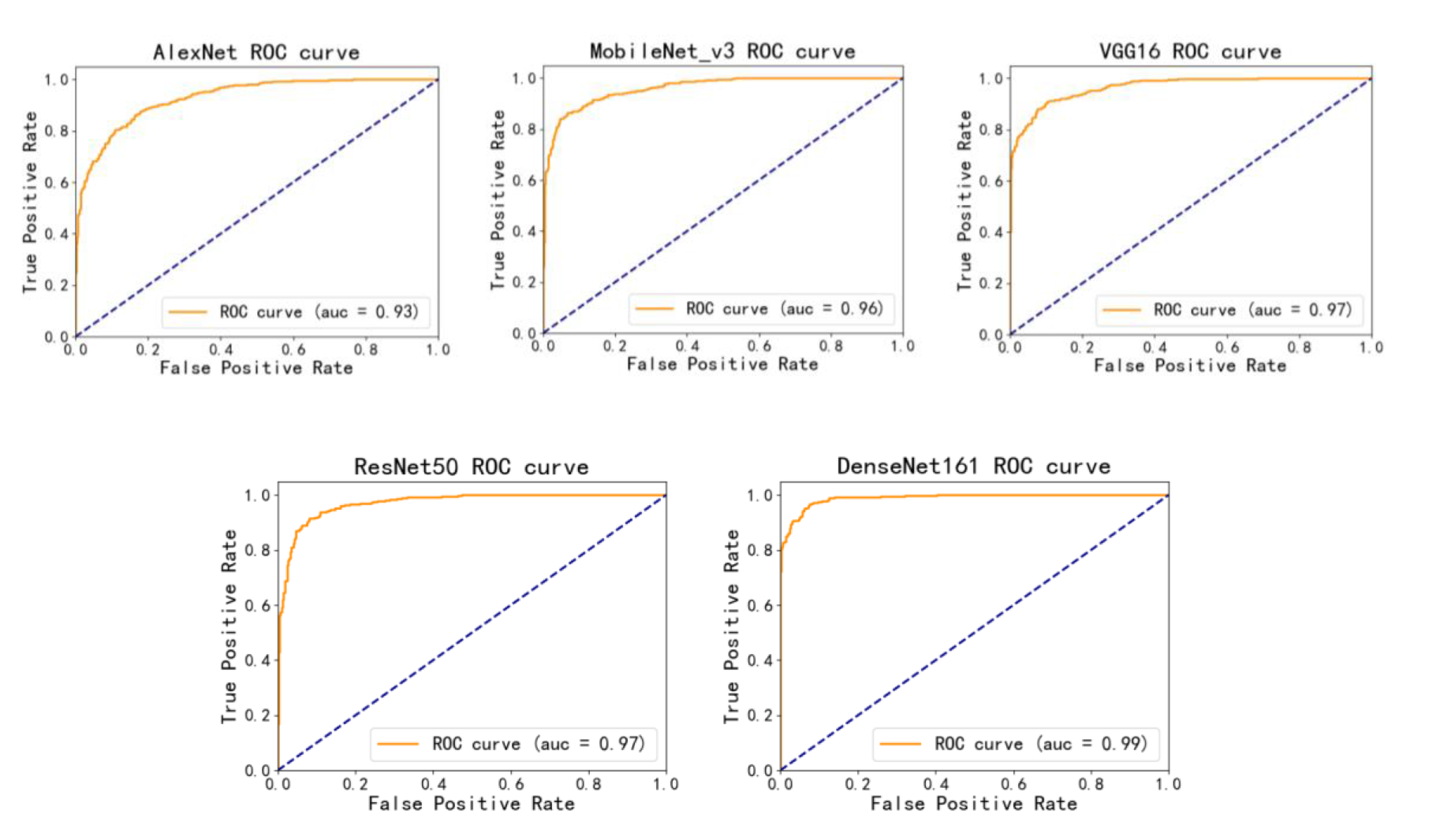
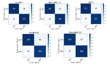
Fig. 3
Confusion matrix of different models
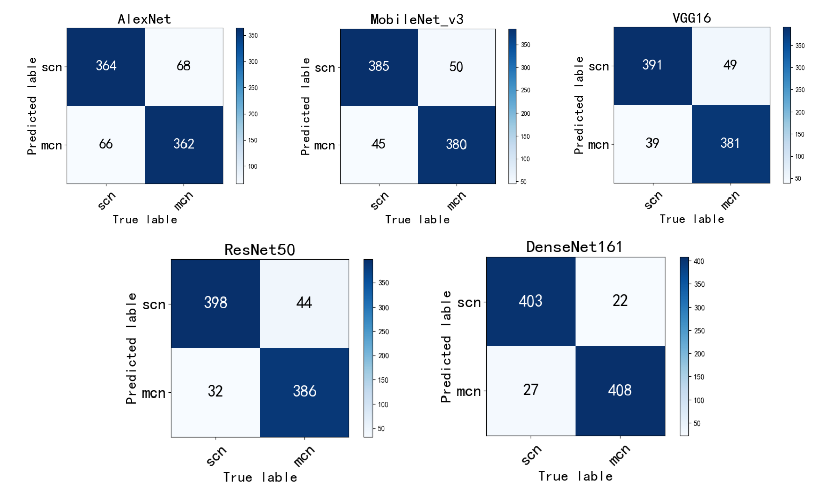
Table 5
Results of MCN and SCN recognition by different methods
| 方法 | 数据 | ACC | |
|---|---|---|---|
| 文献[ | 影像组学特征融合 | 胰腺囊性肿瘤CT图像 | 0.862 |
| 文献[ | CT纹理特征模型+临床影像学特征模型 | SCN与MCN静脉期CT图像 | 0.938 (AUC) |
| 文献[ | 影像组学参数+人工神经网络 | SCN与MCN CT图像 | 0.895 |
| 文献[ | ResNet50+迁移学习 | SCN与MCN内窥镜超声图像 | 0.828 |
| 本文 | DenseNet161+迁移学习 | SCN与MCN T2加权MR图像 | 0.943 |
| [1] |
ARDESHNA D R, CAO T, RODGERS B, et al. Recent advances in the diagnostic evaluation of pancreatic cystic lesions[J]. World J Gastroenterol, 2022, 28(6): 624-634.
doi: 10.3748/wjg.v28.i6.624 |
| [2] | XU X B, CHEN H, SUN B. Progress in diagnosis and treatment of pancreatic cystic tumors[J]. Chinese Journal of Operative Procedures of General Surgery (Electronic Edition), 2020, 14(6): 643-646. |
| 徐西伯, 陈华, 孙备. 胰腺囊性肿瘤的诊治进展[J]. 中华普外科手术学杂志(电子版), 2020, 14(6): 643-646. | |
| [3] |
JANG D K, SONG B J, RYU J K, et al. Preoperative diagnosis of pancreatic cystic lesions: the accuracy of endoscopic ultrasound and cross-sectional imaging[J]. Pancreas, 2015, 44(8): 1329-1333.
doi: 10.1097/MPA.0000000000000396 |
| [4] |
SUN Y, YANG S, QI E, et al. Comparative diagnostic evaluation with contrast-enhanced ultrasound, computed tomography and magnetic resonance imaging in patients with pancreatic cystic neoplasms[J]. Cancer Manag Res, 2020, 12: 2889-2898.
doi: 10.2147/CMAR.S246564 pmid: 32425602 |
| [5] |
BOLLEN T L, WESSELS F J. Radiological workup of cystic neoplasms of the pancreas[J]. Visceral Med, 2018, 34(3): 182-190.
doi: 10.1159/000489674 |
| [6] | 陈海燕. 影像学特征联合增强CT纹理特征鉴别胰腺浆液性囊腺瘤与黏液性囊腺瘤[D]. 浙江大学, 2020. |
| [7] | 袁梦依. 基于影像组学特征融合的胰腺囊性肿瘤分类[D]. 浙江工业大学, 2020. |
| [8] | ZHANG Y F, XU S S, WU J, et al. Value of CT texture analysis in differentiating pancreatic serous cystadenoma from mucinous cystadenoma[J]. Journal of Southeast University (Medical Science Edition), 2022, 41(3): 308-316. |
| 张怡帆, 徐珊珊, 吴锦, 等. CT纹理分析在鉴别胰腺浆液性囊腺瘤与黏液性囊腺瘤中的价值[J]. 东南大学学报(医学版), 2022, 41(3): 308-316. | |
| [9] | CHEN S, CHEN X, CHEN J Y, et al. Application of CT radiomics in differential diagnosis of pancreatic serous and mucinous cystic neoplasm[J]. Chinese Journal of CT and MRI, 2022, 20(10): 92-95. |
| 陈帅, 陈晓, 陈井亚, 等. 基于CT影像组学对胰腺浆液及黏液性囊性肿瘤鉴别诊断[J]. 中国CT和MRI杂志, 2022, 20(10): 92-95. | |
| [10] | YANG Y F, QI Z X, NIE S D. Differentiation of benign and malignant breast lesions based on multimodal MRI and deep learning[J]. Chinese J Magn Reson, 2022, 39(4): 401-412. |
| 杨一风, 祁章璇, 聂生东. 基于多模态MRI与深度学习的乳腺病变良恶性鉴别[J]. 波谱学杂志, 2022, 39(4): 401-412. | |
| [11] | WEI Z H, YAN S J, HAN B S, et al. Diagnosis of alzheimer's disease based on multi-output three-dimensional convolutional neural network[J]. Chinese J Magn Reson, 2021, 38(1): 92-100. |
| 魏志宏, 闫士举, 韩宝三, 等. 基于多输出的3D卷积神经网络诊断阿尔兹海默病[J]. 波谱学杂志, 2021, 38(1): 92-100. | |
| [12] |
NGUON L S, SEO K, LIM J H, et al. Deep learning-based differentiation between mucinous cystic neoplasm and serous cystic neoplasm in the pancreas using endoscopic ultrasonography[J]. Diagnostics, 2021, 11(6): 1052.
doi: 10.3390/diagnostics11061052 |
| [13] |
VILAS-BOAS F, RIBEIRO T, AFONSO J, et al. Deep learning for automatic differentiation of mucinous versus non-mucinous pancreatic cystic lesions: a pilot study[J]. Diagnostics, 2022, 12(9): 2041.
doi: 10.3390/diagnostics12092041 |
| [14] | HUANG G, LIU Z, VAN DER MAATEN L, et al. Densely connected convolutional networks[C]// Proceedings of the IEEE conference on computer vision and pattern recognition, 2017: 4700-4708. |
| [15] |
ZHOU Q, ZHOU Z, CHEN C, et al. Grading of hepatocellular carcinoma using 3D SE-DenseNet in dynamic enhanced MR images[J]. Comput Biol Med, 2019, 107: 47-57.
doi: S0010-4825(19)30032-0 pmid: 30776671 |
| [16] |
WANG Z, LI X, YAO M, et al. A new detection model of microaneurysms based on improved FC-DenseNet[J]. Sci Rep, 2022, 12(1): 1-9.
doi: 10.1038/s41598-021-99269-x |
| [17] |
GUO W, XU Z, ZHANG H. Interstitial lung disease classification using improved DenseNet[J]. Multimed Tools Appl, 2019, 78(21): 30615-30626.
doi: 10.1007/s11042-018-6535-y |
| [18] | YANG Y H, LIU M, WANG X M, et al. Breast cancer image recognition based on DenseNet and transfer learning[J]. Journal of Jilin University, 2022, 40(2): 213-218. |
| 杨雨航, 刘铭, 王新民, 等. 基于DenseNet和迁移学习的乳腺癌图像识别[J]. 吉林大学学报, 2022, 40(2): 213-218. | |
| [19] |
ZHOU Y, ZHANG X, WANG Y, et al. Transfer learning and its application research[J]. J Phys: Conf Ser, 2021, 1920(1): 012058.
doi: 10.1088/1742-6596/1920/1/012058 |
| [20] | LI Y, SONG P H. Review of transfer learning in medical image classification[J]. Journal of Image and Graphics, 2022, 27(3): 672-686. |
| 黎英, 宋佩华. 迁移学习在医学图像分类中的研究进展[J]. 中国图象图形学报, 2022, 27(3): 672-686. | |
| [21] | KINGMA D P, BA J. Adam: A method for stochastic optimization[J]. arXiv preprint arXiv:1412.6980, 2014. |
| [22] | ZEILER M D. Adadelta: an adaptive learning rate method[J]. arXiv preprint arXiv:1212.5701, 2012. |
| [23] | DUCHI J, HAZAN E, SINGER Y. Adaptive subgradient methods for online learning and stochastic optimization[J]. J Mach Learn Res, 2011, 12(7): 2121-2159. |
| [24] | DEAN J, CORADO G, MONGA R, et al. Large scale distributed deep networks[J]. Adv Neural Inf Process Syst, 2012, 25: 1-11. |
| [1] | CAO Fei, XU Qianqian, CHEN Hao, ZU Jie, LI Xiaowen, TIAN Jin, BAO Lei. An Intelligent Diagnosis Method for NIID Based on Cross Self-supervision and DWI [J]. Chinese Journal of Magnetic Resonance, 2025, 42(2): 154-163. |
| [2] | XUE Peiyang, GENG Chen, LI Yuxin, BAO Yifang, LU Yucheng, DAI Yakang. A Classification Method for Cerebral Aneurysms in TOF-MRA Based on Improved 3D ResNet50 Model [J]. Chinese Journal of Magnetic Resonance, 2025, 42(1): 56-66. |
| [3] | NING Xinzhou, HUANG Zhen, CHEN Xiqu, LIU Xinjie, CHEN Gang, ZHANG Zhi, BAO Qingjia, LIU Chaoyang. Research on Transformer Super-Resolution Reconstruction Algorithm for Ultrafast Spatiotemporal Encoding Magnetic Resonance Imaging [J]. Chinese Journal of Magnetic Resonance, 2024, 41(4): 454-468. |
| [4] | YANG Liming, WANG Yuanjun. Research Progress of Denoising Algorithms for Diffusion Tensor Images [J]. Chinese Journal of Magnetic Resonance, 2024, 41(3): 341-361. |
| [5] | Dai Junlong, He Cong, Wu Jie, Bian Yun. Pancreatic Cystic Neoplasms Segmentation Network Combining Dual Decoding and Global Attention Upsampling Modules [J]. Chinese Journal of Magnetic Resonance, 2024, 41(2): 151-161. |
| [6] | YANG Yu, CHEN Bo, WU Liubin, LIN Enping, HUANG Yuqing, CHEN Zhong. Spectrum Reconstruction for Laplace NMR: From Handcraft Regularization to Deep Learning [J]. Chinese Journal of Magnetic Resonance, 2024, 41(2): 191-208. |
| [7] | CHANG Bo, SUN Haoyun, GAO Qingyu, WANG Lijia. Research Progress on Cardiac Segmentation in Different Modal Medical Images by Traditional Methods and Deep Learning [J]. Chinese Journal of Magnetic Resonance, 2024, 41(2): 224-244. |
| [8] | XU Zhenshun, YUAN Xiaohan, HUANG Ziheng, SHAO Chengwei, WU Jie, BIAN Yun. Multi-source Feature Classification Model of Pancreatic Mucinous and Serous Cystic Neoplasms Based on Deep Learning [J]. Chinese Journal of Magnetic Resonance, 2024, 41(1): 19-29. |
| [9] | LAI Jiawen, WANG Yuling, CAI Xiaoyu, ZHOU Lihua. Multidimensional Information Fusion Method for Meniscal Tear Classification Based on CNN-SVM [J]. Chinese Journal of Magnetic Resonance, 2023, 40(4): 423-434. |
| [10] | WANG Hui, WANG Tiantian, WANG Lijia. Squeeze-and-excitation Residual U-shaped Network for Left Myocardium Segmentation Based on Cine Cardiac Magnetic Resonance Images [J]. Chinese Journal of Magnetic Resonance, 2023, 40(4): 435-447. |
| [11] | Li Yijie, YANG Xinyu, YANG Xiaomei. Magnetic Resonance Image Reconstruction of Multi-scale Residual Unet Fused with Attention Mechanism [J]. Chinese Journal of Magnetic Resonance, 2023, 40(3): 307-319. |
| [12] | LU Qiqi, LIAN Zifeng, LI Jialong, SI Wenbin, MAI Zhaohua, FENG Yanqiu. Magnetic Resonance R2* Parameter Mapping of Liver Based on Self-supervised Deep Neural Network [J]. Chinese Journal of Magnetic Resonance, 2023, 40(3): 258-269. |
| [13] | ZHANG Jiajun, LU Yucheng, BAO Yifang, LI Yuxin, GENG Chen, HU Fuyuan, DAI Yakang. An Automatic Segmentation Method of Cerebral Arterial Tree in TOF-MRA Based on DBCNet [J]. Chinese Journal of Magnetic Resonance, 2023, 40(3): 320-331. |
| [14] | HUANG Min,LI Siyi,CHEN Junbo,ZHOU Dao. Progress of Magnetic Resonance Fingerprinting Technology and Its Clinical Application [J]. Chinese Journal of Magnetic Resonance, 2023, 40(2): 207-219. |
| [15] | QIAN Chengyi,WANG Yuanjun. Research Progress on Imaging Classification of Alzheimer’s Disease Based on Deep Learning [J]. Chinese Journal of Magnetic Resonance, 2023, 40(2): 220-238. |
| Viewed | ||||||
|
Full text |
|
|||||
|
Abstract |
|
|||||