

Chinese Journal of Magnetic Resonance >
Association of 129Xe Ventilation Functional MRI with Pulmonary Lesion Types
Received date: 2024-03-23
Online published: 2024-04-09
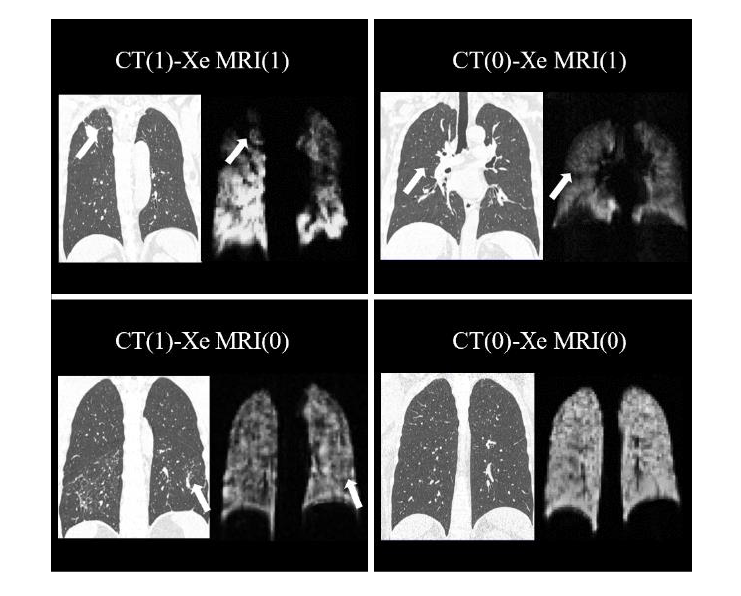
This study investigates the correlation between 129Xe ventilation functional MRI and CT-detected pulmonary lesion types. We performed 129Xe ventilation functional MRI and CT scans on a sample of 143 patients, subsequently analyzing the relationship between lung ventilation function at the lobe level and CT lesion types. The findings suggest that the ventilation function from 129Xe MRI was consistent with CT results in the majority of lung lobes (74.6%). The ventilation function impairment of lung correlates with CT lesion types, particularly in instances of mixed diseases and emphysema/bullae, which are more prone to induce lung ventilation dysfunction. Moreover, distinct ventilation patterns are observed among different lesion types. That is, the data from both 129Xe ventilation functional MRI and CT provide complementary insights. The preliminary finding of this study offers data support for evaluating lung ventilation function caused by lung lesions, and provides more references for clinical imaging diagnosis, thereby contributing to the refinement of diagnostic and therapeutic strategies for pulmonary diseases.
CHEN Qi , LI Haidong , FANG Yuan , SHEN Luyang , LIU Wuji , LUO Ming , LI Yecheng , ZHANG Ming , ZHAO Xiuchao , SHI Lei , ZHOU Qian , HAN Yeqing , ZHOU Xin . Association of 129Xe Ventilation Functional MRI with Pulmonary Lesion Types[J]. Chinese Journal of Magnetic Resonance, 2024 , 41(3) : 276 -285 . DOI: 10.11938/cjmr20243103
| [1] | EMAMI K, KADLECEK S J, WOODBURN J M, et al. Improved technique for measurement of regional fractional ventilation by hyperpolarized 3He MRI[J]. Magn Reson Med, 2010, 63(1): 137-150. |
| [2] | LOPES A J. Advances in spirometry testing for lung function analysis[J]. Expert Rev Respir Med, 2019, 13(6): 559-569. |
| [3] | OGUMA T, HIRAI T, NIIMI A, et al. Limitations of airway dimension measurement on images obtained using multi-detector row computed tomography[J]. PLoS One, 2013, 8(10): e76381. |
| [4] | DE BACKER J W, VOS W G, VINCHURKAR S C, et al. Validation of computational fluid dynamics in CT-based airway models with SPECT/CT[J]. Radiology, 2010, 257(3): 854-862. |
| [5] | KERN A L, VOGEL-CLAUSSEN J. Hyperpolarized gas MRI in pulmonology[J]. Br J Radiol, 2018, 91(1084): 20170647. |
| [6] | WANG G X, YANG H Y, LI J, et al. Overview and progress of X-nuclei magnetic resonance imaging in biomedical studies[J]. Magn Reson Lett, 2023, 3(4): 327-343. |
| [7] | CHEN X M, ZHAO X C, SUN X P, et al. Study on the automatic accumulation-thawing device of hyperpolarized 129Xe[J]. Chinese J Magn Reson, 2022, 39(3): 316-326. |
| 陈小明, 赵修超, 孙献平, 等. 超极化129Xe自动收集-升华装置研究[J]. 波谱学杂志, 2022, 39(3): 316-326. | |
| [8] | DRIEHUYS B, MARTINEZ-JIMENEZ S, CLEVELAND Z I, et al. Chronic obstructive pulmonary disease: safety and tolerability of hyperpolarized 129Xe MR imaging in healthy volunteers and patients[J]. Radiology, 2012, 262(1): 279-289. |
| [9] | SHUKLA Y, WHEATLEY A, KIRBY M, et al. Hyperpolarized 129Xe magnetic resonance imaging: tolerability in healthy volunteers and subjects with pulmonary disease[J]. Acad Radiol, 2012, 19(8): 941-951. |
| [10] | MCINTOSH M J, KOONER H K, EDDY R L, et al. Asthma control, airway mucus, and 129Xe MRI ventilation after a single benralizumab dose[J]. Chest, 2022, 162(3): 520-533. |
| [11] | KOONER H K, MCINTOSH M J, MATHESON A M, et al. Postacute COVID-19 Syndrome: 129Xe MRI Ventilation Defects and Respiratory Outcomes 1 Year Later[J]. Radiology, 2023, 307(2): e222557. |
| [12] | LI H D, ZHAO X C, WANG Y J, et al. Damaged lung gas exchange function of discharged COVID-19 patients detected by hyperpolarized 129Xe MRI[J]. Sci Adv, 2021, 7(1): eabc8180. |
| [13] | ZHOU Q, RAO Q, LI H D, et al. Evaluation of injuries caused by coronavirus disease 2019 using multi-nuclei magnetic resonance imaging[J]. Magn Reson Lett, 2021, 1(1): 2-10. |
| [14] | XIE H Y, ZHAO Z, ZHANG W J, et al. Quantitative analysis of lung function and airway remodeling using ventilation/perfusion single photon emission tomography/computed tomography and HRCT in patients with chronic obstructive pulmonary disease and asthma[J]. Ann Nucl Med, 2023, 37(9): 504-516. |
| [15] | EIBSCHUTZ L S, RABIEE B, ASADOLLAHI S, et al. FDG-PET/CT of COVID-19 and Other Lung Infections[J]. Semin Nucl Med, 2022, 52(1): 61-70. |
| [16] | MATIN T N, RAHMAN N, NICKOL A H, et al. Chronic obstructive pulmonary disease: lobar analysis with hyperpolarized 129Xe MR imaging[J]. Radiology, 2017, 282(3): 857-868. |
| [17] | EDDY R L, SVENNINGSEN S, KIRBY M, et al. Is computed tomography airway count related to asthma severity and airway structure and function?[J]. Am J Respir Crit Care Med, 2020, 201(8): 923-933. |
| [18] | FAIN S B, GONZALEZ-FERNANDEZ G, PETERSON E T, et al. Evaluation of structure-function relationships in asthma using multidetector CT and hyperpolarized 3He MRI[J]. Acad Radiol, 2008, 15(6): 753-762. |
| [19] | AGUSTí A, CELLI B R, CRINER G J, et al. Global initiative for chronic obstructive lung disease 2023 report: GOLD executive summary[J]. Eur Respir J, 2023, 61(4): 2300239. |
| [20] | DE LANGE E E, ALTES T A, PATRIE J T, et al. Evaluation of asthma with hyperpolarized helium-3 MRI: correlation with clinical severity and spirometry[J]. Chest, 2006, 130(4): 1055-1062. |
| [21] | VIRGINCAR R S, CLEVELAND Z I, KAUSHIK S S, et al. Quantitative analysis of hyperpolarized 129Xe ventilation imaging in healthy volunteers and subjects with chronic obstructive pulmonary disease[J]. NMR Biomed, 2013, 26(4): 424-435. |
| [22] | CAREY K J, HOTVEDT P, MUMMY D G, et al. Comparison of hyperpolarized 3He-MRI, CT based parametric response mapping, and mucus scores in asthmatics[J]. Front Physiol, 2023, 14: 1178339. |
| [23] | COCCONCELLI E, BERNARDINELLO N, GIRAUDO C, et al. Characteristics and prognostic factors of pulmonary fibrosis after COVID-19 pneumonia[J]. Front Med (Lausanne), 2021, 8: 823600. |
| [24] | XIE R L, WANG Y, ZHAO Y N, et al. Lung nodule pre-diagnosis and insertion path planning for chest CT images[J]. BMC Med Imaging, 2023, 23(1): 22. |
| [25] | DODD J D, LAVELLE L P, FABRE A, et al. Imaging in cystic fibrosis and non cystic fibrosis bronchiectasis[J]. Semin Resp Crit Care, 2015, 36(2): 194-206. |
/
| 〈 |
|
〉 |