


引言
二型糖尿病(type 2 diabetes mellitus,T2DM)是一种复杂的代谢疾病,主要特征是胰岛素抵抗或/和胰岛素缺乏引起的持续高血糖[1].2021年全球约有5.37亿成年人患糖尿病,其中90%是二型糖尿病,预计到2045年将会上升到7.83亿[2,3].T2DM通常会导致很多严重的并发症,例如血管类疾病、肾病、视网膜病以及周围神经病变等[4].除此之外,持续的高血糖还会引起脑损伤,定义为糖尿病性脑病.长期的高血糖不仅会增加脑萎缩、腔隙性脑梗死的风险还会增加脑白质病变的风险[5].磁共振成像(magnetic resonance imaging,MRI)研究发现T2DM患者大脑的总体积、灰质总体积、白质总体积均较小[6,7],T2DM患者在纹状体、海马、丘脑、杏仁核等区域体积减小[8].白质病变是一种常见的T2DM慢性血管并发症,可能会引起脑卒中、认知能力下降以及年龄相关的功能障碍等[9].
弥散张量成像(diffusion tensor imaging,DTI)是一种高灵敏度的表征脑组织微观结构的MRI技术,常用于白质疾病诊断和预测[5,10].DTI的研究报道T2DM患者在颞叶、额叶、顶叶皮层和皮层下微观结构完整性降低[11].对皮层下结构进一步分析发现,T2DM患者在纹状体[12]、胼胝体[13]、丘脑[14]等区域出现了各向异性分数(fractional anisotropy,FA)显著降低.然而这些临床研究并未进一步分析这些改变可能的病理机制.只有少数临床前研究运用DTI评估了糖尿病动物模型的脑白质结构改变.在之前的研究中报道了链脲佐菌素(streptozotocin,STZ)诱导的一型糖尿病大鼠患病4周后纹状体和皮层区域FA值显著降低[5,15]. 高脂高糖饲养加STZ诱导的T2DM大鼠患病8周后在丘脑区域出现显著的FA下降[16].20周龄的糖尿病模型db/db小鼠(瘦素受体基因突变的一种糖尿病模型小鼠)也出现了皮层、海马、胼胝体区域的FA下降[17].
虽然在临床研究中已发现T2DM患者纹状体微观结构存在异常[12],临床前也有研究报道STZ诱导的一型糖尿病(type 1 diabetes mellitus,T1DM)大鼠在患病4周后出现纹状体区域FA下降[5,15],但目前尚缺乏针对T2DM大鼠发病早期(例如患病4周)的脑白质结构改变的DTI研究.因此,本文旨在通过MRI探索高脂饮食加低剂量STZ诱导的T2DM大鼠发病早期是否会出现脑白质损伤并结合免疫组化方法进一步探索其病理机制.为此,我们通过8周高脂饮食和单次低剂量(30 mg/kg)STZ注射诱导T2DM模型大鼠,在成模4周后通过DTI对其脑白质结构进行可视化分析,通过免疫组化实验探索MRI指标异常的可能机制.
1 实验部分
1.1 实验动物
选用30只4周龄雄性Sprague-Dawley(SD)大鼠(购于北京维通利华实验科技有限公司),体重为104~140 g.正式实验前,进行一周的适应性饲养.所有动物饲养于无特定病原体(specific pathogen free,SPF)动物房的标准聚丙烯笼(2~3只/笼),室温控制(23±1 ℃),明暗循环各12 h.标准维持饲料(武汉市万千佳兴生物科技有限公司)或含有45%脂肪供能的高脂饮食(D12451,武汉市万千佳兴生物科技有限公司)和饮用水充足供应.所有实验流程及操作均符合国家实验动物伦理和使用委员会规定,研究获得中国科学院精密测量科学与技术创新研究院伦理审查委员会的批准(APM25005A).
1.2 实验药品与试剂
本文所用药品与试剂如表1所示.
表1 实验药品与试剂
Table 1
| 试剂名称 | 纯度 | 供应商 |
|---|---|---|
| 链脲佐菌素(STZ) | ≥99% | S0130, Sigma |
| 戊巴比妥钠 | ≥99% | 57-33-0, Sigma |
| 柠檬酸-柠檬酸钠缓冲液 | 0.1 mol/L, pH=4.5 | pH1716,飞净 |
| 0.9%生理盐水 | 0.9% (w/v) | 武汉滨湖双鹤药业有限公司 |
| 丙三醇 | ≥99% | 国药集团化学试剂有限公司 |
| 乙二醇 | ≥99.5% | 国药集团化学试剂有限公司 |
| 多聚甲醛 | ≥95% | 国药集团化学试剂有限公司 |
| 氯化钠 | ≥99.5% | 国药集团化学试剂有限公司 |
| 氯化钾 | ≥99.5% | 国药集团化学试剂有限公司 |
| 十二水合磷酸氢二钾 | ≥99% | 国药集团化学试剂有限公司 |
| 磷酸二氢钾 | ≥99.5% | 国药集团化学试剂有限公司 |
| 氢氧化钠 | ≥99.8% | 国药集团化学试剂有限公司 |
| 羊血清 | ≥99% | 武汉飞弈科技有限公司 |
| 异氟烷 | ≥99% | 深圳市瑞沃德生命科技股份有限公司 |
| 葡萄糖 | ≥99.5% | 国药集团化学试剂有限公司 |
| 蔗糖 | ≥99% | 国药集团化学试剂有限公司 |
| 髓鞘碱性蛋白(MBP)一抗 | ≥95% | Ab218011, sigma |
| 磷酸化神经丝(SMI-31)一抗 | 纯化型 | SMI-31P-100, Covance |
| 二抗羊抗小鼠 Alexa Fluor 594 | 免疫原亲和纯化型 | ab150116, abcam |
| 二抗羊抗兔 Alexa Fluor 488 | 免疫原亲和纯化型 | ab150077, abcam |
文中使用溶液的配置如下:
(1)1%戊巴比妥钠溶液:称取0.1 g戊巴比妥钠,以生理盐水定容至10 mL;
(2)0.01 mol/L磷酸缓冲液(phosphate-buffered solution,PBS)溶液:称取8 g 氯化钠、3.05 g十二水合磷酸氢二钾、0.2 g氯化钾、0.2 g磷酸二氢钾,以蒸馏水定容至1 L;
(3)4%多聚甲醛溶液:称取40 g多聚甲醛,以0.01 mol/L PBS定容至1 L;
(4)防冻液:按体积比为0.01 mol/L PBS : 丙三醇 : 乙二醇 = 5 : 2 : 3配制.
1.3 实验设计
整个实验过程如图1所示.将30只大鼠随机分为生理盐水对照组(CON)和二型糖尿病组(T2DM),每组15只.采用高脂饮食和低剂量STZ诱导T2DM模型.CON组大鼠饲喂正常饲料(即标准维持饲料),T2DM组大鼠饲喂高脂饲料(即D12451).饲养8周后(12周龄),T2DM组大鼠禁食12 h后,将STZ溶解于pH 4.5的柠檬酸缓冲液(10 mg/mL),腹腔注射剂量为30 mg/kg的STZ,CON组注射等剂量的柠檬酸缓冲液(citrate buffer).在16周龄时,所有大鼠均进行口服葡萄糖耐量测试和MRI采集,然后灌流大鼠并取脑组织进行免疫组化评估.每周测量体重,每四周采集一次空腹血糖(fasting blood glucose,FBG).由于实验过程中有两只大鼠死亡,最终实验大鼠数量为28只(CON=14,T2DM=14).
图1
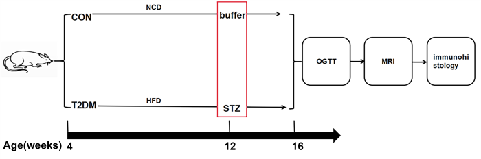
图1
实验流程图. CON:对照组大鼠;T2DM:二型糖尿病组大鼠;NCD:标准维持饲料;HFD:高脂饮食;STZ:链脲佐菌素;OGTT:口服葡萄糖耐量测试
Fig. 1
The pipeline of whole experiment. CON: the saline control group rats; T2DM: the T2DM group rats; NCD: normal chow diet; HFD: high-fat diet; STZ: streptozotocin; OGTT; oral glucose tolerance test
1.4 口服葡萄糖耐量测试
在大鼠16周龄时进行口服葡萄糖耐量实验(oral glucose tolerance test,OGTT).糖耐测试前大鼠禁食12 h.准备40%葡萄糖溶液.用采血笔刺破大鼠尾尖,取尾尖血滴入血糖试纸,使用血糖仪(鱼跃医疗)测量空腹初始血糖值(0 min)并记录.然后按照1.5 g/kg的剂量给大鼠灌胃配好的葡萄糖溶液,检测并记录大鼠灌胃后15、30、60、120 min的血糖值.以血糖值为纵坐标,时间为横坐标,画出折线图,并根据梯形法计算血糖折线图线下面积(area under curve,AUC).
1.5 MRI数据采集和数据分析
28只大鼠的MRI数据均在Bruker 7.0 T/20 cm成像仪(Ettlingen,Germany)上采集,直径为72 mm的体线圈用于发射射频脉冲,直径为40 mm的四通道正交表面线圈用于接收信号.实验过程中,使用与纯氧混合后浓度为1.5%~2.5%异氟烷的气体麻醉大鼠,使用热水循环系统保持大鼠体温为37 ℃,实时监控呼吸频率维持在约50次/分钟[18-
T2加权成像采用快速采集弛豫增强(rapid acquision with relaxation enhancement,RARE)序列,参数设置如下:视野大小(field of view,FOV)= 3.0 cm×3.0 cm,采集矩阵256×256,片厚0.6 mm,重复时间(time of repetition,TR)= 5 250 ms,有效回波时间(effective echo time,TEeff)= 36 ms,RARE因子是4,重复次数为4.
DTI采用6次激发平面回波成像序列,梯度为20个方向,FOV = 3.0 cm×3.0 cm,采集矩阵128×128,片厚0.8 mm,TR = 5 000 ms,TEeff = 26 ms,扩散梯度间隔时间(Δ)= 14 ms,扩散梯度持续时间(δ) = 3 ms,b = 0和800 s/mm2,累加次数为3.
逐个检查图像质量,剔除明显伪影的图像后进行下一步分析.采用基于体素形态学(voxel-based morphometry,VBM)的方法对T2加权的RARE图像进行分析,以评估大鼠脑白质结构的变化.图像处理采用SPM12(
采用基于体素的分析方法(voxel-based analysis, VBA)对DTI数据进行分析[21].首先,将原始的 Bruker数据转化为Nifti格式;其次,利用FSL中的FMRIB扩散工具箱(
1.6 免疫组化
MRI测试结束后,每组5只大鼠腹腔注射1%戊巴比妥钠麻醉后用0.9%的生理盐水和4%的多聚甲醛经心脏灌注,然后取脑固定.脑组织经过20%、30%的蔗糖溶液依次沉糖脱水后使用冰冻切片机(Cryostar NX50,Thermo Fisher)冠状切片,片厚为30 μm.脑片用0.01 mol/L的PBS洗涤3次,每次5 min,然后将脑片放入体积分数为10%的羊血清在37 ℃下封闭1.5 h.用于髓鞘碱性蛋白(myelin basic protein,MBP)染色的脑片与兔源抗MBP抗体(1 : 1 000稀释, Abcam, Cambridge, UK)在4 ℃下孵育过夜.然后用0.01 mol/L的PBS洗涤三次,每次5 min.用二抗羊抗兔Alexa Fluor 488 (1 : 1 000稀释, Abcam, Cambridge, UK)在37 ℃下孵育1 h.用于磷酸化神经丝(phosphorylated neurofilament,SMI-31)染色的脑片与小鼠源抗磷酸化神经丝SMI-31抗体(1 : 1 000稀释, Covance, New Jersey, USA)在4 ℃下孵育过夜.然后用0.01 mol/L的PBS洗涤三次,每次5 min.用二抗羊抗小鼠Alexa Fluor 594(1 : 1 000稀释, Abcam, Cambridge, UK)在37 ℃下孵育1 h.然后用PBS在黑暗中洗涤三次,每次5 min.贴片晾干封片,然后在玻片扫描显微镜下进行荧光显示成像,用Olympus VS120数字切片扫描系统进行拍照. 所有抗体均用PBS缓冲液进行稀释.
1.7 统计分析
数据以均值±标准误的形式表示,采用SPSS 20.0(SPSS Statistics,IBM)进行分析.采用重复测量方差分析体重、空腹血糖水平和口服糖耐量测试.采用双样本t检验对16周龄大鼠的体重和空腹血糖水平、MRI检测、免疫组化进行分析.用Benjamini-Yekutieli方法对多重比较的FDR进行校正.统计显著性水平设置为p < 0.05(FDR校正).
2 结果
2.1 大鼠体重和血糖变化
图2
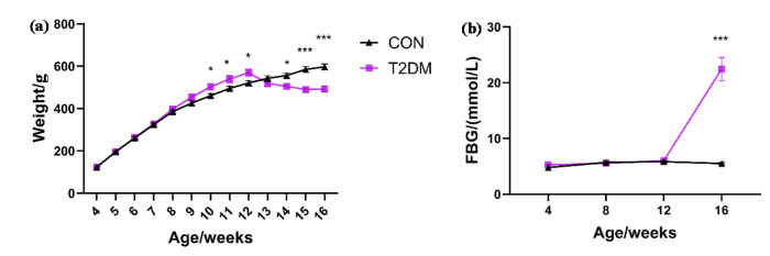
图2
大鼠体重和血糖的变化. (a) 4~16周龄两组大鼠体重变化;(b) 4~16周龄两组大鼠空腹血糖变化. *p < 0.05;***p < 0.001
Fig. 2
The changes in body weight and fasting blood glucose level in rats. (a) Changes in body weight of rats in the two groups from 4 to 16 weeks of age; (b) Changes in fasting blood glucose in the two groups of rats aged 4 to 16 weeks. *p < 0.05; ***p < 0.001
2.2 大鼠口服糖耐量测试结果
图3

图3
大鼠口服糖耐量测试. (a)两组大鼠OGTT测试血糖动态变化曲线;(b)两组大鼠OGTT变化曲线的曲线下面积,***p < 0.001
Fig. 3
Oral glucose tolerance test in rats. (a) The dynamic change curves of blood glucose in the OGTT test of the two groups of rats; (b) The areas under the OGTT change curves of the two groups of rats, with ***p < 0.001
2.3 磁共振影像结果
图4展示了T2DM大鼠4周后脑白质病变结果.采用VBM的方法对两组大鼠RARE图像分析发现T2DM大鼠纹状体的脑白质体积显著萎缩.进一步用VBA的方法对两组大鼠DTI结果进行分析发现:与CON组大鼠相比,T2DM大鼠在纹状体区域FA、MD、AD值显著降低,RD值显著升高.与CON组大鼠相比,T2DM大鼠在运动皮层(M1)和体感皮层(S1FL、S1HL)区域FA、MD、AD、RD值显著降低.对纹状体ROI(如图4中FA所示红色方框)进行DTI定量分析,相对于CON组,T2DM组FA显著降低[0.286±0.011(CON)vs. 0.200±0.009(T2DM), p<0.000 1];MD显著降低[(2.381±0.055)×10-3 mm2/s(CON)vs. (2.225±0.024) ×10-3 mm2/s(T2DM), p=0.025];AD显著降低[(0.972±0.022) ×10-3 mm2/s(CON)vs. (0.931±0.008) ×10-3 mm2/s(T2DM), p=0.025];RD显著增高[(0.627±0.012) ×10-3 mm2/s(CON)vs. (0.711± 0.006) ×10-3 mm2/s(T2DM), p<0.000 1].
图4
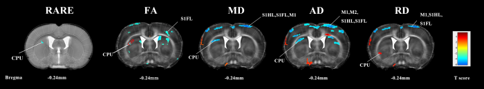
图4
二型糖尿病对大鼠脑白质微观结构的影响. CPU:尾状壳核(纹状体);M1:初级运动皮层;S1FL:初级体感皮层,前肢区域;S1HL:初级体感皮层,后肢区域. p<0.005(FDR校正),RARE的聚类大小为100,DTI的聚类大小为50
Fig. 4
Effect of T2DM on the microstructure of white matter in rats. CPU: caudate putamen (striatum), M1: primary motor cortex, S1FL: primary somatosensory cortex, forelimb area, S1HL: primary somatosensory cortex, hindlimb area. p<0.005 (FDR correction), voxel clusters of RARE =100, voxel clusters of DTI =50
2.4 免疫组化结果
图5
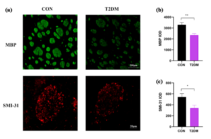
图5
大鼠纹状体免疫组化. (a)两组大鼠纹状体区域的MBP和SMI-31免疫组化染色,MBP免疫反应染色的比例尺为200 μm,SMI-31免疫反应染色的比例尺为20 μm;(b)纹状体MBP的IOD值;(c)纹状体SMI-31的IOD值. * p < 0.05;** p < 0.01
Fig. 5
Immunohistological staining of the striatum in rats. (a) Immunohistochemical staining of MBP and SMI-31 in the striatum region of the two groups of rats. The scale of MBP immunoresponse staining was 200 μm, and the scale of SMI-31 immunoresponse staining was 20 μm; (b) The IOD value of MBP in the striatum region; (c) The IOD value of SMI-31 in the striatum region. * p < 0.05; ** p < 0.01
3 讨论
本研究通过高脂饮食联合低剂量STZ注射成功构建了T2DM大鼠模型,并利用MRI技术结合免疫组化方法,系统探究了T2DM大鼠脑白质结构的改变及其病理机制.本研究发现与CON组相比,造模成功4周后T2DM组大鼠体重降低、空腹血糖升高、口服葡萄糖耐受能力受损,并且T2DM大鼠在纹状体区域表现出显著的白质体积萎缩和微观结构完整性破坏,表现为FA、MD和AD的降低,以及RD的升高.免疫组化结果进一步证实了纹状体区域的轴突损伤和脱髓鞘现象.下面将从模型构建、影像学发现、病理机制以及研究意义等方面展开讨论.
T2DM通常会导致脑萎缩,包括灰质和白质体积的减少[25].临床研究报道肥胖青少年T2DM患者在全脑白质体积明显萎缩[26],伴有轻度认知障碍的T2DM患者在额颞叶和边缘系统出现显著的白质体积萎缩[27]. VBM结果显示T2DM大鼠在纹状体区域白质体积萎缩,这与之前报道的T1DM大鼠纹状体白质体积减少的结果一致[28].在T2DM患者也发现丘脑内侧背侧和纹状体结构萎缩[29],海马到纹状体的功能连通性减少[30]. 纹状体作为基底神经节的重要组成部分,参与运动调节和认知功能,其结构异常可能与T2DM患者常见的运动障碍和认知功能下降有关[31].此外,纹状体也是已知的特别容易受到缺血性脑损伤的区域[32],纹状体主要由大脑动脉供血,对灌注压变化和缺血缺氧极为敏感.而糖尿病是导致缺血性脑卒中预后的主要不利原因[33],T2DM相关的微血管病变可导致脑血流自动调节能力受损和慢性低灌注,因此纹状体白质萎缩可能是糖尿病相关脑血管病变的早期表现之一.
T2DM与中枢神经系统和血管疾病密切相关,被认为是脑白质病变的危险因素[9],大多数T2DM表现为偏心率较高的融合性脑白质损伤[34],且比正常人有更多的脑白质损伤[9].DTI对白质微观结构的检测非常灵敏,在先前的文献中已有报道T2DM会破坏患者脑白质的完整性.大量研究表明T2DM存在广泛的白质异常,例如额叶[26]、颞叶[35]、胼胝体[36]、纹状体[12]等区域FA值显著降低,然而各项研究结果却不尽相同,不同研究的不一致性可能是样本量小、患者的人口统计学特征不同以及技术方法的多样性[37].我们的实验结果表明T2DM大鼠患病4周在纹状体区域出现了白质完整性改变,表现为FA、MD和AD值降低,RD值升高.纹状体FA的下降在T1DM大鼠上有相同的发现[5,15],并且在T2DM小鼠模型中也发现了背侧纹状体FA和MD值下降[38].缺血性脑卒中大鼠也报道了纹状体FA、AD显著下降,RD显著上升[39].
DTI指标的改变可能与轴突变性、髓磷脂破坏有关[40].FA值的减小与轴突损伤、脱髓鞘有关,AD值的减少与轴突损伤密切相关,RD值的增加与脱髓鞘损伤密切相关[41,42],AD和RD有望成为轴突损伤和髓鞘损伤的特异性标志物[42].SMI-31通常染色轴突精细网络,SMI-31神经丝免疫染色减少通常意味着轴突变性[5].髓鞘是由髓磷脂包绕而成的包裹在神经细胞轴突外面的同心圆结构的一层膜,MBP是中枢神经系统髓鞘的主要蛋白,位于髓鞘浆膜表面,维持中枢神经系统髓鞘结构和功能的稳定.MBP免疫染色减少通常意味着脱髓鞘,且在内侧胼胝体RD值与MBP荧光强度显著相关[43].患病4周T1DM大鼠纹状体、运动与体感皮层区域FA显著降低,并且通过Bielschowsky银染色、Klüver-Barrera染色、SMI-31免疫染色、亚甲基蓝染色等组织学染色验证了在这些脑区FA的降低与脱髓鞘、轴突退化和神经元功能障碍有关[5]. 视网膜缺血小鼠DTI结合组织学结果显示了AD和RD的改变分别是轴突和髓鞘损伤的标志[42].20周龄的糖尿病db/db小鼠胼胝体的FA下降,且快蓝染色显示出了脱髓鞘[17].高脂高糖饲养加STZ诱导的T2DM大鼠患病8周后在丘脑区域出现显著的FA下降,少突细胞转录因子和MBP染色说明了T2DM在丘脑区域出现少突胶质细胞损伤和脱髓鞘,解释了其脑白质的病理变化[16].在我们实验中,SMI-31和MBP免疫染色表达显著降低证实了T2DM大鼠在纹状体区域出现了轴突损伤和脱髓鞘,这些病理改变可能是T2DM大鼠纹状体区域DTI指标改变的生物学基础.
运动与体感皮层区域出现FA、MD、AD、RD值降低,可能还涉及细胞毒性水肿、髓鞘肿胀等水分子在所有方向的扩散受限的情况.皮质中的水扩散各向异性主要源于锥形神经元以及轴突的密集排列[44]. 曾有报道患病4周的T1DM大鼠运动/体感皮层区域FA显著降低,且该区域的椎体神经元出现髓鞘完整性受损、树突受损和线粒体异常[5],因此我们猜测T2DM大鼠运动与体感皮层区域FA、MD、AD的降低也与该区域轴突、神经元损伤有关.然而髓鞘损伤通常导致RD升高,而我们实验中T2DM大鼠运动与体感皮层RD却是降低的.重度弥散性轴索损伤的新西兰大白兔脑棋型中,白质RD出现显著降低[45],在辐射损伤的早期模型中也观察到了短暂的RD下降[46].此外在视网膜缺血小鼠模型中也发现了AD值降低、RD值呈相似下降趋势,提示该类损伤仅引发神经轴突退化,未造成髓鞘损伤[42].这种现象被认为是多种原因造成的,包括微管排列紊乱、轴突结构分解产生的细胞碎片、轴突运输受损以及轴突损伤引起的细胞肿胀导致的细胞外水分子向细胞内空间的转移[46].关于T2DM大鼠运动与体感皮层区域出现FA、MD、AD、RD值降低的具体生理机制需进一步探索.
4 结论
本研究利用MRI技术检测了T2DM大鼠模型脑白质结构变化,发现T2DM大鼠纹状体白质显著萎缩,并且免疫组化检测到其微观结构的完整性发生显著改变,可能与轴突损伤、脱髓鞘有关.DTI研究为糖尿病脑白质损伤的早期检测和评估提供了有效工具,未来研究可进一步探索干预措施(如胰岛素增敏剂或抗氧化治疗)对白质损伤的保护作用,为临床防治糖尿病脑病提供理论依据.
致谢
中国科学院精密测量科学与技术创新研究院的方芳、安萍萍对本文动物饲养、行为学实验提供了帮助和建议,在此表示感谢.
利益冲突
无
参考文献
Type 2 diabetes
[J].
DOI:10.1016/S0140-6736(22)01655-5
PMID:36332637
[本文引用: 1]

Type 2 diabetes accounts for nearly 90% of the approximately 537 million cases of diabetes worldwide. The number affected is increasing rapidly with alarming trends in children and young adults (up to age 40 years). Early detection and proactive management are crucial for prevention and mitigation of microvascular and macrovascular complications and mortality burden. Access to novel therapies improves person-centred outcomes beyond glycaemic control. Precision medicine, including multiomics and pharmacogenomics, hold promise to enhance understanding of disease heterogeneity, leading to targeted therapies. Technology might improve outcomes, but its potential is yet to be realised. Despite advances, substantial barriers to changing the course of the epidemic remain. This Seminar offers a clinically focused review of the recent developments in type 2 diabetes care including controversies and future directions.Copyright © 2022 Elsevier Ltd. All rights reserved.
IDF diabetes atlas: Global estimates of undiagnosed diabetes in adults for 2021
[J].DOI:10.1016/j.diabres.2021.109118 URL [本文引用: 1]
Global aetiology and epidemiology of type 2 diabetes mellitus and its complications
[J].
DOI:10.1038/nrendo.2017.151
PMID:29219149
[本文引用: 1]
Globally, the number of people with diabetes mellitus has quadrupled in the past three decades, and diabetes mellitus is the ninth major cause of death. About 1 in 11 adults worldwide now have diabetes mellitus, 90% of whom have type 2 diabetes mellitus (T2DM). Asia is a major area of the rapidly emerging T2DM global epidemic, with China and India the top two epicentres. Although genetic predisposition partly determines individual susceptibility to T2DM, an unhealthy diet and a sedentary lifestyle are important drivers of the current global epidemic; early developmental factors (such as intrauterine exposures) also have a role in susceptibility to T2DM later in life. Many cases of T2DM could be prevented with lifestyle changes, including maintaining a healthy body weight, consuming a healthy diet, staying physically active, not smoking and drinking alcohol in moderation. Most patients with T2DM have at least one complication, and cardiovascular complications are the leading cause of morbidity and mortality in these patients. This Review provides an updated view of the global epidemiology of T2DM, as well as dietary, lifestyle and other risk factors for T2DM and its complications.
The burden and risks of emerging complications of diabetes mellitus
[J].DOI:10.1038/s41574-022-00690-7 [本文引用: 1]
Abnormalities in the brain of streptozotocin-induced type 1 diabetic rats revealed by diffusion tensor imaging
[J].DOI:10.1016/j.nicl.2012.09.004 URL [本文引用: 8]
Association between type 2 diabetes mellitus and brain atrophy: a meta-analysis
[J].
DOI:10.4093/dmj.2021.0189
PMID:35255549
[本文引用: 1]
Type 2 diabetes mellitus (T2DM) is known to be associated with cognitive decline and brain structural changes. This study systematically reviews and estimates human brain volumetric differences and atrophy associated with T2DM. PubMed, PsycInfo, and Cochrane Library were searched for brain imaging studies reporting on brain volume differences between individuals with T2DM and healthy controls. Data were examined using meta-analysis, and association between age, sex, diabetes characteristics and brain volumes were tested using meta-regression. A total of 14,605 entries were identified; after title, abstract and full-text screening applying inclusion and exclusion criteria, 64 studies were included and 42 studies with compatible data contributed to the meta-analysis (n=31,630; mean age 71.0 years; 44.4% male, 26,942 control, 4,688 diabetes). Individuals with T2DM had significantly smaller total brain volume, total grey matter volume, total white matter volume and hippocampal volume (approximately 1% to 4%); meta-analyses of smaller samples focusing on other brain regions and brain atrophy rate in longitudinal investigations also indicated smaller brain volumes and greater brain atrophy associated with T2DM. Meta-regression suggests that diabetes-related brain volume differences start occurring in early adulthood, decreases with age and increases with diabetes duration. T2DM is associated with smaller total and regional brain volume and greater atrophy over time. These effects are substantial and highlight an urgent need to develop interventions to reduce the risk of T2DM for brain health.
Gray and white matter abnormality in patients with T2DM-related cognitive dysfunction: a systemic review and meta-analysis
[J].
DOI:10.1038/s41387-022-00214-2
PMID:35970833
[本文引用: 1]
Brain structure abnormality in patients with type 2 diabetes mellitus (T2DM)-related cognitive dysfunction (T2DM-CD) has been reported for decades in magnetic resonance imaging (MRI) studies. However, the reliable results were still unclear. This study aimed to make a systemic review and meta-analysis to find the significant and consistent gray matter (GM) and white matter (WM) alterations in patients with T2DM-CD by comparing with the healthy controls (HCs).Published studies were systemically searched from PubMed, MEDLINE, Cochrane Library and Web of Science databases updated to November 14, 2021. Studies reporting abnormal GM or WM between patients with T2DM-CD and HCs were selected, and their significant peak coordinates (x, y, z) and effect sizes (z-score or t-value) were extracted to perform a voxel-based meta-analysis by anisotropic effect size-signed differential mapping (AES-SDM) 5.15 software.Total 15 studies and 16 datasets (1550 participants) from 7531 results were involved in this study. Compared to HCs, patients with T2DM-CD showed significant and consistent decreased GM in right superior frontal gyrus, medial orbital (PFCventmed. R, BA 11), left superior temporal gyrus (STG. L, BA 48), and right calcarine fissure / surrounding cortex (CAL. R, BA 17), as well as decreased fractional anisotropy (FA) in right inferior network, inferior fronto-occipital fasciculus (IFOF. R), right inferior network, longitudinal fasciculus (ILF. R), and undefined area (32, -60, -42) of cerebellum. Meta-regression showed the positive relationship between decreased GM in PFCventmed.R and MoCA score, the positive relationship between decreased GM in STG.L and BMI, as well as the positive relationship between the decreased FA in IFOF.R and age or BMI.T2DM impairs the cognitive function by affecting the specific brain structures. GM atrophy in PFCventmed. R (BA 11), STG. L (BA 48), and CAL. R (BA 17), as well as WM injury in IFOF. R, ILF. R, and undefined area (32, -60, -42) of cerebellum. And those brain regions may be valuable targets for future researches. Age, BMI, and MoCA score have a potential influence on the altered GM or WM in T2DM-CD.© 2022. The Author(s).
Structural characteristics of amygdala subregions in type 2 diabetes mellitus
[J].DOI:10.1016/j.bbr.2024.114992 URL [本文引用: 1]
A narrative review of multimodal imaging of white matter lesions in type-2 diabetes mellitus
[J].
DOI:10.21037/apm-21-3299
PMID:35016461
[本文引用: 3]
To discuss the relevant studies about the structural and functional changes of the brain in patients with type-2 DM (T2DM) and white matter lesion (WML) in recent years, and to summarize them.T2DM is a common metabolic disease with increasing prevalence worldwide. This disease is closely related to central nervous system and vascular disease, and is considered a risk factor for white matter lesions in the brain. Compared to healthy individuals, WML patients with T2DM exhibit changes in brain perfusion, functional networks, nerve fiber structure, and brain tissue metabolism.We analyzed recent studies related to structural and functional changes in the brain of patients suffering from T2DM and WML and summarized them.Multimodal magnetic resonance imaging (MRI) utilizes noninvasive and sensitive imaging techniques to provide multiparametric information in patients with T2DM to help in clinical practice. It features non-invasively and with high sensitivity assess the histomorphological and functional abnormalities of white matter in patients with T2DM using various parameters. We can use multimodal MRI methods to reflect the microscopic damage of neuromyelin structures and pathological changes of neuronal metabolic functions in WML in T2DM patients, and thus speculate the disease progression. This approach can be helpful for the early diagnosis and treatment of patients with such a disease who do not exhibit neurological deficit, to effectively improve their prognosis.
Diffusion tensor imaging and beyond
[J].DOI:10.1002/mrm.v65.6 URL [本文引用: 1]
Investigating brain microstructural alterations in type 1 and type 2 diabetes using diffusion tensor imaging: a systematic review
[J].
DOI:10.3390/brainsci11020140
URL
[本文引用: 1]
Type 1 and type 2 diabetes mellitus have an impact on the microstructural environment and cognitive functions of the brain due to its microvascular/macrovascular complications. Conventional Magnetic Resonance Imaging (MRI) techniques can allow detection of brain volume reduction in people with diabetes. However, conventional MRI is insufficiently sensitive to quantify microstructural changes. Diffusion Tensor Imaging (DTI) has been used as a sensitive MRI-based technique for quantifying and assessing brain microstructural abnormalities in patients with diabetes. This systematic review aims to summarise the original research literature using DTI to quantify microstructural alterations in diabetes and the relation of such changes to cognitive status and metabolic profile. A total of thirty-eight published studies that demonstrate the impact of diabetes mellitus on brain microstructure using DTI are included, and these demonstrate that both type 1 diabetes mellitus and type 2 diabetes mellitus may affect cognitive abilities due to the alterations in brain microstructures.
Brain microstructural alterations in type 2 diabetes: diffusion kurtosis imaging provides added value to diffusion tensor imaging
[J].
DOI:10.1007/s00330-018-5746-y
PMID:30338363
[本文引用: 3]
To investigate brain microstructural changes in white matter and gray matter of type 2 diabetes mellitus (T2DM) patients using diffusion kurtosis imaging.Diffusion kurtosis imaging (b values = 0, 1250, and 2500 s/mm) was performed for 30 T2DM patients and 28 controls. FMRIB Software Library with tract-based spatial statistics was used to analyze intergroup differences in fractional anisotropy (FA), mean diffusivity (MD), mean kurtosis (MK), axial kurtosis (K), and radial kurtosis (K) of multiple white matter regions. Atlas-based ROI analysis was conducted in gray matter structures and some fiber tracts. Correlations between MK changes and clinical measurements were determined.In whole-brain tract-based spatial statistics analysis, T2DM patients exhibited abnormalities in 29.6%, 30.4%, 35.4%, 10.5%, and 26.0% of white matter regions as measured by FA, MD, MK, K, and K, respectively, when compared to the controls. MK reduction was contributed more by the decreased K. In atlas-based analysis, MK detected more ROIs (27/48) with white matter microstructural changes than FA (13/48) and MD (17/48). MK decreased in bilateral thalamus and caudate, while FA showed statistically significant difference only in the left caudate. MK values negatively correlated with disease duration in the genu of corpus callosum and anterior corona radiata (R = -0.512 and -0.459) and positively correlated with neuropsychological scores in the cingulum (hippocampus) (R = 0.466 and 0.440).Diffusion kurtosis imaging detects more brain regions with white matter and gray matter microstructural alterations of T2DM patients than DTI metrics. It provides valuable information for studying the pathology of diabetic encephalopathy and may lead to better imaging biomarkers for monitoring disease progression.• Diffusion kurtosis imaging detects more brain regions with microstructural alterations in white matter and gray matter of T2DM patients than DTI. • Mean kurtosis changes are associated with disease severity and impaired neuropsychological function in T2DM. • Diffusion kurtosis imaging demonstrates potential to assess cognitive impairment in T2DM patients and predict disease progression.
Abnormalities of brain white matter in type 2 diabetes mellitus: a meta-analysis of diffusion tensor imaging
[J].
DOI:10.3389/fnagi.2021.693890
URL
[本文引用: 1]
Aims: The study aimed to conduct a meta-analysis to determine the abnormalities of white matter in patients with type 2 diabetes mellitus (T2DM) by identifying the consistency of diffusion tensor imaging (DTI).
Micro-structural white matter abnormalities in type 2 diabetic patients: a DTI study using TBSS analysis
[J].Patients with type 2 diabetes mellitus (T2DM) have usually been found cognitive impairment associated with brain white matter (WM) abnormalities. However, findings have varied across studies, and any potential relationship with Alzheimer's disease (AD) remains unclear. The aim of this study was to assess the whole-brain WM integrity of T2DM patients and to compare our findings with those of published AD cases.In this study, we used diffusion tensor imaging (DTI) combined with tract-based spatial statistics (TBSS) to investigate whole-brain WM abnormalities in 48 T2DM patients and 48 healthy controls. The effects of age and gender were also evaluated.In our study, significantly decreasing FA and increasing MD and DA values (P<0.05) were found in some WM regions closely related to the default mode network (DMN), including cingulum, the right frontal lobe involving the right uncinate fasciculus (UF), bilateral parietal lobes involving the superior longitudinal fasciculus (SLF) and the inferior longitudinal fasciculus (ILF), and the right middle temporal gyrus (MTG) involving the UF and the ILF. We also found abnormalities in the thalamus involving the fornix (FX), anterior thalamic radiation (ATR), and posterior thalamic radiation (PTR). The damaged regions above are similar to those found in patients with AD, as reported in previous studies.The present study not only provides useful information about the WM regions and tracts affected by T2DM but also offers insight into the underlying neuropathological process in T2DM patients and the relationship between T2DM and AD.
Reproducibility of diffusion tensor imaging-derived parameters: implications for the streptozotocin-induced type 1 diabetic rats
[J].DOI:10.1007/s10334-022-01048-w [本文引用: 3]
Presence of white matter lesions associated with diabetes-associated cognitive decline in male rat models of pre-type 2 diabetes
[J].DOI:10.12659/MSM.918557 URL [本文引用: 2]
Magnetic resonance imaging detects cerebral gray and white matter injury correlated with cognitive impairments in diabetic db/db mice
[J].DOI:10.1016/j.bbr.2023.114510 URL [本文引用: 2]
Magnetic resonance imaging the brain structures involved in nicotine susceptibility in rats
[J].
尼古丁易感的脑结构特征的磁共振成像研究
[J].
DOI:10.11938/cjmr20212890
[本文引用: 1]
本文旨在利用磁共振成像手段探究尼古丁易感个体的脑结构特征,即脑结构特性对尼古丁依赖程度的预测.选用成年雄性SD大鼠进行纵向研究,利用基于微型渗透压泵的间歇性给药方式对大鼠进行腹腔注射尼古丁14天,随后强制戒断14天.于第0、15、29天进行躯体戒断行为测试以量化其尼古丁依赖严重程度.对第1天的脑结构图像与戒断行为评分进行回归分析,结果发现尼古丁依赖严重程度与双侧前边缘皮层、左侧颗粒状岛叶皮层灰质体积和双侧丘脑白质体积呈负相关,与右侧海马CA1脑区和左侧丘脑灰质体积呈正相关.以上脑区的结构特征,能够作为尼古丁易感的生物标志物,在个体接触尼古丁之前预测其尼古丁依赖风险,对易感人群进行有针对性的早期干预.
Effects of seizure-inducing doses nicotine on hippocampal structure in adolescent female rats
[J].
致痫剂量尼古丁对青少年雌性大鼠海马结构的影响
[J].
STZ-induced progressive brain atrophy studied by magnetic resonance imaging and histochemical staining
[J].
STZ诱导大鼠1型糖尿病进行性脑萎缩的磁共振成像及组织化学研究
[J].
DOI:10.11938/cjmr20150305
[本文引用: 1]
1型糖尿病(T1DM)是一种慢性代谢疾病,主要表现为胰岛素分泌量较正常情况下降,会对人体的多个器官和系统造成持续性的损伤.关于糖尿病的横向研究发现糖尿病患者相比于正常人存在着显著的脑萎缩,但关于糖尿病引起的脑萎缩随时间发生进行性改变的研究比较少见.实验采用腹腔注射链脲佐菌素(STZ)来诱导建立大鼠的1型糖尿病模型,运用磁共振成像(MRI)的方法对萎缩的脑区进行定位并在造模后12周和20周两个时间点对脑萎缩的程度进行对比分析,然后运用组织化学染色的方法观察在MRI上出现进行性萎缩的脑区中的神经元所发生的病理改变.MRI的结果表明:STZ诱导的T1DM大鼠相比于正常对照组大鼠出现了显著性的全脑体积、灰质体积和白质体积的萎缩,并且在多个白质脑区和灰质脑区均出现了萎缩程度随着病程的延长而逐渐加重.组织化学染色的结果发现,STZ诱导的T1DM大鼠相对于正常对照组大鼠在体感皮层、运动皮层和海马CA3区,均出现明显的神经元萎缩现象.
Brain structural plasticity in rats subjected to early binocular enucleation characterized by high resolution anatomical magnetic resonance imaging and diffusion tensor imaging
[J].
DOI:10.1016/j.mrl.2022.10.001
PMID:40919275
[本文引用: 1]
Visual deprivation leads to structural neuroplasticity in the blind subjects, including gray matter (GM) and white matter (WM) atrophy and alterations in structural connectivity. The rat model of binocular enucleation (BE) is a frequently used animal model for studying brain plasticity induced by early blindness. Yet few neuroimaging studies have been performed on this model to investigate whether or not the BE rats have image phenotypes similar to or comparable to, those observed in the early blind subjects. The current study aimed to assess brain structural plasticity in BE rats using anatomical magnetic resonance imaging (MRI) and diffusion tensor imaging (DTI). The results demonstrated that early BE at postnatal day 4 (P4) caused almost complete degeneration of optic nerve (ON) and optic chiasma (OCH), atrophy in a number of visual and non-visual structures, including optic tract (OT), dorsal lateral geniculate nucleus (DLG) and corpus callosum (CC). The BE rats also exhibited impairments of WM microstructural integrity in the OT, and reduction of structural connectivity between the normal-appearing visual cortex (VC) and somatosensory/motor cortices at 4 months of age, likely as manifestations of deafferentation-induced maldevelopment. The structural neuroplasticity in BE rats observable to structural MRI parallels largely with what has been reported in blind subjects, suggesting that longitudinal neuroimaging studies on animal models of sensory deprivation can provide insights into how the brain changes its wiring and function during development/adaption in response to the lack of sensory stimuli.© 2022 The Authors.
High-fat diet impairs the effects of a single bout of endurance exercise on glucose transport and insulin sensitivity in rat skeletal muscle
[J].DOI:10.1016/j.metabol.2007.07.017 URL [本文引用: 1]
Combination of high-fat diet-fed and low-dose streptozotocin-treated rat: a model for type 2 diabetes and pharmacological screening
[J].
DOI:10.1016/j.phrs.2005.05.004
PMID:15979893
[本文引用: 1]
The objective of the present study was to develop a rat model that replicates the natural history and metabolic characteristics of human type 2 diabetes and is also suitable for pharmacological screening. Male Sprague-Dawley rats (160-180 g) were divided into two groups and fed with commercially available normal pellet diet (NPD) (12% calories as fat) or in-house prepared high-fat diet (HFD) (58% calories as fat), respectively, for a period of 2 weeks. The HFD-fed rats exhibited significant increase in body weight, basal plasma glucose (PGL), insulin (PI), triglycerides (PTG) and total cholesterol (PTC) levels as compared to NPD-fed control rats. Besides, the HFD rats showed significant reduction in glucose disappearance rate (K-value) on intravenous insulin glucose tolerance test (IVIGTT). Hyperinsulinemia together with reduced glucose disappearance rate (K-value) suggested that the feeding of HFD-induced insulin resistance in rats. After 2 weeks of dietary manipulation, a subset of the rats from both groups was injected intraperitoneally with low dose of streptozotocin (STZ) (35 mg kg(-1)). Insulin-resistant HFD-fed rats developed frank hyperglycemia upon STZ injection that, however, caused only mild elevation in PGL in NPD-fed rats. Though there was significant reduction in PI level after STZ injection in HFD rats, the reduction observed was only to a level that was comparable with NPD-fed control rats. In addition, the levels of PTG and PTC were further accentuated after STZ treatment in HFD-fed rats. In contrast, STZ (35 mg kg(-1), i.p.) failed to significantly alter PI, PTG and PTC levels in NPD-fed rats. Thus, these fat-fed/STZ-treated rats simulate natural disease progression and metabolic characteristics typical of individuals at increased risk of developing type 2 diabetes because of insulin resistance and obesity. Further, the fat-fed/STZ-treated rats were found to be sensitive for glucose lowering effects of insulin sensitizing (pioglitazone) as well as insulinotropic (glipizide) agents. Besides, the effect of pioglitazone and glipizide on the plasma lipid parameters (PTG and PTC) was shown in these diabetic rats. The present study represents that the combination of HFD-fed and low-dose STZ-treated rat serves as an alternative animal model for type 2 diabetes simulating the human syndrome that is also suitable for testing anti-diabetic agents for the treatment of type 2 diabetes.
Retinopathy in a diet-induced type 2 diabetic rat model and role of epigenetic modifications
[J].
DOI:10.2337/db19-1009
PMID:31949005
[本文引用: 1]
Type 2 diabetes accounts for 90% of the population with diabetes, and these patients are generally obese and hyperlipidemic. In addition to hyperglycemia, hyperlipidemia is also closely related with diabetic retinopathy. The aim was to investigate retinopathy in a model closely mimicking the normal progression and metabolic features of the population with type 2 diabetes and elucidate the molecular mechanism. Retinopathy was evaluated in rats fed a 45% kcal as fat diet for 8 weeks before administering streptozotocin, 30 mg/kg body weight (T2D), and compared with age- and duration-matched type 1 diabetic rats (T1D) (60 mg/kg streptozotocin). The role of epigenetic modifications in mitochondrial damage was evaluated in retinal microvasculature. T2D rats were obese and severely hyperlipidemic, with impaired glucose and insulin tolerance compared with age-matched T1D rats. While at 4 months of diabetes, T1D rats had no detectable retinopathy, T2D rats had significant retinopathy, their mitochondrial copy numbers were lower, and mtDNA and promoter DNA methylation was exacerbated. At 6 months, retinopathy was comparable in T2D and T1D rats, suggesting that obesity exaggerates hyperglycemia-induced epigenetic modifications, accelerating mitochondrial damage and diabetic retinopathy. Thus, maintenance of good lifestyle and BMI could be beneficial in regulating epigenetic modifications and preventing/retarding retinopathy in patients with diabetes.© 2020 by the American Diabetes Association.
Brain atrophy in type 2 diabetes: regional distribution and influence on cognition
[J].
DOI:10.2337/dc13-0143
PMID:23939539
[本文引用: 1]
Type 2 diabetes (T2DM) is associated with brain atrophy and cerebrovascular disease. We aimed to define the regional distribution of brain atrophy in T2DM and to examine whether atrophy or cerebrovascular lesions are feasible links between T2DM and cognitive function.This cross-sectional study used magnetic resonance imaging (MRI) scans and cognitive tests in 350 participants with T2DM and 363 participants without T2DM. With voxel-based morphometry, we studied the regional distribution of atrophy in T2DM. We measured cerebrovascular lesions (infarcts, microbleeds, and white matter hyperintensity [WMH] volume) and atrophy (gray matter, white matter, and hippocampal volumes) while blinded to T2DM status. With use of multivariable regression, we examined for mediation or effect modification of the association between T2DM and cognitive measures by MRI measures.T2DM was associated with more cerebral infarcts and lower total gray, white, and hippocampal volumes (all P < 0.05) but not with microbleeds or WMH. T2DM-related gray matter loss was distributed mainly in medial temporal, anterior cingulate, and medial frontal lobes, and white matter loss was distributed in frontal and temporal regions. T2DM was associated with poorer visuospatial construction, planning, visual memory, and speed (P ≤ 0.05) independent of age, sex, education, and vascular risk factors. The strength of these associations was attenuated by almost one-half when adjusted for hippocampal and total gray volumes but was unchanged by adjustment for cerebrovascular lesions or white matter volume.Cortical atrophy in T2DM resembles patterns seen in preclinical Alzheimer disease. Neurodegeneration rather than cerebrovascular lesions may play a key role in T2DM-related cognitive impairment.
Preliminary evidence for brain complications in obese adolescents with type 2 diabetes mellitus
[J].
DOI:10.1007/s00125-010-1857-y
PMID:20668831
[本文引用: 2]
Central nervous system abnormalities, including cognitive and brain impairments, have been documented in adults with type 2 diabetes who also have multiple co-morbid disorders that could contribute to these observations. Assessing adolescents with type 2 diabetes will allow the evaluation of whether diabetes per se may adversely affect brain function and structure years before clinically significant vascular disease develops.Eighteen obese adolescents with type 2 diabetes and 18 obese controls without evidence of marked insulin resistance, matched on age, sex, school grade, ethnicity, socioeconomic status, body mass index and waist circumference, completed MRI and neuropsychological evaluations.Adolescents with type 2 diabetes performed consistently worse in all cognitive domains assessed, with the difference reaching statistical significance for estimated intellectual functioning, verbal memory and psychomotor efficiency. There were statistical trends for executive function, reading and spelling. MRI-based automated brain structural analyses revealed both reduced white matter volume and enlarged cerebrospinal fluid space in the whole brain and the frontal lobe in particular, but there was no obvious grey matter volume reduction. In addition, assessments using diffusion tensor imaging revealed reduced white and grey matter microstructural integrity.This is the first report documenting possible brain abnormalities among obese adolescents with type 2 diabetes relative to obese adolescent controls. These abnormalities are not likely to result from education or socioeconomic bias and may result from a combination of subtle vascular changes, glucose and lipid metabolism abnormalities and subtle differences in adiposity in the absence of clinically significant vascular disease. Future efforts are needed to elucidate the underlying pathophysiological mechanisms.
White matter atrophy in type 2 diabetes mellitus patients with mild cognitive impairment
[J].
DOI:10.3389/fnins.2020.602501
URL
[本文引用: 1]
Type 2 diabetes mellitus (T2DM) patients are highly susceptible to developing dementia, especially for those with mild cognitive impairment (MCI), but its underlying cause is still unclear. In this study, we performed a battery of neuropsychological tests and high-resolution sagittal T1-weighted structural imaging to explore how T2DM affects white matter volume (WMV) and cognition in 30 T2DM-MCI patients, 30 T2DM with normal cognition (T2DM-NC) patients, and 30 age-, sex-, and education-matched healthy control (HC) individuals. The WMV of the whole brain was obtained with automated segmentation methods. Correlations between the WMV of each brain region and neuropsychological tests were analyzed in the T2DM patients. The T2DM-NC patients and HC individuals did not reveal any significant differences in WMV. Compared with the T2DM-NC group, the T2DM-MCI group showed statistically significant reduction in the WMV of seven brain regions, mainly located in the frontotemporal lobe and limbic system, five of which significantly correlated with Montreal Cognitive Assessment (MoCA) scores. Subsequently, we evaluated the discriminative ability of these five regions for MCI in T2DM patients. The WMV of four regions, including left posterior cingulate, precuneus, insula, and right rostral middle frontal gyrus had high diagnostic value for MCI detection in T2DM patients (AUC &gt; 0.7). Among these four regions, left precuneus WMV presented the best diagnostic value (AUC: 0.736; sensitivity: 70.00%; specificity: 73.33%; Youden index: 0.4333), but with no significant difference relative to the minimum AUC. In conclusion, T2DM could give rise to the white matter atrophy of several brain regions. Each WMV of left posterior cingulate, precuneus, insula, and right rostral middle frontal gyrus could be an independent imaging biomarker to detect cognitive impairment at the early stage in T2DM patients and play an important role in its pathophysiological mechanism.
Voxel-based morphology analysis of STZ-induced type 1 diabetes mellitus rats with and without cognitive impairment
[J].
DOI:S0304-3940(18)30555-X
PMID:30125641
[本文引用: 1]
Incidence of diabetes has increased dramatically. Consequently, diabetes-induced cognitive impairment has attracted increasing attention. This study aimed to explore the changes in brain structure in the diabetic rats with and without cognitive impairment. Morris water maze method was used for screening the diabetic rats with/without cognitive impairment. These diabetic rats and controls were imaged using magnetic resonance imaging that segmented into gray and white matter, which was further analyzed using voxel-based morphology (VBM) and regions of interest (ROI) based image retrieval. The ROI results showed that the whole brain volume decreased in diabetic rats with/without cognitive impairment as compared to the control (P < 0.05). The VBM results showed differences in the caudate putamen and prefrontal cortex in the diabetic rats with/without cognitive impairment. The change in the brain of rats with cognitive impairment occurred primarily in the area associated with cognition, such as caudate putamen and hippocampus, and the bi-directional change occurred in the different area of hippocampus. The current results provided important imaging information for early diagnosis and timely treatment of diabetic cognitive impairment.Copyright © 2018 Elsevier B.V. All rights reserved.
Abnormal subcortical nuclei shapes in patients with type 2 diabetes mellitus
[J].
DOI:10.1007/s00330-017-4790-3
PMID:28374074
[本文引用: 1]
Type 2 diabetes mellitus (T2DM) increases the risk of brain atrophy and dementia. We aimed to elucidate deep grey matter (GM) structural abnormalities and their relationships with T2DM cognitive deficits by combining region of interest (ROI)-based volumetry, voxel-based morphometry (VBM) and shape analysis.We recruited 23 T2DM patients and 24 age-matched healthy controls to undergo T1-weighted structural MRI scanning. Images were analysed using the three aforementioned methods to obtain deep GM structural shapes and volumes. Biochemical and cognitive assessments were made and were correlated with the resulting metrics.Shape analysis revealed that T2DM is associated with focal atrophy in the bilateral caudate head and dorso-medial part of the thalamus. ROI-based volumetry only detected thalamic volume reduction in T2DM when compared to the controls. No significant between-group differences were found by VBM. Furthermore, a worse performance of cognitive processing speed correlated with more severe GM atrophy in the bilateral dorso-medial part of the thalamus. Also, the GM volume in the bilateral dorso-medial part of the thalamus changed negatively with HbA.Shape analysis is sensitive in identifying T2DM deep GM structural abnormalities and their relationships with cognitive impairments, which may greatly assist in clarifying the neural substrate of T2DM cognitive dysfunction.• Type 2 diabetes mellitus is accompanied with brain atrophy and cognitive dysfunction • Deep grey matter structures are essential for multiple cognitive processes • Shape analysis revealed local atrophy in the dorso-medial thalamus and caudatum in patients • Dorso-medial thalamic atrophy correlated to cognitive processing speed slowing and high HbA1c. • Shape analysis has advantages in unraveling neural substrates of diabetic cognitive deficits.
Alterations of white matter integrity and hippocampal functional connectivity in type 2 diabetes without mild cognitive impairment
[J].DOI:10.3389/fnana.2018.00021 URL [本文引用: 1]
Diabetic striatal disease: clinical presentation, neuroimaging, and pathology
[J].DOI:10.2169/internalmedicine.48.1996 URL [本文引用: 1]
Temporal profile of magnetic resonance imaging changes following forebrain ischemia in the gerbil
[J].Quantitative T2 magnetic resonance (MR) imaging was used to examine gerbil brains 1, 3, 10, and 30 days after 5 min forebrain ischemia. T2 was increased in the dorsal-lateral striatum 1 and 3 days post-ischemia, and in the hippocampus 3 days post-ischemia. T2 was normal 10 days post-ischemia, and decreased in the hippocampus and dorsal-lateral striatum 30 days post-ischemia. Neuronal counts in the dorsal-lateral striatum and CA1 hippocampal region were uniformly decreased 30 days post-ischemia. The increase in T2 shortly after ischemia is attributed to brain edema localized to regions where neuronal injury developed. The late decrease in T2 may be due to decreased water in gliotic tissue, or to ferritin-positive microglia, following forebrain ischemia. Tissue atrophy at later times gave enlarged ventricles on MR images.
STZ-induced diabetes exacerbates neurons ferroptosis after ischemic stroke by upregulating LCN2 in neutrophils
[J].DOI:10.1016/j.expneurol.2024.114797 URL [本文引用: 1]
White matter hyperintensity shape and location feature analysis on brain MRI; proof of principle study in patients with diabetes
[J].
DOI:10.1038/s41598-018-20084-y
PMID:29382936
[本文引用: 1]
Cerebral small vessel disease is a heterogeneous disease in which various underlying etiologies can lead to different types of white matter hyperintensities (WMH). WMH shape features might aid in distinguishing these different types. In this proof of principle study in patients with type 2 diabetes mellitus (T2DM), we present a novel approach to assess WMH using shape features. Our algorithm determines WMH volume and different WMH shape and location features on 3T MRI scans. These features were compared between patients with T2DM (n = 60) and a matched control group (n = 54). Although a more traditional marker (WMH volume) was not significantly different between groups (natural log transformed Beta (95% CI): 0.07 (-0.11 <-> 0.24)), patients with T2DM showed a larger number of non-punctuate WMH (median (10th-90th percentile), patients: 40 lesions per person (16-86); controls: 26 (5-58)) and a different shape (eccentricity) of punctuate deep WMH (Beta (95% CI): 0.40 (0.23 <-> 0.58)) compared to controls. In conclusion, our algorithm identified WMH features that are not part of traditional WMH assessment, but showed to be distinguishing features between patients with T2DM and controls. Future studies could address these features to further unravel the etiology and functional impact of WMH.
Nepsilon-(carboxymethyl)-lysine, white matter, and cognitive function in diabetes patients
[J].
DOI:10.1017/cjn.2015.398
URL
[本文引用: 1]
Objective: To study the relationship of Nε-(carboxymethyl)-lysine level (CML) with microstructure changes of white matter (WM), and cognitive impairment in patients with type 2 diabetes mellitus (T2DM) and to discuss the potential mechanism underlying T2DM-associated cognitive impairment. Methods: The study was performed in T2DM patients (n=22) with disease course ≥5 years and age ranging from 65 to 75 years old. A control group consisted of 25 sex- and age-matched healthy volunteers. Fractional anisotropy (FA) of several WM regions was analyzed by diffusion tensor imaging scan. Plasma CML levels were measured by enzyme-linked immunosorbent assay, and cognitive function was assessed by Mini-Mental State Examination and Montreal cognitive assessment (MoCA). Results: The total Mini-Mental State Examination score in the patient group (25.72±3.13) was significantly lower than the control group (28.16±2.45) (p<0.05). In addition, the total MoCA score in the patient group (22.15±3.56) was significantly lower than the control group 25.63±4.12) (p<0.01). In the patient group, FA values were significantly decreased in the corpus callosum, cingulate fasciculus, inferior fronto-occipital fasciculus, parietal WM, hippocampus, and temporal lobes relative to corresponding regions of healthy controls (p<0.05). Plasma CML level was negatively correlated with average FA values in the global brain (r=−0.58, p<0.01) and MoCA scores (r=−0.47, p<0.05). Conclusions: In T2DM, WM microstructure changes occur in older patients, and elevations in CML may play a role in the development of cognitive impairment.
Brain changes in overweight/obese and normal-weight adults with type 2 diabetes mellitus
[J].
DOI:10.1007/s00125-017-4266-7
PMID:28447116
[本文引用: 1]
Overweight and obesity may significantly worsen glycaemic and metabolic control in type 2 diabetes. However, little is known about the effects of overweight and obesity on the brains of people with type 2 diabetes. Here, we investigate whether the presence of overweight or obesity influences the brain and cognitive functions during early stage type 2 diabetes.This study attempted to uncouple the effects of overweight/obesity from those of type 2 diabetes on brain structures and cognition. Overweight/obese participants with type 2 diabetes had more severe and progressive abnormalities in their brain structures and cognition during early stage type 2 diabetes compared with participants with normal weight. Relationships between each of these measures and disease duration were also examined.Global mean cortical thickness was lower in the overweight/obese type 2 diabetes group than in the normal-weight type 2 diabetes group (z = -2.96, p for group effect = 0.003). A negative correlation was observed between disease duration and global mean white matter integrity (z = 2.42, p for interaction = 0.02) in the overweight/obese type 2 diabetes group, but not in the normal-weight type 2 diabetes group. Overweight/obese individuals with type 2 diabetes showed a decrease in psychomotor speed performance related to disease duration (z = -2.12, p for interaction = 0.03), while normal-weight participants did not.The current study attempted to uncouple the effects of overweight/obesity from those of type 2 diabetes on brain structures and cognition. Overweight/obese participants with type 2 diabetes had more severe and progressive abnormalities in brain structures and cognition during early stage type 2 diabetes compared with normal-weight participants.
Altered white matter microstructures in type 2 diabetes mellitus: a coordinate-based meta-analysis of diffusion tensor imaging studies
[J].
DOI:10.3389/fendo.2021.658198
URL
[本文引用: 1]
Type 2 diabetes mellitus (T2DM) is often accompanied by cognitive decline and depressive symptoms. Numerous diffusion tensor imaging (DTI) studies revealed microstructural white matter (WM) abnormalities in T2DM but the findings were inconsistent. The present study aimed to conduct a coordinate‐based meta‐analysis (CBMA) to identify statistical consensus of DTI studies in T2DM.
Converging evidence points towards a role of insulin signaling in regulating compulsive behavior
[J].
DOI:10.1038/s41398-019-0559-6
[本文引用: 1]
Obsessive–compulsive disorder (OCD) is a neuropsychiatric disorder with childhood onset, and is characterized by intrusive thoughts and fears (obsessions) that lead to repetitive behaviors (compulsions). Previously, we identified insulin signaling being associated with OCD and here, we aim to further investigate this link in vivo. We studied TALLYHO/JngJ (TH) mice, a model of type 2 diabetes mellitus, to (1) assess compulsive and anxious behaviors, (2) determine neuro-metabolite levels by 1 H magnetic resonance spectroscopy (MRS) and brain structural connectivity by diffusion tensor imaging (DTI), and (3) investigate plasma and brain protein levels for molecules previously associated with OCD (insulin, Igf1, Kcnq1, and Bdnf) in these subjects. TH mice showed increased compulsivity-like behavior (reduced spontaneous alternation in the Y-maze) and more anxiety (less time spent in the open arms of the elevated plus maze). In parallel, their brains differed in the white matter microstructure measures fractional anisotropy (FA) and mean diffusivity (MD) in the midline corpus callosum (increased FA and decreased MD), in myelinated fibers of the dorsomedial striatum (decreased FA and MD), and superior cerebellar peduncles (decreased FA and MD). MRS revealed increased glucose levels in the dorsomedial striatum and increased glutathione levels in the anterior cingulate cortex in the TH mice relative to their controls. Igf1 expression was reduced in the cerebellum of TH mice but increased in the plasma. In conclusion, our data indicates a role of (abnormal) insulin signaling in compulsivity-like behavior.
Effects of Xiaoshuan enteric-coated capsule on white and gray matter injury evaluated by diffusion tensor imaging in ischemic stroke
[J].
DOI:10.1177/0963689718802755
URL
[本文引用: 1]
\n Xiaoshuan enteric-coated capsule (XSECC) is a drug approved by the Chinese State Food and Drug Administration for the treatment of stroke. This study was to investigate the effects of XSECC on white and gray matter injury in a rat model of ischemic stroke by diffusion tensor imaging (DTI) and histopathological analyses. The ischemia was induced by middle cerebral artery occlusion (MCAO). The cerebral blood flow measured by arterial spin labeling was improved by treatment with XSECC on the 3rd, 7th, 14th and 30th days after MCAO. Spatiotemporal white and gray matter changes in MCAO rats were examined with DTI-derived parameters (fractional anisotropy, FA; apparent diffusion coefficient, ADC; axial diffusivity, λ\n //\n ; radial diffusivity, λ\n ⊥\n ). The increased FA was found in the XSECC treatment group in the corpus callosum, external capsule and internal capsule, linked with the decreased λ\n //\n, λ\n ⊥\n and ADC on the 3rd day and reduced ADC on the 30th day in the external capsule, suggesting XSECC reduced the axon and myelin damage in white matter after stroke. The relative FA in the striatum, cortex and thalamus in XSECC treatment group was significantly increased on the 3rd, 7th, 14th and 30th days accompanied by the increased λ\n //\n on the 3rd day and reduced relative ADC and λ\n ⊥\n on the 30th day, indicating that XSECC attenuated cell swelling and membrane damage in the early stage and tissue liquefaction necrosis in the late stage in gray matter after stroke. Additionally, XSECC-treated rats exhibited increased mean fiber length assessed by diffusion tensor tractography. Moreover, histopathological analyses provided evidence that XSECC relieved nerve cell and myelin damage in white and gray matter after stroke. Our research reveals that XSECC could alleviate white and gray matter injury, especially reducing nerve cell damage and promoting the repair of axon and myelin after ischemic stroke.\n
Analysis of white matter tract integrity using diffusion kurtosis imaging reveals the correlation of white matter microstructural abnormalities with cognitive impairment in type 2 diabetes mellitus
[J].
DOI:10.3389/fendo.2024.1327339
URL
[本文引用: 1]
This study aimed to identify disruptions in white matter integrity in type 2 diabetes mellitus (T2DM) patients by utilizing the white matter tract integrity (WMTI) model, which describes compartment-specific diffusivities in the intra- and extra-axonal spaces, and to investigate the relationship between WMTI metrics and clinical and cognitive measurements.
White matter pathways as both a target and mediator of health behaviors
[J].DOI:10.1111/nyas.2018.1428.issue-1 URL [本文引用: 1]
Diffusion tensor imaging detects and differentiates axon and myelin degeneration in mouse optic nerve after retinal ischemia
[J].DOI:10.1016/j.neuroimage.2003.07.005 URL [本文引用: 4]
Correlations of quantitative MRI metrics with myelin basic protein (MBP) staining in a murine model of demyelination
[J].DOI:10.1002/nbm.v32.9 URL [本文引用: 1]
Diffusion-tensor MR imaging of gray and white matter development during normal human brain maturation
[J].
Establishment of an acute diffuse axonal injury model and early diffusion tensor imaging manifestations
[J].
急性弥漫性轴索损伤模型的建立与早期弥散张量成像表现
[J].
Longitudinal diffusion tensor magnetic resonance imaging study of radiation-induced white matter damage in a rat model
[J].
DOI:10.1158/0008-5472.CAN-08-2661
PMID:19155304
[本文引用: 2]
Radiation-induced white matter (WM) damage is a major side effect of whole brain irradiation among childhood cancer survivors. We evaluate longitudinally the diffusion characteristics of the late radiation-induced WM damage in a rat model after 25 and 30 Gy irradiation to the hemibrain at 8 time points from 2 to 48 weeks postradiation. We hypothesize that diffusion tensor magnetic resonance imaging (DTI) indices including fractional anisotropy (FA), trace, axial diffusivity (lambda(//)), and radial diffusivity (lambda( perpendicular)) can accurately detect and monitor the histopathologic changes of radiation-induced WM damage, measured at the EC, and that these changes are dose and time dependent. Results showed a progressive reduction of FA, which was driven by reduction in lambda(//) from 4 to 40 weeks postradiation, and an increase in lambda( perpendicular) with return to baseline in lambda(//) at 48 weeks postradiation. Histologic evaluation of irradiated WM showed reactive astrogliosis from 4 weeks postradiation with reversal at 36 weeks, and demyelination, axonal degeneration, and necrosis at 48 weeks postradiation. Moreover, changes in lambda(//) correlated with reactive astrogliosis (P < 0.01) and lambda( perpendicular) correlated with demyelination (P < 0.01). Higher radiation dose (30 Gy) induced earlier and more severe histologic changes than lower radiation dose (25 Gy), and these differences were reflected by the magnitude of changes in lambda(//) and lambda( perpendicular). DTI indices reflected the histopathologic changes of WM damage and our results support the use of DTI as a biomarker to noninvasively monitor radiation-induced WM damage.
