
Chinese Journal of Magnetic Resonance ›› 2021, Vol. 38 ›› Issue (3): 367-380.doi: 10.11938/cjmr20212883
• Articles • Previous Articles Next Articles
Lu HUO1,2,Xiao-xin HU3,Qin XIAO3,Ya-jia GU3,Xu CHU1,4,Luan JIANG1,*( )
)
Received:2021-01-14
Published:2021-09-05
Online:2021-03-12
Contact:
Luan JIANG
E-mail:jiangl@sari.ac.cn
CLC Number:
Lu HUO,Xiao-xin HU,Qin XIAO,Ya-jia GU,Xu CHU,Luan JIANG. Automatic Segmentation of Breast and Fibroglandular Tissues in DCE-MR Images Based on nnU-Net[J]. Chinese Journal of Magnetic Resonance, 2021, 38(3): 367-380.
Add to citation manager EndNote|Reference Manager|ProCite|BibTeX|RefWorks
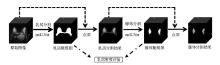
Fig.1
The framework for breast and FGT segmentation in fat-suppressed breast DCE-MR images
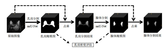
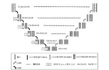
Fig.2
Network architecture in this study
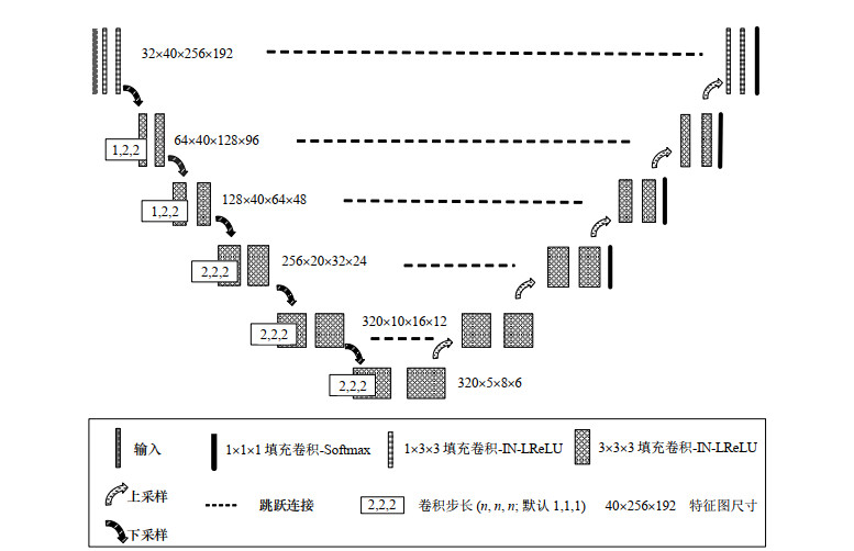

Fig.3
Comparison between the images (a) before and (b) after CLAHE processing
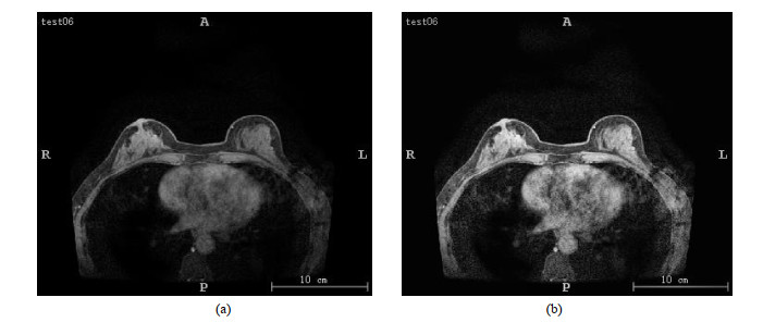
Table 1
Performance metrics for segmentation
| DSC | Acc | Sen | Spec | ASD/mm | |
| 乳房分割 | 0.969±0.007 | 0.995±0.006 | 0.961±0.027 | 0.994±0.002 | 0.181±0.032 |
| 腺体分割 | 0.893±0.054 | 0.997±0.003 | 0.926±0.017 | 0.998±0.002 | 0.240±0.021 |
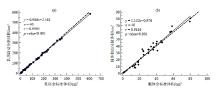
Fig.4
The correlation between the automatic segmentation volume obtained using the proposed model and the ground truth volume. (a) Breast; (b) FGT
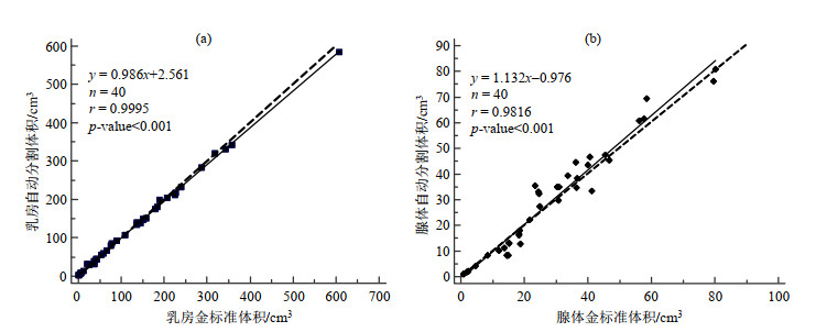
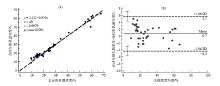
Fig.5
(a) The correlation and (b) Bland-Altman plot between the automatic segmentation breast densities obtained using the proposed model and the ground truth breast density
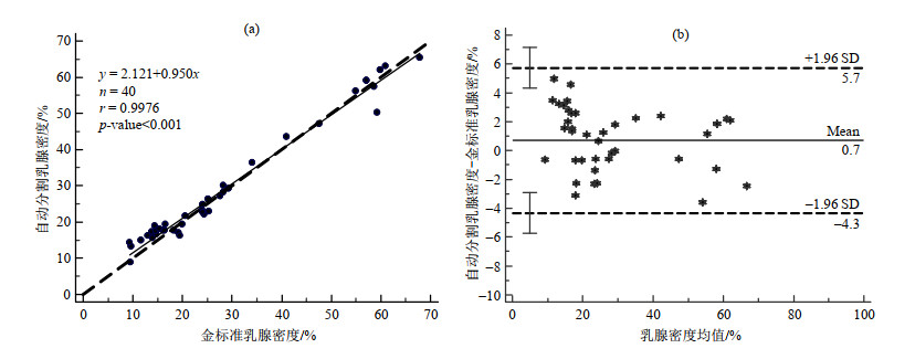
Table 2
DSC values of breast segmentation and FGT segmentation for two groups with different imaging parameters
| 乳房分割 | 腺体分割 | |
| 组1 | 0.973±0.011 | 0.903±0.055 |
| 组2 | 0.977±0.026 | 0.946±0.042 |
Table 3
DSC values of breast segmentation and FGT segmentation for four groups with different breast density ratings
| 乳房分割 | 腺体分割 | |
| Ⅰ类 | 0.989±0.004 | 0.906±0.048 |
| Ⅱ类 | 0.980±0.008 | 0.951±0.021 |
| Ⅲ类 | 0.941±0.029 | 0.917±0.031 |
| Ⅳ类 | 0.956±0.010 | 0.868±0.037 |

Fig.6
Segmentation examples of two groups with different breast DCE-MR imaging parameters. The top and bottom lines are Group 1 and Group 2. From left to right: the original image, the ground truth of whole breast, the segmentation mask of whole breast, the ground truth of FGT, and the segmentation mask of FGT
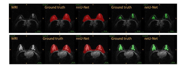
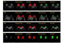
Fig.7
Segmentation examples of four groups with different breast density ratings. From top down: Category I, Category Ⅱ, Category Ⅲ and Category Ⅳ (Ⅰ - fatty: < 25%; Ⅱ - scattered: 25% ~ 50%; Ⅲ - heterogeneously dense: 50% ~ 75%; Ⅳ - dense: > 75%). From left to right: the original image, the ground truth of whole breast, the segmentation mask of whole breast, the ground truth of FGT, and the segmentation mask of FGT

Table 4
Performance metrics for segmentation during cross validation (taking DSC as an example)
| 乳房分割 | 腺体分割 | |||
| 验证集 | Fold 1 | 0.970±0.049 | 0.941±0.061 | |
| Fold 2 | 0.982±0.014 | 0.945±0.046 | ||
| Fold 3 | 0.971±0.044 | 0.942±0.047 | ||
| Fold 4 | 0.982±0.016 | 0.946±0.052 | ||
| Fold 5 | 0.975±0.012 | 0.942±0.031 | ||
| 平均值±标准差 | 0.976±0.027 | 0.944±0.047 | ||
| 测试集 | 0.969±0.007 | 0.893±0.054 | ||
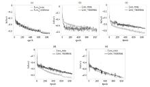
Fig.8
Loss curves of the breast segmentation model on the training (validation) set. Figures (a) ~ (e) represent the loss curves of each model of five-fold cross-validation, respectively
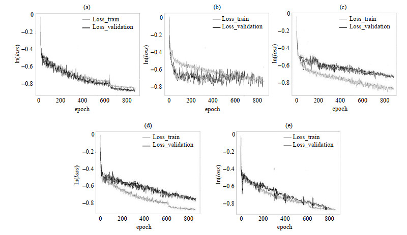
Fig.9
Loss curves of the FGT segmentation model on the training (validation) set. Figures (a) ~ (e) represent the loss curves of each model of five-fold cross-validation, respectively
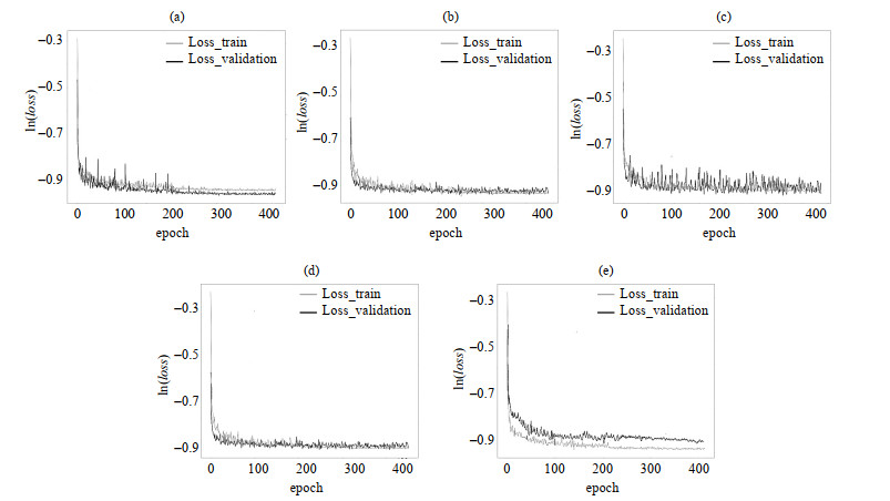
Table 5
Comparison between DSC values obtained by the proposed method and literatures
| 方法 | 测试样本数 | 乳房分割 | 腺体分割 |
| 文献[ | 27 | 0.94±0.03 | 0.80±0.13 |
| 文献[ | 37 | 0.96±0.02 | 0.83±0.06 |
| 文献[ | 28 | 0.86±0.05 | 0.83±0.06 |
| 文献[ | 22 | 0.94±0.03 | 0.81±0.11 |
| 本文方法 | 40 | 0.97±0.01 | 0.89±0.05 |
| 1 | BOYD N F , GUO H , MARTIN L J , et al. Mammographic density and the risk and detection of breast cancer[J]. New Eng J Med, 2007, 356 (1): 227- 236. |
| 2 |
KLIFA C , CARBALLIDO-GAMIO J , WILMES L , et al. Magnetic resonance imaging for secondary assessment of breast density in a high-risk cohort[J]. Magn Reson Imaging, 2010, 28 (1): 8- 15.
doi: 10.1016/j.mri.2009.05.040 |
| 3 |
WU S D , WEINSTEIN S P , CONANT E F , et al. Automated chest wall line detection for whole-breast segmentation in sagittal breast MR images[J]. Med Phys, 2013, 40 (4): 042301.
doi: 10.1118/1.4793255 |
| 4 |
NIE K , CHEN J H , CHAN S , et al. Development of a quantitative method for analysis of breast density based on three-dimensional automated segmentation of breast in 3-D MR images[J]. Med Phys, 2008, 35 (12): 5253- 5262.
doi: 10.1118/1.3002306 |
| 5 |
GUBERN-MÉRIDA A , KALLENBERG M , MANN R M , et al. Breast segmentation and density estimation in breast MRI: A fully automatic framework[J]. IEEE J Biomed Health, 2015, 19 (1): 349- 357.
doi: 10.1109/JBHI.2014.2311163 |
| 6 |
IVANOVSKA T , LAQUA R , WANG L , et al. A level set based framework for quantitative evaluation of breast tissue density from MRI data[J]. PLoS One, 2014, 9 (11): e112709.
doi: 10.1371/journal.pone.0112709 |
| 7 |
WU S D , WEINSTEIN S P , CONANT E F , et al. Automated fibroglandular tissue segmentation and volumetric density estimation in breast MRI using an atlas-aided fuzzy C-means method[J]. Med Phys, 2013, 40 (12): 122302.
doi: 10.1118/1.4829496 |
| 8 | KOREZ R, LIKAR B, PERNUŠ F, et al. Model-based segmentation of vertebral bodies from MR images with 3D CNNs[C]//Medical Image Computing and Computer-Assisted Intervention-MICCAI 2016. Cham, Switzerland: Springer International Publishing, 2016: 433-441. |
| 9 | MOESKOPS P, WOLTERINK J M, VAN DER VELDEN B H M, et al. Deep learning for multi-task medical image segmentation in multiple modalities[C]//Medical Image Computing and Computer-Assisted Intervention-MICCAI 2016. Cham, Switzerland: Springer International Publishing, 2016: 478-486. |
| 10 | RODRIGUEZ-RUIZ A, TEUWEN J, CHUNG K, et al. Pectoral muscle segmentation in breast tomosynthesis with deep learning[C]//Medical Imaging 2018: Computer-Aided Diagnosis. Bellingham, WA: SPIE, 2018: 564-570. |
| 11 |
ZHANG J , GAO Y Z , PARK S H , et al. Structured learning for 3-D perivascular space segmentation using vascular features[J]. IEEE T Biomed Eng, 2017, 64 (12): 2803- 2812.
doi: 10.1109/TBME.2016.2638918 |
| 12 | CHRIST P, ETTLINGER F, GRÜN F, et al. Automatic liver and tumor segmentation of CT and MRI volumes using cascaded fully convolutional neural networks[EB/OL]. (2017-2-23)[2020-12-19]. https://arxiv.org/pdf/1505.04597v1. |
| 13 | RONNEBERGER O, FISCHER P, BROX T. U-Net: Convolutional networks for biomedical image segmentation[C]//Medical Image Computing and Computer-Assisted Intervention-MICCAI 2015. Cham, Switzerland: Springer International Publishing, 2015: 234-241. |
| 14 | ZHAO S Y , WANG Y J . Classification of Alzheimer's disease patients based on magnetic resonance images and an improved UNet++ model[J]. Chinese J Magn Reson, 2020, 37 (3): 321- 331. |
| 赵尚义, 王远军. 基于磁共振图像和改进的UNet++模型区分阿尔茨海默症患者和健康人群[J]. 波谱学杂志, 2020, 37 (3): 321- 331. | |
| 15 |
KALLENBERG M , PETERSEN K , NIELSEN M , et al. Unsupervised deep learning applied to breast density segmentation and mammographic risk scoring[J]. IEEE Trans Med Imaging, 2016, 35 (5): 1322- 1331.
doi: 10.1109/TMI.2016.2532122 |
| 16 | LIU P , ZHONG Y M , WANG L J . Automatic segmentation of right ventricle in cine cardiac magnetic resonance image based on a dense and multi-scale u-net method[J]. Chinese J Magn Reson, 2020, 37 (4): 456- 468. |
| 刘鹏, 钟玉敏, 王丽嘉. 基于密集多尺度U-net网络的电影心脏磁共振图像右心室自动分割[J]. 波谱学杂志, 2020, 37 (4): 456- 468. | |
| 17 | XIAO L , LOU Y K , ZHOU H Y . A U-Net network-based rapid construction of knee models for specific absorption rate estimation[J]. Chinese J Magn Reson, 2020, 37 (2): 144- 151. |
| 肖亮, 娄煜堃, 周航宇. 用于SAR估计的基于U-Net网络的快速膝关节模型重建[J]. 波谱学杂志, 2020, 37 (2): 144- 151. | |
| 18 |
ZHANG Y , CHEN J H , CHANG K T , et al. Automatic breast and fibroglandular tissue segmentation in breast MRI using deep learning by a fully-convolutional residual neural network U-Net[J]. Acad Radiol, 2019, 26 (11): 1526- 1535.
doi: 10.1016/j.acra.2019.01.012 |
| 19 | DALMIŞ M U , LITJENS G , HOLLAND K , et al. Using deep learning to segment breast and fibroglandular tissue in MRI volumes[J]. Med Phys, 2017, 44 (12): 533- 546. |
| 20 | PIANTADOSI G, SANSONE M, SANSONE C. Breast segmentation in MRI via U-Net deep convolutional neural networks[C]//Proceedings of 2018 International Conference on Pattern Recognition (ICPR). Piscataway, NJ: IEEE press, 2018: 3917-3922. |
| 21 |
JIANG L , HU X X , XIAO Q , et al. Fully automated segmentation of whole breast using dynamic programming in dynamic contrast enhanced MR images[J]. Med Phys, 2017, 44 (6): 2400- 2414.
doi: 10.1002/mp.12254 |
| 22 | ISENSEE F, PETERSEN J, KLEIN A, et al. nnU-Net: Self-adapting framework for U-Net-based medical image segmentation[EB/OL]. (2018-9-27)[2020-12-19]. https://arxiv.org/abs/1809.10486v1 |
| 23 |
HELLER N , ISENSEE F , MAIER-HEIN K H , et al. The state of the art in kidney and kidney tumor segmentation in contrast-enhanced CT imaging: Results of the KiTS19 challenge[J]. Med Image Anal, 2021, 67, 101821.
doi: 10.1016/j.media.2020.101821 |
| 24 | MA J, WANG Y X, AN X L, et al. Toward data-efficient learning: A benchmark for COVID-19 CT lung and infection segmentation[J]. Med Phys, 2020. https://doi.org/10.1002/mp.14676. |
| 25 | PAUL S H . Graphics gems Ⅳ[M]. San Francisco: Margan Kaufmann, 1994, 474- 485. |
| 26 |
YUSHKEVICH P A , PIVEN J , HAZLETT H C , et al. User-guided 3D active contour segmentation of anatomical structures: Significantly improved efficiency and reliability[J]. NeuroImage, 2006, 31 (3): 1116- 1128.
doi: 10.1016/j.neuroimage.2006.01.015 |
| 27 | LÓPEZ-LINARES ROMÁN K , GARCÍA OCAÑA M I , LETE URZELAI N , et al. Medical image segmentation using deep learning[J]. Intelligent Systems Reference Library, 2020, 171, 17- 31. |
| [1] | CAO Fei, XU Qianqian, CHEN Hao, ZU Jie, LI Xiaowen, TIAN Jin, BAO Lei. An Intelligent Diagnosis Method for NIID Based on Cross Self-supervision and DWI [J]. Chinese Journal of Magnetic Resonance, 2025, 42(2): 154-163. |
| [2] | XUE Peiyang, GENG Chen, LI Yuxin, BAO Yifang, LU Yucheng, DAI Yakang. A Classification Method for Cerebral Aneurysms in TOF-MRA Based on Improved 3D ResNet50 Model [J]. Chinese Journal of Magnetic Resonance, 2025, 42(1): 56-66. |
| [3] | NING Xinzhou, HUANG Zhen, CHEN Xiqu, LIU Xinjie, CHEN Gang, ZHANG Zhi, BAO Qingjia, LIU Chaoyang. Research on Transformer Super-Resolution Reconstruction Algorithm for Ultrafast Spatiotemporal Encoding Magnetic Resonance Imaging [J]. Chinese Journal of Magnetic Resonance, 2024, 41(4): 454-468. |
| [4] | YANG Liming, WANG Yuanjun. Research Progress of Denoising Algorithms for Diffusion Tensor Images [J]. Chinese Journal of Magnetic Resonance, 2024, 41(3): 341-361. |
| [5] | Dai Junlong, He Cong, Wu Jie, Bian Yun. Pancreatic Cystic Neoplasms Segmentation Network Combining Dual Decoding and Global Attention Upsampling Modules [J]. Chinese Journal of Magnetic Resonance, 2024, 41(2): 151-161. |
| [6] | YANG Yu, CHEN Bo, WU Liubin, LIN Enping, HUANG Yuqing, CHEN Zhong. Spectrum Reconstruction for Laplace NMR: From Handcraft Regularization to Deep Learning [J]. Chinese Journal of Magnetic Resonance, 2024, 41(2): 191-208. |
| [7] | CHANG Bo, SUN Haoyun, GAO Qingyu, WANG Lijia. Research Progress on Cardiac Segmentation in Different Modal Medical Images by Traditional Methods and Deep Learning [J]. Chinese Journal of Magnetic Resonance, 2024, 41(2): 224-244. |
| [8] | XU Zhenshun, YUAN Xiaohan, HUANG Ziheng, SHAO Chengwei, WU Jie, BIAN Yun. Multi-source Feature Classification Model of Pancreatic Mucinous and Serous Cystic Neoplasms Based on Deep Learning [J]. Chinese Journal of Magnetic Resonance, 2024, 41(1): 19-29. |
| [9] | LAI Jiawen, WANG Yuling, CAI Xiaoyu, ZHOU Lihua. Multidimensional Information Fusion Method for Meniscal Tear Classification Based on CNN-SVM [J]. Chinese Journal of Magnetic Resonance, 2023, 40(4): 423-434. |
| [10] | WANG Hui, WANG Tiantian, WANG Lijia. Squeeze-and-excitation Residual U-shaped Network for Left Myocardium Segmentation Based on Cine Cardiac Magnetic Resonance Images [J]. Chinese Journal of Magnetic Resonance, 2023, 40(4): 435-447. |
| [11] | Li Yijie, YANG Xinyu, YANG Xiaomei. Magnetic Resonance Image Reconstruction of Multi-scale Residual Unet Fused with Attention Mechanism [J]. Chinese Journal of Magnetic Resonance, 2023, 40(3): 307-319. |
| [12] | ZHANG Jiajun, LU Yucheng, BAO Yifang, LI Yuxin, GENG Chen, HU Fuyuan, DAI Yakang. An Automatic Segmentation Method of Cerebral Arterial Tree in TOF-MRA Based on DBCNet [J]. Chinese Journal of Magnetic Resonance, 2023, 40(3): 320-331. |
| [13] | LU Qiqi, LIAN Zifeng, LI Jialong, SI Wenbin, MAI Zhaohua, FENG Yanqiu. Magnetic Resonance R2* Parameter Mapping of Liver Based on Self-supervised Deep Neural Network [J]. Chinese Journal of Magnetic Resonance, 2023, 40(3): 258-269. |
| [14] | TIAN Hui, WU Jie, BIAN Yun, ZHANG Zhiwei, SHAO Chengwei. Classification of Pancreatic Cystic Tumors Based on DenseNet and Transfer Learning [J]. Chinese Journal of Magnetic Resonance, 2023, 40(3): 270-279. |
| [15] | HUANG Min,LI Siyi,CHEN Junbo,ZHOU Dao. Progress of Magnetic Resonance Fingerprinting Technology and Its Clinical Application [J]. Chinese Journal of Magnetic Resonance, 2023, 40(2): 207-219. |
| Viewed | ||||||
|
Full text |
|
|||||
|
Abstract |
|
|||||