
Chinese Journal of Magnetic Resonance ›› 2026, Vol. 43 ›› Issue (2): 223-240.doi: 10.11938/cjmr20253196cstr: 32225.14.cjmr20253196
• Review Articles • Previous Articles
XIE Xinyi, WANG Yuanjun*( )
)
Received:2025-12-29
Published:2026-06-05
Online:2026-04-10
Contact:
WANG Yuanjun
E-mail:yjusst@126.com
CLC Number:
XIE Xinyi, WANG Yuanjun. Research Progress on Super-resolution Reconstruction of Brain Diffusion Magnetic Resonance Images[J]. Chinese Journal of Magnetic Resonance, 2026, 43(2): 223-240.
Add to citation manager EndNote|Reference Manager|ProCite|BibTeX|RefWorks
Table 1
The main tasks involved in the reconstruction application of dMRI
| 分类 | 典型模型 | 典型采集参数 | 目标输出 | 架构发展 |
|---|---|---|---|---|
| 基础扩散指标重建 | DTI:估计主扩散方向及 各向异性特征 | b=1000 s/mm2, 6~30个方向 | 张量参数FA、MD、AD、RD等 | 主要是数据驱动,也有考虑旋转等变性 |
| 高阶微结构指标重建 | NODDI:区分神经突密度、取向分散和自由水 | b=700 s/mm2,90个方向 b=2000 s/mm2,60个方向 | 微结构指标NDI、ODI、fISO等 | 数据驱动、模型驱动、旋转等变性 |
| fODF分辨率提升重建 | fODF:解析交叉纤维等 复杂纤维构型 | b=3000 s/mm2, 60~90个方向 | fODF估计、下游任务有纤维追踪、脑连接组等 | 替代球面反卷积计算,或在单壳重建fODF上增强 |

Fig. 1
Overview map of the dMRI super-resolution task and method system
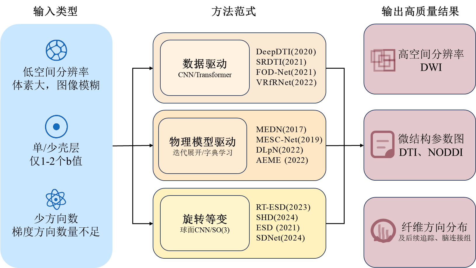

Fig. 2
Classification schematic of reconstruction methods for enhancing fODF resolution

Table 2
Classification table of evaluation indicators
| 类别 | 指标 | 中文名称 | 含义 | 说明 | ||
|---|---|---|---|---|---|---|
| 图像保真度指标 | Peak Signal-to-Noise Ratio (PSNR)[ | 峰值信噪比 | 衡量重建图像与参考图像之间的全局误差,值越高表示重建质量越好 | 基于均方误差计算,对误差敏感,但可能与视觉感知不完全一致,常用于评估DWI或参数图的重建质量 | ||
| Structural Similarity Index (SSIM)[ | 结构相似性指数 | 从亮度、对比度、结构三方面综合评估图像之间的相似度,更符合人眼视觉感知 | 值域为[0, 1],值越接近1表示相似度越高,适用于评估空间超分辨率结果的结构保真度 | |||
| 微结构标量误差指标 | Mean Absolute Error (MAE)[ | 平均绝对误差 | 计算预测标量值与真实值之间绝对误差的平均值 | 最基础的误差度量 | ||
| Mean Squared Error (MSE)[ | 均方误差 | 计算预测值与真实值之间平方误差的平均值 | 对较大误差更敏感 | |||
| Normalized Mean Absolute Error (NMAE)[ | 归一化平均绝对误差 | 对MAE进行归一化处理,便于比较不同量级的数据 | 具体归一化方式需在上下文中明确,通常除以真实值的范围或均值 | |||
| Normalized Mean Squared Error (NMSE)[ | 归一化均方误差 | 对MSE进行归一化处理,便于比较不同量级的数据 | 具体归一化方式需在上下文中明确,通常除以真实值的范围或均值 | |||
| 纤维方向重建评估指标 | Angular Correlation Coefficient (ACC)[ | 角相关系数 | 评估预测的纤维方向与参考真实方向在空间上的一致性 | 评估纤维方向估计整体准确性的核心指标 | ||
| Mean Angular Error (MAE)[ | 平均角度误差 | 衡量预测纤维主峰方向与参考真实方向之间的平均角度差 | 此MAE专指角度误差,与上文“平均绝对误差”含义不同 | |||
| Peak Error (PE)[ | 峰值误差 | 衡量预测纤维主峰幅度与参考真实幅度之间的峰值差 | 反映对纤维强度估计的准确性 | |||
| Proportion of Correct Peaks (PCP)[ | 正确峰比例 | 统计正确识别出的纤维方向的比例 | 一种基于分类正确率的评估方式,计算时设有阈值 | |||
| Earth Mover‘s Distance (EMD)[ | 推土机距离 | 衡量整个fODF分布与参考真实分布之间差异 | 评估分布层面相似性的核心指标,反映整体匹配度,综合考量角度和幅度 | |||
| 纤维追踪评估指标 | Valid Streamlines[ | 有效流线比例 | 在所有生成的流线中,属于任一真实纤维束的流线比例 | 反映追踪结果的纯净度,值低说明假阳性高 | ||
| Bundle Overlap[ | 纤维束重叠度 | 估计的纤维束与真实纤维束体素交集与真实集的比值 | 衡量灵敏度,即找回真实纤维束的能力 | |||
| Bundle Overreach[ | 纤维束过度延伸度 | 估计的纤维束超出真实纤维束的部分与真实集的比值 | 衡量特异度,值高表示假阳性多 | |||
| Valid Bundles[ | 有效纤维束数量 | 被正确识别出的、与真实解剖结构相符的纤维束数量 | 衡量算法重建特定神经通路的能力 | |||
| Dice Coefficient[ | Dice系数 | 两个二元分割结果的空间重叠程度 | 衡量纤维束分割结果 | |||
| 重测信度指标 | Coefficient of Variation (CV)[ | 变异系数 | 计算同一受试者经多次扫描的某指标的标准差与均值的比值 | 反映数据相对于其平均值的离散程度,值越低表示稳定性越好 | ||
| Weighted Mean Coefficient of Variation (wmCoV)[ | 加权平均变异系数 | 针对连接组矩阵,对每个连接边的CV进行加权平均求变异系数,权重取决于连接强度 | 由于连接组数据通常具有高度偏态分布,wmCoV通过对强连接赋予更高权重,更能代表整体连接组的重测信度 | |||
| Intraclass Correlation Coefficient (ICC)[ | 组内相关系数 | 量化重测信度的核心统计量,评估同一受试者多次扫描结果的一致性程度 | ICC > 0.75通常被认为信度优秀,是比简单相关性更严格的指标 | |||
Table 3
Commonly used public dMRI datasets
| 全称 | 缩写 | 主要人群/研究重点 | 特点 | 数据量 | 主要采集参数(b值非0) |
|---|---|---|---|---|---|
| The Rotterdam Study[ | RDS | 荷兰鹿特丹地区老年人慢性病的发病率、预后 | 大型长期随访队列 | MRI约8000人 | b = 1000 s/mm2,25 个方向 |
| The Rhineland Study[ | RLS | 德国莱茵地区人群的深度表型研究 | 人群基线广、影像丰富 | 千例以上 | b范围270 ~ 6800 s/mm2, 112个方向 |
| Pediatric Imaging Neurocognition and Genetics[ | PING | 儿科影像、认知发展与遗传关联 | 儿童、青少年群体 | 约1493名 | b = 1000 s/mm2, 约30个方向 |
| developing Human Connectome Project[ | dHCP | 典型与非典型早期脑发育 | 新生儿、早期发育阶段 | 1173名 | b = 400 s/mm2,64个方向; b = 1000 s/mm2,88个方向; b = 2600 s/mm2,128个方向 |
| Baby Connectome Project[ | BCP | 婴儿脑连接发育 | 婴幼儿期 | 500名 | b = 500 ~ 3000 s/mm2,总约144个方向 |
| Chinese Connectome Project[ | CHCP | 中国人群脑连接组 | 亚洲人群样本 | 366名 | b = 1000 s/mm2,93个方向; b = 2000 s/mm2,92个方向 |
| Amsterdam Open MRI Collection[ | AOMIC | 健康年轻成人,情感与社会认知的神经机制 | 模态丰富,附带行为与心理学量表数据 | 约1370名 | b = 1000 s/mm2,32个方向 |
| ISMRM 2015 Tractometer Challenge Dataset[ | / | 纤维束追踪算法的标准评估与对比 | 追踪算法验证与性能比较的常用基准 | 1名 | b = 1000 s/mm2, 90个方向; b = 2000 s/mm2, 90个方向; b = 3000 s/mm2,90个方向 |
| [1] | MENG J X, WANG Y J. Research progress on tractography of superficial white matter based on diffusion magnetic resonance imaging[J]. Chinese J Magn Reson, 2025, 42(2): 205-220. |
|
孟靖欣, 王远军. 基于扩散磁共振的大脑浅表白质纤维束研究进展[J]. 波谱学杂志, 2025, 42(2): 205-220.
doi: 10.11938/cjmr20243126 |
|
| [2] | JIANG B, ZHANG Z, LIN D, et al. Semi-supervised learning with graph learning-convolutional networks[C]// Proceedings of the IEEE/CVF conference on computer vision and pattern recognition, Long Beach, CA. Piscataway: IEEE, 2019: 11313-11320. |
| [3] | COHEN T S, GEIGER M, KÖHLER J, et al. Spherical CNNs[C]// International Conference on Learning Representations (ICLR), Vancouver, Canada: International Conference on Learning Representations, 2018: 1-15. |
| [4] |
FAIYAZ A, DOYLEY M M, SCHIFITTO G, et al. Artificial intelligence for diffusion MRI-based tissue microstructure estimation in the human brain: an overview[J]. Front Neurol, 2023, 14: 1168833.
doi: 10.3389/fneur.2023.1168833 |
| [5] |
BASSER P J, MATTIELLO J, LEBIHAN D. MR diffusion tensor spectroscopy and imaging[J]. Biophys J, 1994, 66(1): 259-267.
doi: 10.1016/S0006-3495(94)80775-1 pmid: 8130344 |
| [6] |
JENSEN J H, HELPERN J A. MRI quantification of non-Gaussian water diffusion by kurtosis analysis[J]. NMR Biomed, 2010, 23(7): 698-710.
doi: 10.1002/nbm.1518 pmid: 20632416 |
| [7] |
ZHANG H, SCHNEIDER T, WHEELER-KINGSHOTT C A, et al. NODDI: practical in vivo neurite orientation dispersion and density imaging of the human brain[J]. NeuroImage, 2012, 61(4): 1000-1016.
doi: 10.1016/j.neuroimage.2012.03.072 pmid: 22484410 |
| [8] |
TOURNIER J D, CALAMANTE F, CONNELLY A. Robust determination of the fibre orientation distribution in diffusion MRI: non-negativity constrained super-resolved spherical deconvolution[J]. NeuroImage, 2007, 35(4): 1459-1472.
doi: 10.1016/j.neuroimage.2007.02.016 pmid: 17379540 |
| [9] |
TIAN Q, BILGIC B, FAN Q, et al. DeepDTI: High-fidelity six-direction diffusion tensor imaging using deep learning[J]. NeuroImage, 2020, 219: 117017.
doi: 10.1016/j.neuroimage.2020.117017 |
| [10] |
LI H, LIANG Z, ZHANG C, et al. SuperDTI: Ultrafast DTI and fiber tractography with deep learning[J]. Magn Reson Med, 2021, 86(6): 3334-3347.
doi: 10.1002/mrm.28937 pmid: 34309073 |
| [11] |
SABIDUSSI E R, KLEIN S, JEURISSEN B, et al. dtiRIM: A generalisable deep learning method for diffusion tensor imaging[J]. NeuroImage, 2023, 269: 119900.
doi: 10.1016/j.neuroimage.2023.119900 |
| [12] |
ZHANG L, HE J, LI W, et al. Diff-DTI: Fast diffusion tensor imaging using a feature-enhanced joint diffusion model[J]. IEEE J Biomed Health Inform, 2026, 30(2): 1300-1313.
doi: 10.1109/JBHI.2024.3523532 |
| [13] |
MARTIN P, ALTBACH M, BILGIN A. Conditional generative diffusion deep learning for accelerated diffusion tensor and kurtosis imaging[J]. Magn Reson Imaging, 2025, 117: 110309.
doi: 10.1016/j.mri.2024.110309 |
| [14] | TIAN Q, LI Z, FAN Q, et al. SRDTI: Deep learning-based super-resolution for diffusion tensor MRI[PP/OL]. arXiv (2021-02-17) [2025-12-28]. https://arxiv.org/abs/2102.09069. |
| [15] | MA W, PENG L. Image quality transfer with auto-encoding applied to dMRI super-resolution[C]// 2021 4th International Conference on Advanced Electronic Materials, Computers and SoftwareEngineering AEMCSE, Changsha, China. Piscataway: IEEE, 2021: 828-831. |
| [16] |
KARIMI D, JAIMES C, MACHADO-RIVAS F, et al. Deep learning-based parameter estimation in fetal diffusion-weighted MRI[J]. NeuroImage, 2021, 243: 118482.
doi: 10.1016/j.neuroimage.2021.118482 |
| [17] |
KARIMI D, GHOLIPOUR A. Diffusion tensor estimation with transformer neural networks[J]. Artif Intell Med, 2022, 130: 102330.
doi: 10.1016/j.artmed.2022.102330 |
| [18] | FAIYAZ A, UDDIN M N, SCHIFITTO G. Angular upsampling in diffusion MRI using contextual hemihex sub-sampling in q-space[PP/OL]. arXiv (2022-11-01) [2025-12-28]. https://arxiv.org/abs/2211.00240. |
| [19] |
EWERT C, KUGLER D, STIRNBERG R, et al. Geometric deep learning for diffusion mri signal reconstruction with continuous samplings (discus)[J]. Imaging Neurosci, 2024, 2: 1-18.
doi: 10.1162/imag_a_00344 |
| [20] |
ALTMANN S, GRAUHAN N F, MERCADO M A A, et al. Deep learning accelerated brain diffusion-weighted MRI with super resolution processing[J]. Acad Radiol, 2024, 31(10): 4171-4182.
doi: 10.1016/j.acra.2024.02.049 pmid: 38521612 |
| [21] |
LE BIHAN D. What can we see with IVIM MRI?[J]NeuroImage, 2019, 187: 56-67.
doi: S1053-8119(17)31086-8 pmid: 29277647 |
| [22] |
GOLKOV V, DOSOVITSKIY A, SPERL J I, et al. Q-space deep learning: twelve-fold shorter and model-free diffusion MRI scans[J]. IEEE Trans Med Imaging, 2016, 35(5): 1344-1351.
doi: 10.1109/TMI.2016.2551324 |
| [23] |
GIBBONS E K, HODGSON K K, CHAUDHARI A S, et al. Simultaneous NODDI and GFA parameter map generation from subsampled q-space imaging using deep learning[J]. Magn Reson Med, 2019, 81(4): 2399-2411.
doi: 10.1002/mrm.27568 pmid: 30426558 |
| [24] | NATH V, RAMADASS K, SCHILLING K G, et al. DW-MRI microstructure model of models captured via single-shell bottleneck deep learning[C]// Computational Diffusion MRI: International MICCAI Workshop, Lima, Peru. Cham: Springer, 2021: 147-157. |
| [25] | CHEN G, HONG Y, ZHANG Y, et al. Estimating tissue microstructure with undersampled diffusion data via graph convolutional neural networks[C]// International Conference on Medical Image Computing and Computer-Assisted Intervention, Lima, Peru. Cham: Springer, 2020: 280-290. |
| [26] | CHEN G, JIANG H, LIU J, et al. Hybrid graph transformer for tissue microstructure estimation with undersampled diffusion MRI data[C]// International Conference on Medical Image Computing and Computer-Assisted Intervention, Singapore. Cham: Springer, 2022: 113-122. |
| [27] |
YE C. Tissue microstructure estimation using a deep network inspired by a dictionary-based framework[J]. Med Image Anal, 2017, 42: 288-299.
doi: S1361-8415(17)30132-9 pmid: 28910696 |
| [28] |
YE C, LI X, CHEN J. A deep network for tissue microstructure estimation using modified LSTM units[J]. Med Image Anal, 2019, 55: 49-64.
doi: S1361-8415(18)30557-7 pmid: 31022640 |
| [29] |
YE C, LI Y, ZENG X. An improved deep network for tissue microstructure estimation with uncertainty quantification[J]. Med Image Anal, 2020, 61: 101650.
doi: 10.1016/j.media.2020.101650 |
| [30] |
FAIYAZ A, DOYLEY M, SCHIFITTO G, et al. Single-shell NODDI using dictionary-learner-estimated isotropic volume fraction[J]. NMR Biomed, 2022, 35(2): e4628.
doi: 10.1002/nbm.v35.2 |
| [31] | ZHENG T, ZHENG W, SUN Y, et al. An adaptive network with extragradient for diffusion MRI-Based microstructure estimation[C]// International Conference on Medical Image Computing and Computer-Assisted Intervention, Singapore. Cham: Springer, 2022: 153-162. |
| [32] |
ZHENG T, YAN G, LI H, et al. A microstructure estimation Transformer inspired by sparse representation for diffusion MRI[J]. Med Image Anal, 2023, 86: 102788.
doi: 10.1016/j.media.2023.102788 |
| [33] |
LECUN Y, BOTTOU L, BENGIO Y, et al. Gradient-based learning applied to document recognition[J]. Proc IEEE, 2002, 86(11): 2278-2324.
doi: 10.1109/5.726791 |
| [34] | ESTEVES C, ALLEN-BLANCHETTE C, MAKADIA A, et al. Learning SO(3) equivariant representations with spherical CNNs[C]// European Conference on Computer Vision (ECCV), Munich, Germany. Cham: Springer, 2018: 52-68. |
| [35] | KONDOR R, LIN Z, TRIVEDI S. Clebsch-gordan nets: a fully fourier space spherical convolutional neural network[J]. Adv Neural Inf Process Syst, 2018: 31. |
| [36] | SEDLAR S, ALIMI A, PAPADOPOULO T, et al. A spherical convolutional neural network for white matter structure imaging via dMRI[C]// International Conference on Medical Image Computing and Computer-Assisted Intervention, Strasbourg, France. Cham: Springer, 2021: 529-539. |
| [37] | ELALDI A, DEY N, KIM H, et al. Equivariant spherical deconvolution: Learning sparse orientation distribution functions from spherical data[C]// International Conference on Information Processing in Medical Imaging, Bornholm, Denmark. Cham: Springer, 2021: 267-278. |
| [38] |
CONSAGRA W, NING L, RATHI Y. A deep learning approach to multi-fiber parameter estimation and uncertainty quantification in diffusion MRI[J]. Med Image Anal, 2025, 102: 103537.
doi: 10.1016/j.media.2025.103537 |
| [39] | YE C, QIN Y, LIU C, et al. Super-resolved q-space deep learning[C]// International Conference on Medical Image Computing and Computer-Assisted Intervention, Shenzhen, China. Cham: Springer, 2019: 582-589. |
| [40] |
QIN Y, LIU Z, LIU C, et al. Super-Resolved q-Space deep learning with uncertainty quantification[J]. Med Image Anal, 2021, 67: 101885.
doi: 10.1016/j.media.2020.101885 |
| [41] |
QIN Y, LI Y, ZHUO Z, et al. Multimodal super-resolved q-space deep learning[J]. Med Image Anal, 2021, 71: 102085.
doi: 10.1016/j.media.2021.102085 |
| [42] | DHOLLANDER T, MITO R, RAFFELT D, et al. Improved white matter response function estimation for 3-tissue constrained spherical deconvolution[C]// Proceedings of the International Society for Magnetic Resonance in Medicine (ISMRM) 2019. Montreal, Quebec, Canada: ISMRM, 2019: 555. |
| [43] |
JEURISSEN B, TOURNIER J D, DHOLLANDER T, et al. Multi-tissue constrained spherical deconvolution for improved analysis of multi-shell diffusion MRI data[J]. NeuroImage, 2014, 103: 411-426.
doi: S1053-8119(14)00644-2 pmid: 25109526 |
| [44] | KOPPERS S, MERHOF D. Direct estimation of fiber orientations using deep learning in diffusion imaging[C]// International Workshop on Machine Learning in Medical Imaging, Athens, Greece. Cham: Springer, 2016: 53-60. |
| [45] |
LIN Z, GONG T, WANG K, et al. Fast learning of fiber orientation distribution function for MR tractography using convolutional neural network[J]. Med Phys, 2019, 46(7): 3101-3116.
doi: 10.1002/mp.13555 pmid: 31009085 |
| [46] |
NATH V, SCHILLING K G, PARVATHANENI P, et al. Deep learning reveals untapped information for local white-matter fiber reconstruction in diffusion-weighted MRI[J]. Magn Reson Imaging, 2019, 62: 220-227.
doi: S0730-725X(19)30171-7 pmid: 31323317 |
| [47] | NATH V, PATHAK S K, SCHILLING K G, et al. Deep learning estimation of multi-tissue constrained spherical deconvolution with limited single shell DW-MRI[C]// Medical Imaging 2020: Image Processing, Bel Houston, Texas, USA. Bellingham: SPIE, 2020, 11313: 162-171. |
| [48] |
JHA R R, PATHAK S K, NATH V, et al. VRfRNet: Volumetric ROI fODF reconstruction network for estimation of multi-tissue constrained spherical deconvolution with only single shell dMRI[J]. Magn Reson Imaging, 2022, 90: 1-16.
doi: 10.1016/j.mri.2022.03.004 pmid: 35341904 |
| [49] |
BARTLETT J J, DAVEY C E, JOHNSTON L A, et al. Recovering high-quality fiber orientation distributions from a reduced number of diffusion-weighted images using a model-driven deep learning architecture[J]. Magn Reson Med, 2024, 92(5): 2193-2206.
doi: 10.1002/mrm.30187 pmid: 38852179 |
| [50] | YAO T, NEWLIN N, KANAKARAJ P, et al. A unified learning model for estimating fiber orientation distribution functions on heterogeneous multi-shell diffusion-weighted MRI[C]// International Workshop on Computational Diffusion MRI, Vancouver, BC, Canada. Cham: Springer, 2023: 13-22. |
| [51] |
CONSAGRA W, NING L, RATHI Y. Neural orientation distribution fields for estimation and uncertainty quantification in diffusion MRI[J]. Med Image Anal, 2024, 93: 103105.
doi: 10.1016/j.media.2024.103105 |
| [52] | SEDLAR S, PAPADOPOULO T, DERICHE R, et al. Diffusion MRI fiber orientation distribution function estimation using voxel-wise spherical U-net[C]// Computational Diffusion MRI: International MICCAI Workshop, Lima, Peru. Cham: Springer, 2021: 95-106. |
| [53] | ELALDI A, GERIG G, DEY N. E(3) × so(3)-equivariant networks for spherical deconvolution in diffusion MRI[C]// Medical Imaging with Deep Learning, Paris, France. Cambridge: PMLR, 2024: 301-319. |
| [54] | ELALDI A, GERIG G, DEY N. Equivariant spatio-hemispherical networks for diffusion MRI deconvolution[J]. Adv Neural Inf Process Syst, 2024, 37: 52095-52126. |
| [55] | GAO X, LIN R, FENG J, et al. UFO-3: unsupervised three-compartment learning for fiber orientation distribution function estimation[C]// International Conference on Medical Image Computing and Computer-Assisted Intervention, Daejeon, South Korea. Cham: Springer, 2025: 638-649. |
| [56] | SNOUSSI H, KARIMI D. Equivariant spherical CNNs for accurate fiber orientation distribution estimation in neonatal diffusion MRI with reduced acquisition time[PP/OL]. arXiv (2025-04-02) [2025-12-28]. https://arxiv.org/abs/2504.01925. |
| [57] |
ZENG R, LV J, WANG H, et al. FOD-Net: A deep learning method for fiber orientation distribution angular super resolution[J]. Med Image Anal, 2022, 79: 102431.
doi: 10.1016/j.media.2022.102431 |
| [58] | DA SILVA M O, SANTANA C P, DO CARMO D S, et al. FOD-Swin-Net:angular super resolution of fiber orientation distribution using a transformer-based deep model[C]// 2024 IEEE International Symposium on Biomedical Imaging (ISBI), Athens, Greece. Piscataway: IEEE, 2024: 1-5. |
| [59] |
LI J, AI L, YAO R. NVAM-Net: deep learning networks for reconstructing high-quality fiber orientation distributions[J]. Neuroradiology, 2024, 66(7): 1177-1187.
doi: 10.1007/s00234-024-03341-y pmid: 38563964 |
| [60] | YANG L M, WANG Y J. Research progress of denoising algorithms for diffusion tensor images[J]. Chinese J Magn Reson, 2024, 41(3): 341-361. |
|
杨黎明, 王远军. 扩散张量图像去噪算法研究进展[J]. 波谱学杂志, 2024, 41(3): 341-361.
doi: 10.11938/cjmr20243087 |
|
| [61] |
RUBNER Y, TOMASI C, GUIBAS L J. The earth mover's distance as a metric for image retrieval[J]. Int J Comput Vision, 2000, 40(2): 99-121.
doi: 10.1023/A:1026543900054 |
| [62] |
RENAULD E, THÉBERGE A, PETIT L, et al. Validate your white matter tractography algorithms with a reappraised ISMRM 2015 Tractography Challenge scoring system[J]. Sci Rep, 2023, 13(1): 2347.
doi: 10.1038/s41598-023-28560-w pmid: 36759653 |
| [63] | SENTHIL KUMAR V S, SHAHRAZ S. Intraclass correlation for reliability assessment: the introduction of a validated program in SAS (ICC6)[J]. Health Serv Outcome, 2024, 24(1): 1-13. |
| [64] |
VAN ESSEN D C, SMITH S M, BARCH D M, et al. The WU-Minn human connectome project: an overview[J]. NeuroImage, 2013, 80: 62-79.
doi: 10.1016/j.neuroimage.2013.05.041 pmid: 23684880 |
| [65] |
IKRAM M A, BRUSSELLE G, GHANBARI M, et al. Objectives, design and main findings until 2020 from the Rotterdam Study[J]. Eur J Epidemiol, 2020, 35(5): 483-517.
doi: 10.1007/s10654-020-00640-5 pmid: 32367290 |
| [66] | BRETELER M M B, STÖCKER T, PRACHT E, et al. IC-P-165: MRI in the Rhineland study: a novel protocol for population neuroimaging[J]. Alzheimer's & Dementia, 2014, 10: P92-P92. |
| [67] |
JERNIGAN T L, BROWN T T, HAGLER JR D J, et al. The pediatric imaging, neurocognition, and genetics (PING) data repository[J]. NeuroImage, 2016, 124: 1149-1154.
doi: S1053-8119(15)00357-2 pmid: 25937488 |
| [68] | EDWARDS A D, RUECKERT D, SMITH S M, et al. The developing human connectome project neonatal data release[J]. Front Neurol, 2022, 16: 886772. |
| [69] |
HOWELL B R, STYNER M A, GAO W, et al. The UNC/UMN Baby Connectome Project (BCP): An overview of the study design and protocol development[J]. NeuroImage, 2019, 185: 891-905.
doi: S1053-8119(18)30259-3 pmid: 29578031 |
| [70] | GE J, YANG G, HAN M, et al. Increasing diversity in connectomics with the Chinese Human Connectome Project[J]. Nat Neurosci, 2023, 26(1): 163-172. |
| [71] |
SNOEK L, VAN DER MIESEN M M, BEEMSTERBOER T, et al. The amsterdam open MRI collection, a set of multimodal MRI datasets for individual difference analyses[J]. Sci Data, 2021, 8(1): 85.
doi: 10.1038/s41597-021-00870-6 pmid: 33741990 |
| [72] |
MAIER-HEIN K H, NEHER P F, HOUDE J C, et al. The challenge of mapping the human connectome based on diffusion tractography[J]. Nat Commun, 2017, 8(1): 1349.
doi: 10.1038/s41467-017-01285-x |
| [73] | YANG J C, WANG Y J. Improved constrained spherical deconvolution model for brain gray matter microstructure imaging[J]. Chinese J Magn Reson, 2025, 42(1): 67-79. |
|
杨佳铖, 王远军. 改进约束球面反卷积模型的脑灰质微结构成像[J]. 波谱学杂志, 2025, 42(1): 67-79.
doi: 10.11938/cjmr20243117 |
|
| [74] | AJA-FERNÁNDEZ S, MARTÍN-MARTÍN C, PLANCHUELO-GÓMEZ Á, et al. Validation of deep learning techniques for quality augmentation in diffusion MRI for clinical studies[J]. NeuroImage, 2023, 39: 103483. |
| [1] | LI Yinghao, WANG Lihui, WANG Sucheng, ZHU Zhongqi, HUANG Changdong, LI Renfeng, CAO Kaiming, HU Haiyang, JIA Yiming, LIANG Songtao, YANG Guang, LU Qing, WANG Hongzhi. Study on Pancreas Automatic Segmentation, Regional Quantification, and Diabetes Assessment [J]. Chinese Journal of Magnetic Resonance, 2025, 42(4): 378-389. |
| [2] | GAO Zhaoyao, ZHANG Zhan, HU Liangliang, XU Guangyu, ZHOU Sheng, HU Yuxin, LIN Zijie, ZHOU Chao. PMRI Image Reconstruction Method Based on Virtual Coils and GRAPPA-enhanced Network [J]. Chinese Journal of Magnetic Resonance, 2025, 42(4): 390-401. |
| [3] | ZHANG Mingyu, XIAO Sa, SHI Shengjie, ZHANG Xuecheng, ZHOU Xin. Research on a Multi-modal Enhanced Denoising Diffusion Model for Hyperpolarized 129Xe MRI [J]. Chinese Journal of Magnetic Resonance, 2025, 42(4): 364-377. |
| [4] | CAO Fei, XU Qianqian, CHEN Hao, ZU Jie, LI Xiaowen, TIAN Jin, BAO Lei. An Intelligent Diagnosis Method for NIID Based on Cross Self-supervision and DWI [J]. Chinese Journal of Magnetic Resonance, 2025, 42(2): 154-163. |
| [5] | YANG Jiacheng, WANG Yuanjun. Improved Constrained Spherical Deconvolution for Microstructural Imaging of Brain Gray Matter [J]. Chinese Journal of Magnetic Resonance, 2025, 42(1): 67-79. |
| [6] | XUE Peiyang, GENG Chen, LI Yuxin, BAO Yifang, LU Yucheng, DAI Yakang. A Classification Method for Cerebral Aneurysms in TOF-MRA Based on Improved 3D ResNet50 Model [J]. Chinese Journal of Magnetic Resonance, 2025, 42(1): 56-66. |
| [7] | NING Xinzhou, HUANG Zhen, CHEN Xiqu, LIU Xinjie, CHEN Gang, ZHANG Zhi, BAO Qingjia, LIU Chaoyang. Research on Transformer Super-Resolution Reconstruction Algorithm for Ultrafast Spatiotemporal Encoding Magnetic Resonance Imaging [J]. Chinese Journal of Magnetic Resonance, 2024, 41(4): 454-468. |
| [8] | YANG Liming, WANG Yuanjun. Research Progress of Denoising Algorithms for Diffusion Tensor Images [J]. Chinese Journal of Magnetic Resonance, 2024, 41(3): 341-361. |
| [9] | Dai Junlong, He Cong, Wu Jie, Bian Yun. Pancreatic Cystic Neoplasms Segmentation Network Combining Dual Decoding and Global Attention Upsampling Modules [J]. Chinese Journal of Magnetic Resonance, 2024, 41(2): 151-161. |
| [10] | YANG Yu, CHEN Bo, WU Liubin, LIN Enping, HUANG Yuqing, CHEN Zhong. Spectrum Reconstruction for Laplace NMR: From Handcraft Regularization to Deep Learning [J]. Chinese Journal of Magnetic Resonance, 2024, 41(2): 191-208. |
| [11] | CHANG Bo, SUN Haoyun, GAO Qingyu, WANG Lijia. Research Progress on Cardiac Segmentation in Different Modal Medical Images by Traditional Methods and Deep Learning [J]. Chinese Journal of Magnetic Resonance, 2024, 41(2): 224-244. |
| [12] | XU Zhenshun, YUAN Xiaohan, HUANG Ziheng, SHAO Chengwei, WU Jie, BIAN Yun. Multi-source Feature Classification Model of Pancreatic Mucinous and Serous Cystic Neoplasms Based on Deep Learning [J]. Chinese Journal of Magnetic Resonance, 2024, 41(1): 19-29. |
| [13] | LAI Jiawen, WANG Yuling, CAI Xiaoyu, ZHOU Lihua. Multidimensional Information Fusion Method for Meniscal Tear Classification Based on CNN-SVM [J]. Chinese Journal of Magnetic Resonance, 2023, 40(4): 423-434. |
| [14] | WANG Hui, WANG Tiantian, WANG Lijia. Squeeze-and-excitation Residual U-shaped Network for Left Myocardium Segmentation Based on Cine Cardiac Magnetic Resonance Images [J]. Chinese Journal of Magnetic Resonance, 2023, 40(4): 435-447. |
| [15] | Li Yijie, YANG Xinyu, YANG Xiaomei. Magnetic Resonance Image Reconstruction of Multi-scale Residual Unet Fused with Attention Mechanism [J]. Chinese Journal of Magnetic Resonance, 2023, 40(3): 307-319. |
| Viewed | ||||||
|
Full text |
|
|||||
|
Abstract |
|
|||||