


引言
近期研究发现,青少年在使用电子烟后出现癫痫发作行为和自主意识丧失,可能与摄入电子烟中过量的尼古丁有关[9
我们前期的研究已证实,反复暴露于致痫剂量尼古丁可导致青少年雄性大鼠出现显著的大脑结构改变[26]. 而证据表明,尼古丁对青少年雌性海马的影响可能较雄性更为显著.在尼古丁诱导的点燃-癫痫行为模型中,雌性大鼠较雄性更快诱发癫痫,且其海马区域表现出更强的氧化应激反应[27].一项针对出生后28天(Postnatal Day,PND 28)至42天(PND 42)大鼠的研究发现,经过15天高剂量(1.4 mg/kg/day)尼古丁暴露后,仅在雌性大鼠中观察到持续1个月以上的记忆功能损伤[7].慢性尼古丁暴露诱导的海马基因/蛋白质表达改变在青少年雌性和雄性大鼠之间也存在显著差异[28,29].本研究旨在探讨重复致痫剂量尼古丁注射对青少年雌性大鼠大脑和行为的影响,重点关注海马区域相关的变化.
1 实验部分
1.1 实验动物和方法
本研究的动物实验方案获得中国科学院精密测量科学与技术创新研究院伦理审查委员会的批准(批准号:APM21004).实验所用妊娠期雌性Sprague-Dawley(SD)大鼠购自湖南斯莱克景达实验动物有限公司(中国长沙).妊娠期雌鼠单独饲养,所产雌性后代于PND 21断奶,随后以每笼2~3只的密度饲养.饲养环境为无特定病原体(Specific Pathogen Free, SPF)级动物房,室温恒定在25±1 ℃,并维持12小时光照/黑暗循环. 实验期间,所有动物可自由获取标准颗粒饲料及饮用水.
实验所用尼古丁(CAS: 54-11-5, 纯度≥98%, 购自成都德斯特生物有限公司)为游离碱形式,使用生理盐水(武汉滨湖双鹤药业责任有限公司)配制成1 mg/mL的尼古丁溶液.于PND 28将雌性SD大鼠随机分为尼古丁组(Nic组)和生理盐水对照组(Sal组).从PND 28~42,每日在给药前对大鼠进行称重并记录数据,然后分别给予Nic组大鼠2 mg/kg尼古丁溶液腹腔注射,Sal组大鼠则注射等量生理盐水.给药时间选择在PND 28~42,因为这段时期是啮齿动物中较为保守的青少年期[30],在这段时间的重复尼古丁注射范式在前人尼古丁对青少年啮齿动物影响的研究中使用过[31,32].大约PND 90被认为是啮齿动物大脑成熟的时间节点[2].为检验青少年尼古丁暴露对其成年期的影响,本文选择了一个成年时间节点,即PND 103.
实验设计分为三个独立部分:第一部分用于PND 43和PND 103时间点的MRI分析(Sal组: 样本量(n)=16; Nic组: n=18);第二部分用于给药期间癫痫行为观察及PND 103时间点的新物体识别实验(Sal组: n=9; Nic组: n=9);第三部分用于PND 43时间点的苏木精-伊红(Hematoxylin-Eosin, HE)染色(Sal组: n=3; Nic组: n=3)和免疫组织化学染色(Sal组: n=5; Nic组: n=5).
1.2 磁共振图像采集和数据分析
所有MRI实验均在7.0 T动物扫描仪(Bruker BioSpec 70/20 USR)上完成.该扫描系统配备72 mm直径的射频发射体线圈和40 mm正交表面接收线圈.实验过程中,采用1.8%~2.5%的异氟烷(自瑞沃德生命科技有限公司)维持大鼠的麻醉状态,通过恒温系统维持动物体温在37 ℃,同时监测呼吸频率.
采用快速弛豫增强序列(Rapid Acquisition with Relaxation Enhancement,RARE)获取T2加权成像.参数设置如下:视野大小3 cm×3 cm,片厚0.6 mm,连续采集20片,矩阵大小128×128,重复时间4 000 ms,有效回波时间36 ms,RARE因子4,重复8次.图像分析采用基于体素的形态测量学(Voxel-Based Morphometry,VBM)方法.对图像进行灰度不均匀校正后,利用SPM12软件中的Old Segment选项将每只大鼠的全脑T2加权图像分割为灰质(Grey Matter,GM)、白质(White Matter, WM)和脑脊液(Cerebrospinal Fluid,CSF).再利用DARTEL算法将其配准至空间分辨率为125 μm×125 μm×125 μm的组织概率模板.通过平均所有样本的配准结果生成GM/WM/CSF模板.随后,使用半高宽为0.375 mm的高斯核对每个样本的GM/WM/CSF图像进行空间平滑处理.
每只大鼠的全脑GM/WM/CSF体积通过对前囟(Bregma)5.64 mm~-8.28 mm之间的冠状切片中的体素GM/WM/CSF体积求和来计算,三部分体积相加作为总体积(total).感兴趣区域(Region of Interest,ROI)是通过肉眼比对Paxinos Watson大鼠脑图谱与全脑T2模板的冠状面解剖结构,将图谱叠加到全脑T2模板上,根据图谱标注的位置判断其解剖结构名称后,使用MRIcro软件手动绘制的.
1.3 尼古丁引起的癫痫行为评估
在给药第1、8、12、15天记录大鼠的癫痫发作评分.尼古丁注射后立即将动物放入45 cm×45 cm×45 cm透明丙烯酸盒中观察10 min.根据癫痫发作的5分量表对动物进行评分:0为无影响;1为轻度头部震颤和竖尾;2为轻度震颤扩展到全身;3为剧烈震颤伴狂奔;4为阵挛性癫痫发作;5为强直性或强直-阵挛性癫痫发作.4分及以上被认为是惊厥性癫痫发作.
1.4 新物体识别实验(Novel Object Recognition Task,NORT)
NORT被广泛应用于评估啮齿类动物的识别记忆能力.实验装置为一个60 cm×60 cm×45 cm的黑色哑光箱体.实验流程包括三个连续阶段:(i)适应阶段:大鼠每日在测试箱体中自由探索5 min;(ii)训练阶段:在测试箱体底部对称位置固定两个测试物体,允许大鼠自由探索5 min;(iii)测试阶段:随机将其中一个物体替换为体积相似但形状颜色不同的新物体,每只大鼠进行5 min的自由探索.行为学判定标准为:当大鼠的鼻尖与物体距离小于2 cm时判定为探索行为.采用ANY-maze视频追踪系统记录并分析大鼠对各物体的探索时间.识别指数(Recognition Index,RI)通过以下公式计算:RI = 新物体探索时间 /(新物体探索时间+熟悉物体探索时间).为消除气味干扰,每次测试结束后均使用75%乙醇对测试箱体及物体进行清洁.
1.5 组织化学/免疫组织化学染色
大鼠经腹腔注射5%水合氯醛(7 mL/kg)麻醉后,采用500 mL生理盐水及250 mL 4%多聚甲醛(溶于0.01 mol/L磷酸盐缓冲液, PBS, pH 7.4)进行心脏灌注固定.随后取出脑组织,脱水处理依次在20%和30%蔗糖溶液中进行,直至组织完全沉底.水合氯醛、多聚甲醛、配置磷酸盐缓冲液的氯化钠、十二水合磷酸氢二钠、氯化钾、磷酸二氢钾、蔗糖均购自国药集团化学试剂有限公司.
对于HE染色,脑组织经石蜡包埋后,使用旋转切片机(RM2016, Leica, Germany)进行冠状面连续切片,厚度为2.5 μm.经二甲苯脱蜡后,通过梯度乙醇溶液进行再水化处理,蒸馏水冲洗.随后,苏木精染色5 min,流水冲洗,梯度乙醇脱水,伊红染色5 min.最后,二甲苯透明化处理后,中性树脂封片.包埋石蜡购自阿拉丁试剂有限公司,乙醇、二甲苯购自国药集团化学试剂有限公司,苏木精-伊红来源于购自碧云天的C0105型HE染色试剂盒,中性树脂购自博士德生物工程有限公司.
胶质纤维酸性蛋白(Glial Fibrillary Acidic Protein,GFAP)和离子钙结合适配器分子1(Ionized Calcium Binding Adapter Molecule 1,IBA1)分别是星形胶质细胞和小胶质细胞标志物,其免疫组织化学染色采用冷冻切片机(Leica Biosystems Inc, Wetzlar,Germany)制备的30 μm厚冠状面切片.染色流程如下:切片经0.01 mol/L PBS漂洗5 min,于含0.5% Triton X-100和10%正常山羊血清的封闭液中室温孵育60 min.随后,分别与兔抗GFAP一抗(1 : 1000; Invitrogen, PA110019)或兔抗IBA1一抗(1 : 1000; Wako, 019-19741)在4 ℃孵育24 h.PBS漂洗后,与兔荧光二抗(1 : 1000; Abcam, ab150077)在37 ℃孵育90 min.染色完成后,经PBS漂洗、风干,中性树脂封片.所有染色切片均在Olympus BX-UCB显微镜下观察采集图像.采用Image-Pro Plus 7软件分析IBA1免疫反应产物的积分光密度(Integrated Optical Density,IOD),并使用ImageJ软件定量分析小胶质细胞胞体面积. 每个脑切片随机选取4个视野(0.16 mm²/视野)进行统计分析.
1.6 统计学方法
所有实验数据均以均值±标准差表示.采用SPSS和Matlab软件进行统计分析.为评估给药处理、年龄因素及其交互作用对青少年雌性大鼠海马体积的影响,分别在体素水平和ROI水平进行双因素方差分析(Two-way ANOVA).采用独立样本t检验分析特定时间点尼古丁处理对ROI水平海马体积的影响,以及评估尼古丁处理对RI、IOD以及小胶质细胞胞体面积的影响.对于体重数据的纵向分析,采用重复测量方差分析(Repeated Measures ANOVA,RM-ANOVA),其中时间作为组内因素,尼古丁处理作为组间因素. 经错误发现率(False Discovery Rate,FDR)校正的t检验用于评估尼古丁处理对每日体重变化的影响.癫痫发作评分的时间效应分析采用Friedman检验.
2 结果
2.1 体重、癫痫、NORT结果
在各实验部分中,体重数据的分析结果呈现一致性特征.文中仅展示MRI部分大鼠体重变化趋势(图1(a))及统计分析结果.独立样本t检验显示,在PND 28, Sal组和Nic组大鼠的体重无显著性差异(p > 0.05).RM-ANOVA结果表明,在给药期间青少年大鼠体重变化的给药效应及时间×给药效应均未达到统计学显著水平,但时间效应显著[F(2.452, 78.455) = 1 260.23, p < 0.001].图1(b)展示了Nic组在第1、8、12、15天达到惊厥性癫痫发作标准(评分 ≥ 4)和未达到发作标准的大鼠数量分布.首次尼古丁注射后,67%(6/9, 中位数: 5)的大鼠表现出惊厥性癫痫发作.Friedman检验结果显示,Nic组大鼠惊厥性癫痫发作数量未呈现显著的时间效应.图1(c)显示,在PND 103进行的NORT中,Nic组大鼠的识别指数(RI)较Sal组显著降低约18%,表明尼古丁处理损害了大鼠的新物体识别记忆能力(p < 0.003).
图1

图1
尼古丁对青少年雌性大鼠(a)体重、(b)癫痫发作评分、(c)新物体识别测试的影响,*代表p < 0.05
Fig. 1
Nicotine effects on (a) body weight, (b) seizures score, and (c) NORT in adolescent female rats, * : p < 0.05
2.2 磁共振成像结果
双因素ANOVA结果(图2(a))显示全脑GM、WM、CSF、total体积的给药效应与给药×时间效应均不显著. 然而,时间效应在所有脑区体积指标中均呈现显著差异[GM: F(1, 64) = 72.89, p < 0.001; WM: F(1, 64) = 342.83, p < 0.001; CSF: F(1, 64) = 5.14, p < 0.028; total: F(1, 64) = 174.74, p < 0.001].图2(b)展示了以全脑灰质体积作为协变量,在体素水平上进行灰质体积的双因素方差分析结果.一个显著的体素簇覆盖了部分的背侧海马阿蒙角1区与背侧齿状回(dCA1/DG),其灰质体积的时间效应及给药×时间效应均未达到统计学显著性水平,而给药效应显著[F(1, 64) = 12, p < 0.001, 未校正, 体素簇 ≥ 100].
图2
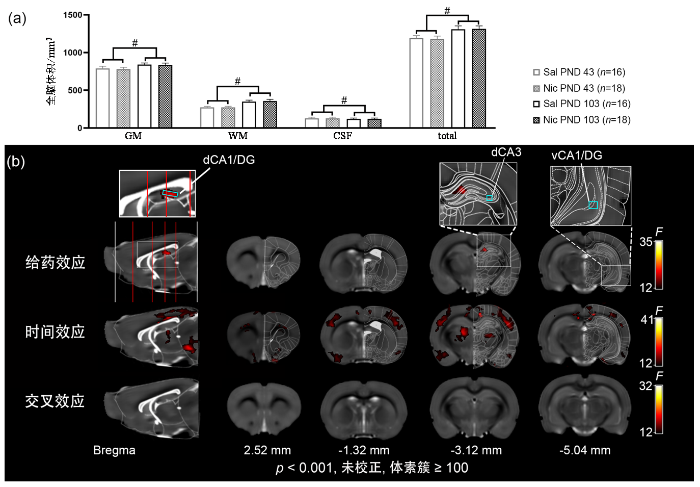
图2
(a)尼古丁对青少年雌性大鼠脑体积的影响,#代表时间效应p < 0.05;(b)体素水平全脑灰质体积的双因素方差分析结果
Fig. 2
(a) Nicotine effects on brain volume of adolescent female rats. # : time effects, p < 0.05; (b) the two-way ANOVA results of the whole brain GM volume at voxel level
图3分别展示了ROI水平海马亚区[dCA1/DG, 背侧海马阿蒙角3区(dCA3), 腹侧海马阿蒙角1区与腹侧齿状回(vCA1/DG)]的GM、WM、CSF体积.双因素方差分析结果显示尼古丁给药在dCA1/DG亚区的GM体积中呈现显著性效应[F (1, 64) = 14.25, p < 0.001]. 经FDR校正的事后检验表明,在年龄相同的雌性大鼠中,Nic组dCA1/DG的GM体积相较于Sal组显著增加(p < 0.02, FDR校正).然而,在年龄相同的雌性大鼠中,Nic组和Sal组的WM、CSF体积均无显著性差异.此外,在相同年龄的雌性大鼠中,尼古丁给药对dCA3和vCA1/DG亚区的GM、WM、CSF体积均无显著性影响,表明尼古丁对海马亚区体积的影响具有区域特异性.
图3
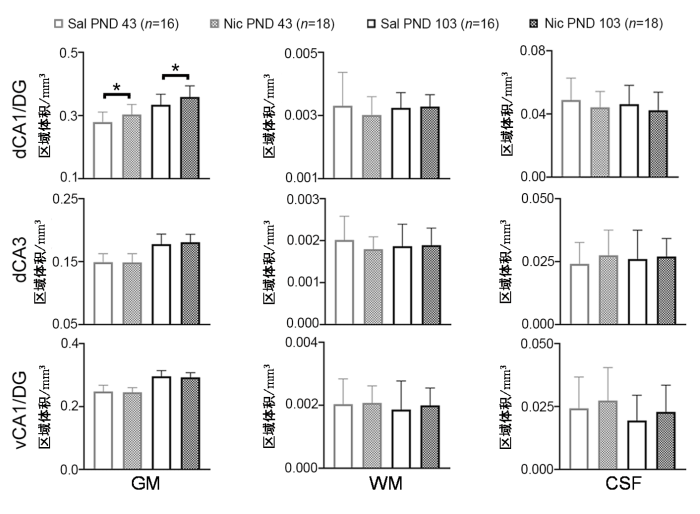
图3
尼古丁对海马亚区体积的短期和长期影响,*代表p < 0.05
Fig. 3
Short- and long-term nicotine effects on volume of hippocampal subregions, * : p < 0.05
2.3 组织化学和免疫组织化学染色结果
图4展示了PND43雌性大鼠背侧海马的组织学染色结果.通过对海马HE染色和GFAP染色观察,未发现Sal组和Nic组有明显的区别.然而,IBA1染色结果则显示,在dCA1亚区Nic组的IOD显著高于Sal组(p < 0.006),同时小胶质细胞胞体面积亦显著增大(p < 0.001).在dDG亚区中,Nic组的IOD较Sal组显著升高(p < 0.05),小胶质细胞胞体面积无显著性差异.在dCA3亚区中,Sal组和Nic组的IOD和小胶质细胞胞体面积均无显著差异.这些结果表明,尼古丁暴露对背侧海马小胶质细胞的激活可能具有区域选择性.
图4
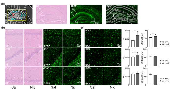
图4
尼古丁对青少年雌性大鼠背侧海马亚区短期内组织学/免疫组织学的影响,*代表p < 0.05. (a) GM增大区域以及HE、GFAP、IBA1染色的背侧海马冠状面展示,正方形方框代表亚区的位置;(b)背侧海马亚区的HE、GFAP染色代表图;(c)背侧海马亚区IBA1染色代表图以及IOD与小胶质细胞胞体面积的统计结果
Fig. 4
Short-term nicotine effects on histology / immunohistology of dorsal hippocampal subregions in adolescent female rats, * : p < 0.05. (a) Dorsal hippocampal coronal sections of the GM enlarged regions and HE, GFAP, and IBA1staining, with square boxes indicating the locations of subregions; (b) representative images of HE and GFAP staining in subregions of the dorsal hippocampus; (c) representative images of IBA1 staining in subregions of the dorsal hippocampus, along with statistical results of IOD and microglial soma area
3 讨论
这一现象说明,观察到的大鼠脑结构变化和行为学改变并非继发于全身性紊乱,而是尼古丁直接神经调控的结果.图1(b)中白色部分大鼠的癫痫评分均为3分.也就是说,2 mg/kg的尼古丁给药在所有青少年雌性大鼠中诱导了3~5级癫痫发作,且癫痫发作强度未随时间变化呈现致敏性或耐受趋势,说明其癫痫效应是一过性的.这与我们团队在青少年雄性大鼠中观察到的结果一致[26],暗示本研究中尼古丁诱发的癫痫行为可能不受性别因素的显著调节.在认知功能方面,本研究通过NORT发现Nic组大鼠在PND 103时表现出新物体识别能力显著下降,这一结果与人类吸烟者和青少年成瘾剂量尼古丁暴露的动物展现出海马依赖性记忆缺陷的结果一致[5,8].特别指出的是,Mateos等人[7]采用PND 28~42尼古丁暴露方案,在PND 76检测到雌性大鼠的新物体识别损伤.而本研究将观测时间延长至成年早期(PND 103),说明这种认知功能障碍具有时间延续性.
从机制层面,MRI可观测的GM体积改变可能源于多重组织学因素,例如神经元和神经胶质细胞的数量、形态(分支数、长度、树突棘密度)等[36-
小胶质细胞参与神经发育过程中的突触修剪和神经环路重塑[47,48].发育时期暴露于烟草或尼古丁诱发的大脑结构改变被认为可能与小胶质细胞功能有关[5,49].在本研究PND43发现,尼古丁诱导了雌性大鼠dCA1/DG小胶质细胞的激活,表现为IBA1表达增加和小胶质细胞胞体面积增加,这与先前的研究结果一致[26,50].这种激活可能通过双重机制影响神经发育:青少年时期尼古丁诱导的小胶质细胞多数发挥促炎功能,表现为吞噬与修剪作用[49];另一方面,小胶质细胞能够分泌前体BDNF和成熟BDNF,促进青少年小鼠运动皮层的树突棘的形成[48,51].这种动态拮抗效应可能是海马灰质体积代偿性增大的结构基础.
4 结论
本研究证实,15天的尼古丁重复暴露可显著诱导青少年雌性大鼠持续性的海马GM体积的增大.其GM体积增大可能与青少年时期小胶质细胞的异常激活和成年期识别记忆功能障碍有关.这些发现揭示了尼古丁暴露通过小胶质细胞介导的神经调控作用干扰雌性大鼠海马正常发育的潜在机制,特别凸显了青少年雌性大鼠海马对尼古丁的高度敏感性.该结果为青少年吸烟者海马异常的结构与功能提供了重要的实验依据,提示靶向小胶质细胞稳态可能成为干预尼古丁所致神经发育障碍的新策略.
致谢
感谢中国科学院精密测量科学与技术创新研究院的安萍萍老师对动物饲养提供的帮助.感谢同单位司腾霄博士对行为学实验提供的帮助和建议.
利益冲突
无
参考文献
Protecting children and adolescents from tobacco and nicotine
[J].
The structural reorganization of the prefrontal cortex during adolescence as a framework for vulnerability to the environment
[J].DOI:10.1016/j.pbb.2020.173044 URL [本文引用: 2]
Development of the motivational system during adolescence, and its sensitivity to disruption by nicotine
[J].DOI:10.1016/j.dcn.2011.05.010 URL [本文引用: 1]
Cigarette use, anxiety, and insomnia from adolescence to early adulthood: A longitudinal indirect effects test
[J].DOI:10.1016/j.addbeh.2021.106981 URL [本文引用: 1]
Nicotine use during late adolescence and young adulthood is associated with changes in hippocampal volume and memory performance
[J].
Lasting synaptic changes underlie attention deficits caused by nicotine exposure during adolescence
[J].
DOI:10.1038/nn.2770
PMID:21336271
[本文引用: 1]

Tobacco smoking and nicotine exposure during adolescence interfere with prefrontal cortex (PFC) development and lead to cognitive impairments in later life. The molecular and cellular underpinnings of these consequences remain elusive. We found that adolescent nicotine exposure induced lasting attentional disturbances and reduced mGluR2 protein and function on presynaptic terminals of PFC glutamatergic synapses. Restoring mGluR2 activity in vivo by local infusion of a group II mGluR agonist in adult rats that received nicotine as adolescents rescued attentional disturbances.
Adolescent exposure to nicotine and/or the cannabinoid agonist CP 55,940 induces gender-dependent long-lasting memory impairments and changes in brain nicotinic and CB(1) cannabinoid receptors
[J].
DOI:10.1177/0269881110370503
PMID:20562169
[本文引用: 2]
We have analysed the long-term effects of adolescent (postnatal day 28-43) exposure of male and female rats to nicotine (NIC, 1.4 mg/kg/day) and/or the cannabinoid agonist CP 55,940 (CP, 0.4 mg/kg/day) on the following parameters measured in the adulthood: (1) the memory ability evaluated in the object location task (OL) and in the novel object test (NOT); (2) the anxiety-like behaviour in the elevated plus maze; and (3) nicotinic and CB(1) cannabinoid receptors in cingulated cortex and hippocampus. In the OL, all pharmacological treatments induced significant decreases in the DI of females, whereas no significant effects were found among males. In the NOT, NIC-treated females showed a significantly reduced DI, whereas the effect of the cannabinoid agonist (a decrease in the DI) was only significant in males. The anxiety-related behaviour was not changed by any drug. Both, nicotine and cannabinoid treatments induced a long-lasting increase in CB(1) receptor activity (CP-stimulated GTPγS binding) in male rats, and the nicotine treatment also induced a decrease in nicotinic receptor density in the prefrontal cortex of females. The results show gender-dependent harmful effects of both drugs and long-lasting changes in CB(1) and nicotinic receptors.
Sex differences in adult cognitive deficits after adolescent nicotine exposure in rats
[J].
DOI:10.1016/j.ntt.2013.05.001
PMID:23673345
[本文引用: 2]
This study was designed to determine whether deficits in adult serial pattern learning caused by adolescent nicotine exposure persist as impairments in asymptotic performance, whether adolescent nicotine exposure differentially retards learning about pattern elements that are inconsistent with "perfect" pattern structure, and whether there are sex differences in rats' response to adolescent nicotine exposure as assessed by a serial multiple choice task. The current study replicated the results of our initial report (Fountain et al., 2008) using this task by showing that adolescent nicotine exposure (1.0mg/kg/day nicotine for 35days) produced a specific cognitive impairment in male rats that persisted into adulthood at least a month after adolescent nicotine exposure ended. In addition, sex differences were observed even in controls, with additional evidence that adolescent nicotine exposure significantly impaired learning relative to same-sex controls for chunk boundary elements in males and for violation elements in females. All nicotine-induced impairments were overcome by additional training so that groups did not differ at asymptote. An examination of the types of errors rats made indicated that adolescent nicotine exposure slowed learning without affecting rats' cognitive strategy in the task. This data pattern suggests that exposure to nicotine in adolescence may have impaired different aspects of adult stimulus-response discrimination learning processes in males and females, but left abstract rule learning processes relatively spared in both sexes. These effects converge with other findings in the field and reinforce the concern that adolescent nicotine exposure poses an important threat to cognitive capacity in adulthood. Copyright © 2013 Elsevier Inc. All rights reserved.
Nicotine arms race: JUUL and the high-nicotine product market
[J].
DOI:10.1136/tobaccocontrol-2018-054796
PMID:30733312
[本文引用: 2]
Until recently, purveyors of vaping products marketed e-liquids in the 1%-3% range of nicotine concentration with those at 3% described as 'super high' intended for two packs/day smokers. The goal of this study is to evaluate the degree to which JUUL, with its 5% nicotine and 75% US market share, has spurred other e-liquid vendors to raise the nicotine levels of their products.Online search to identify brands offering e-liquid in exceptionally high nicotine concentration (≥5%) and to catalogue the appearance of devices which emulate JUUL.JUUL compatible pods (14) and JUUL knock off devices (39) were identified which offer equal or higher nicotine than JUUL. More than 70 e-liquid brands sell high-nicotine products (≥5%) in bulk (≥30 mL) equivalent to >40 cigarette packs. All of these products come in multiple youth appealing sweet and fruity flavours. It was noted that nicotine percentage is inconsistently reported (eg, JUUL is 5% by weight vs 5.9% by volume).JUUL has triggered a widespread rush among aerosol purveyors to market e-liquid in unprecedentedly high nicotine concentrations. The rapidly rising popularity of high-nicotine e-liquids threatens to addict a generation of youth. When sold in large quantity bottles (eg, 30 mL) they represent a childhood poisoning risk. Labelling of nicotine concentration in e-liquids needs to be standardised to avoid consumer confusion. The addictiveness and toxicity of these products makes it imperative that regulators act swiftly to enact protective measures.© Author(s) (or their employer(s)) 2019. No commercial re-use. See rights and permissions. Published by BMJ.
Seizures after vaping nicotine in youth: A canary or a red herring?
[J].DOI:10.1016/j.jadohealth.2019.10.016 URL
Severe acute toxicity of inhaled nicotine and e-cigarettes
[J].DOI:10.1016/j.chest.2019.10.008 URL
Adverse experience reports of seizures in youth and young adult electronic nicotine delivery systems users
[J].DOI:10.1016/j.jadohealth.2019.10.002 URL [本文引用: 1]
Nicotine elicits convulsive seizures by activating amygdalar neurons
[J].
DOI:10.3389/fphar.2017.00057
PMID:28232801
[本文引用: 2]
Nicotinic acetylcholine (nACh) receptors are implicated in the pathogenesis of epileptic disorders; however, the mechanisms of nACh receptors in seizure generation remain unknown. Here, we performed behavioral and immunohistochemical studies in mice and rats to clarify the mechanisms underlying nicotine-induced seizures. Treatment of animals with nicotine (1-4 mg/kg, i.p.) produced motor excitement in a dose-dependent manner and elicited convulsive seizures at 3 and 4 mg/kg. The nicotine-induced seizures were abolished by a subtype non-selective nACh antagonist, mecamylamine (MEC). An alpha 7 nACh antagonist, methyllycaconitine, also significantly inhibited nicotine-induced seizures whereas an alpha 4 beta 2 nACh antagonist, dihydro-beta-erythroidine, affected only weakly. Topographical analysis of Fos protein expression, a biological marker of neural excitation, revealed that a convulsive dose (4 mg/kg) of nicotine region-specifically activated neurons in the piriform cortex, amygdala, medial habenula, paratenial thalamus, anterior hypothalamus and solitary nucleus among 48 brain regions examined, and this was also suppressed by MEC. In addition, electric lesioning of the amygdala, but not the piriform cortex, medial habenula and thalamus, specifically inhibited nicotine-induced seizures. Furthermore, microinjection of nicotine (100 and 300 mu g/side) into the amygdala elicited convulsive seizures in a dose-related manner. The present results suggest that nicotine elicits convulsive seizures by activating amygdalar neurons mainly via alpha 7 nACh receptors.
Relationship between nicotine-induced seizures and hippocampal nicotinic receptors
[J].A controversy has existed for several years concerning the physiological relevance of the nicotinic receptor measured by alpha-bungarotoxin binding. Using mice derived from a classical F2 and backcross genetic design, a relationship between nicotine-induced seizures and alpha-bungarotoxin nicotinic receptor concentration was found. Mice sensitive to the convulsant effects of nicotine had greater alpha-bungarotoxin binding in the hippocampus than seizure insensitive mice. The binding sites from seizure sensitive and resistant mice were equally affected by treatment with dithiothreitol, trypsin or heat. Thus it appears that the difference between seizure sensitive and insensitive animals may be due to a difference in hippocampal nicotinic receptor concentration as measured with alpha-bungarotoxin binding.
Self-administration of nicotine and cigarette smoke extract in adolescent and adult rats
[J].
DOI:S0028-3908(16)30279-9
PMID:27346207
[本文引用: 1]
Although smoking initiation typically occurs during adolescence, most preclinical studies of tobacco use involve adult animals. Furthermore, their focus is largely on nicotine alone, even though cigarette smoke contains thousands of constituents. The present study therefore aimed to determine whether aqueous constituents in cigarette smoke affect acquisition of nicotine self-administration during adolescence in rats. Adolescent and adult male rats, aged postnatal day (P) 25 and 85, respectively, were food trained on a fixed ratio 1 (FR1) schedule, then allowed to self-administer one of 5 doses of nicotine (0, 3.75, 7.5, 15, or 30 μg/kg) or aqueous cigarette smoke extract (CSE) with equivalent nicotine content. Three progressively more difficult schedules of reinforcement, FR1, FR2, and FR5, were used. Both adolescent and adult rats acquired self-administration of nicotine and CSE. Nicotine and CSE similarly increased non-reinforced responding in adolescents, leading to enhanced overall drug intake as compared to adults. When data were corrected for age-dependent alterations in non-reinforced responding, adolescents responded more for low doses of nicotine and CSE than adults at the FR1 reinforcement schedule. No differences in adolescent responding for the two drugs were seen at this schedule, whereas adults had fewer responses for CSE than for nicotine. However, when the reinforcement schedule was increased to FR5, animals dose-dependently self-administered both nicotine and CSE, but no drug or age differences were observed. These data suggest that non-nicotine tobacco smoke constituents do not influence the reinforcing effect of nicotine in adolescents.Published by Elsevier Ltd.
Adolescent vs. adult-onset nicotine self-administration in male rats: Duration of effect and differential nicotinic receptor correlates
[J].Adolescence is the life stage when tobacco addiction typically begins. Adolescent neurobehavioral development may be altered by nicotine self-administration in a way that persistently potentiates addiction. Previously, we showed that female adolescent rats self-administer more nicotine than do adults and that the increased nicotine intake then persists through the transition to adulthood [E.D. Levin, A. Rezvani, D. Montoya, J. Rose, H. Swartzwelder, Adolescent-onset nicotine self-administration modeled in female rats, Psychopharmacology 169 (2003) 141-149.]. In the current study, male Sprague-Dawley rats were given access to nicotine via the standard operant IV self-administration procedure (nicotine bitartrate dose of 0.03 mg/kg/infusion). One group of male rats started during adolescence the other group started in young adulthood. After the end of the four-week period of self-administration brain regions of the rats were assessed for alpha4beta2 nicotinic receptor binding. We found that male rats, like females, show higher nicotine self-administration when starting during adolescence as compared to starting in adulthood (p<0.001). Indeed, the effect in adolescent males was even greater than that in females, with more than triple the rate of nicotine self-administration vs. the adult-onset group during the first 2 weeks. The adolescent onset nicotine-self-administering rats also had significantly greater high affinity nicotinic receptor binding in the midbrain and the striatum, whereas hippocampal binding did not differ between the age groups. Striatal values significantly correlated with nicotine self-administration during the first 2 weeks in the adult-onset group but not the adolescent-onset rats, suggesting that the differences in self-administration may depend in part on underlying disparities in synaptic responses to nicotine. After the initial 2 weeks, nicotine self-administration in male rats declined toward adult-like levels, as the adolescent rats approached adulthood. This study showed that adolescent male rats self-administer significantly more nicotine than do male adult rats, but that adolescent-onset nicotine self-administration in male rats declines over weeks of continued use to approach adult-onset levels. In a previous study, we found that female rats also show greater nicotine self-administration with adolescent onset vs. adult onset, but that the females continued higher rates of self-administration into adulthood. Our results thus reinforce the concept that the adolescent brain is unusually receptive to the effects of nicotine in a manner that reinforces the potential for addiction.
Age differences in conditioned place preferences and taste aversions to nicotine
[J].
DOI:10.1002/dev.21400
PMID:27027859
[本文引用: 2]
Adolescents and adults differ in their behavioral sensitivities to drugs of abuse, including nicotine. Studies have shown that both rewarding and aversive properties of drugs of abuse can exist concomitantly. The present study investigated the ontogeny of these opposing qualities across a range of doses using a combined conditioned taste aversion and place preference paradigm in pair-housed rats that were not deprived of food or water. Results indicated that adolescents were more sensitive to the rewarding properties of nicotine than adults. In contrast, although all doses produced a taste aversion at both ages in the same rats, the aversion was weaker at lower than high doses in adolescents whereas adults showed strong aversion at all doses, suggesting modest attenuation in nicotine's aversive properties among adolescents relative to adults. Thus, attenuated aversive and accented appetitive sensitivities of adolescents to nicotine can be experienced simultaneously in the same animals. © 2016 Wiley Periodicals, Inc. Dev Psychobiol 58: 660-666, 2016.© 2016 Wiley Periodicals, Inc.
Enhanced vulnerability to the rewarding effects of nicotine during the adolescent period of development
[J].
DOI:10.1016/j.pbb.2008.05.009
PMID:18571223
[本文引用: 1]
This study compared the rewarding and aversive effects of nicotine in adolescent, adult, and adult rats preexposed to nicotine during adolescence. Prior to conditioning, the rats were tested for their initial preference for either of 2 distinct compartments. Adolescent and adult rats then received various nicotine doses in their initially non-preferred side on one day and saline in the other side on alternate days. This 2-day procedure was repeated over 8 consecutive days. Following conditioning, rats were re-tested for their preference. Another cohort of adolescent and adult rats were conditioned with various doses of D-amphetamine. Nicotine produced CPP in an inverted U-shaped manner in both age groups. However, adolescents displayed a larger upward shift in CPP that was significant across a wider dose range relative to adults. There were no developmental differences to CPP produced by D-amphetamine. In a final study, adolescents were prepared with pumps that delivered nicotine for 14 days. These rats were conditioned later as adults using the same procedures used previously. Pre-exposure to nicotine during adolescence diminished the aversive effects produced by the highest nicotine dose in naive adults. Taken together, these studies provide a basis for enhanced vulnerability to nicotine during adolescence.
Nicotine as a modulator of behavior: beyond the inverted U
[J].Nicotine is the crucial component in tobacco that underlies smoking behavior; however, the effects of nicotine can vary in both human and animal studies. Recent data from knockout mouse studies, neurotransmitter release studies and electrophysiological experiments support the hypothesis that conflicting behavioral effects elicited by nicotine can result from the activation of different subtypes of nicotinic acetylcholine receptors and the stimulation of antagonistic neuronal pathways. Thus, small differences in the activation state, connectivity or sensitivity of neuronal pathways among individuals might result in large differences in behavioral responses to nicotine. An understanding of the molecular and cellular processes that oppose nicotine reinforcement will be crucial for the development of new interventions to initiate smoking cessation or to prevent the transition from occasional smoking to dependence.
Magnetic resonance imaging the brain structures involved in nicotine susceptibility in rats
[J].
尼古丁易感的脑结构特征的磁共振成像研究
[J].
DOI:10.11938/cjmr20212890
[本文引用: 1]
本文旨在利用磁共振成像手段探究尼古丁易感个体的脑结构特征,即脑结构特性对尼古丁依赖程度的预测.选用成年雄性SD大鼠进行纵向研究,利用基于微型渗透压泵的间歇性给药方式对大鼠进行腹腔注射尼古丁14天,随后强制戒断14天.于第0、15、29天进行躯体戒断行为测试以量化其尼古丁依赖严重程度.对第1天的脑结构图像与戒断行为评分进行回归分析,结果发现尼古丁依赖严重程度与双侧前边缘皮层、左侧颗粒状岛叶皮层灰质体积和双侧丘脑白质体积呈负相关,与右侧海马CA1脑区和左侧丘脑灰质体积呈正相关.以上脑区的结构特征,能够作为尼古丁易感的生物标志物,在个体接触尼古丁之前预测其尼古丁依赖风险,对易感人群进行有针对性的早期干预.
Strain comparison of nicotine-induced seizure sensitivity and nicotinic receptors
[J].Nicotine-induced seizure sensitivity was assessed in 19 inbred mouse strains. Two routes of drug administration were utilized: acute intravenous (IV) infusion and intraperitoneal (IP) injection. Dose-response curves for sensitivity to IP nicotine-induced seizures were constructed for the 19 inbred strains and a heterogeneous stock (HS/Ibg) of mice. Differences were observed among the strains both in ED50 values and slopes of the dose-response curves following IP injection of nicotine. ST/bJ mice were the most sensitive having an ED50 of 2.34 +/- 0.09 mg/kg nicotine. DBA/1J mice were the most resistant strain with an ED50 value of 6.16 +/- 0.02 m/kg nicotine. Latency to clonic seizure was measured in the 19 inbred mouse strains using the acute IV route of drug administration. Again, ST/bJ mice were the most sensitive and DBA/2Ibg were the most resistant to IV nicotine-induced seizures. Significant correlations were observed between latency to IV nicotine-induced seizures and both ED50 values and the slope of the dose-response curves for IP nicotine-induced seizures. However, the pattern of results differed between the two routes of drug administration. The relationship between seizure sensitivity and nicotinic receptor concentration in three brain regions, cortex, midbrain and hippocampus, was also assessed using alpha-bungarotoxin (BTX) as the ligand. A significant relationship was observed between BTX binding in the three brain regions and sensitivity to IV nicotine-induced seizures such that strains with greater concentrations of BTX binding sites were more sensitive to nicotine-induced seizures than were strains with lower concentrations of BTX binding sites.(ABSTRACT TRUNCATED AT 250 WORDS)
Dynamics of hippocampal acetylcholine release during lithium-pilocarpine-induced status epilepticus in rats
[J].
DOI:10.1111/jnc.12787
PMID:24909269
[本文引用: 1]
The lithium-pilocarpine model is a rat model of epilepsy that mimics status epilepticus in humans. Here, we report changes of acetylcholine (ACh) release in the hippocampus before, during and after status epilepticus as monitored by microdialysis in unanesthetized rats. Administration of pilocarpine (30 mg/kg s.c.) to rats pretreated with lithium chloride (127 mg/kg i.p.) caused a massive, six-fold increase of hippocampal ACh release, paralleling the development of tonic seizures. When seizures were stopped by administration of diazepam (10 mg/kg i.p.) or ketamine (75 mg/kg i.p.), ACh levels returned to normal. Extracellular concentrations of glutamate remained unchanged during this procedure. Administration of atropine (1 mg/kg i.p.) 2 h after pilocarpine caused a further increase of ACh but did not affect seizures, whereas injection of mecamylamine (5 mg/kg i.p.) reduced ACh levels and seizures in a delayed fashion. Local infusion of tetrodotoxin, 1 μM locally) or hemicholinium (10 μM locally) strongly reduced ACh release and had delayed effects on seizures. Administration of glucose or inositol (250 mg/kg each i.p.) had no visible consequences. In parallel experiments, lithium-pilocarpine-induced status epilepticus also enhanced striatal ACh release, and hippocampal ACh levels equally increased when status epilepticus was induced by kainate (30 mg/kg i.p.). Taken together, our results demonstrate that seizure development in status epilepticus models is accompanied by massive increases of extracellular ACh, but not glutamate, levels. Treatments that reduce seizure activity also reliably reduce extracellular ACh levels.© 2014 International Society for Neurochemistry.
Extracellular levels of ATP and acetylcholine during lithium-pilocarpine induced status epilepticus in rats
[J].
DOI:10.1016/j.neulet.2015.11.028
PMID:26610905
[本文引用: 1]
Acetylcholine (ACh) and ATP are rapidly acting neurotransmitters with a putative role in epileptic seizures. In the present study we investigated extracellular concentrations of both neurotransmitters in parallel by microdialysis in rat hippocampus. We found that infusion of neostigmine increases, while calcium-free perfusion and infusion of tetrodotoxin (TTX) decreases, ACh levels. Calcium-free perfusion also decreased ATP levels which were, however, not affected by neostigmine or TTX. During status epilepticus, ACh levels were increased threefold but returned to baseline after the termination of seizures by diazepam. ATP levels were unchanged during status epilepticus but a several-fold increase was seen when AOPCP, an inhibitor of 5'-endonucleotidase, was infused. The results demonstrate an increase of ATP levels during epileptic seizures which, however, was not of neuronal origin. Copyright © 2015 Elsevier Ireland Ltd. All rights reserved.
Mecamylamine inhibits seizure-like activity in CA1-CA3 hippocampus through antagonism to nicotinic receptors
[J].
Repeated exposure to high-dose nicotine induces prefrontal gray matter atrophy in adolescent male rats
[J].DOI:10.1016/j.neuroscience.2024.11.059 URL [本文引用: 3]
Differences in vulnerability to nicotine-induced kindling between female and male periadolescent rats
[J].DOI:10.1007/s00213-012-2799-5 URL [本文引用: 1]
An animal model of adolescent nicotine exposure: effects on gene expression and macromolecular constituents in rat brain regions
[J].Nearly all smokers begin tobacco use in adolescence, and approximately 25% of US teenagers are daily smokers. Prenatal nicotine exposure is known to produce brain damage, to alter synaptic function and to cause behavioral anomalies, but little or no work has been done to determine if the adolescent brain is also vulnerable. We examined the effect of adolescent nicotine exposure on indices of cell damage in male and female rats with an infusion paradigm designed to match the plasma levels found in human smokers or in users of the transdermal nicotine patch. Measurements were made of DNA and protein as well as expression of mRNAs encoding genes involved in differentiation and apoptosis (p53, c-fos) in cerebral cortex, midbrain and hippocampus. Following nicotine treatment from postnatal days 30-47.5, changes in macromolecular constituents indicative of cell loss (reduced DNA) and altered cell size (protein/DNA ratio) were seen across all three brain regions. In addition, expression of p53 showed region- and gender-selective alterations consistent with cell damage; c-fos, which is constitutively overexpressed after gestational nicotine exposure, was unaffected with the adolescent treatment paradigm. Although these measures indicate that the fetal brain is more vulnerable to nicotine than is the adolescent brain, the critical period for nicotine-induced developmental neurotoxicity clearly extends into adolescence. Effects on gene expression and cell number, along with resultant or direct effects on synaptic function, may contribute to increased addictive properties and long-term behavioral deficits.
Sex-selective hippocampal alterations after adolescent nicotine administration: Effects on neurospecific proteins
[J].Nicotine is a neuroteratogen that targets cell development and synaptic function into adolescence, when smoking typically commences. We used a rat model of adolescent nicotine exposure to characterize the types of cells involved in hippocampal alterations. Nicotine was given to adolescent rats by minipump infusions from postnatal day (PN) 30 to PN47.5, using a dose rate (6 mg/kg/day) that replicates the plasma nicotine levels found in smokers. We examined specific neuronal and astrocyte proteins in the posttreatment period (PN50, PN60), when deficits in neurotransmission first appear: glial fibrillary acidic protein (GFAP), a marker for astrocytes; neurofilament 68-kDa protein (NF68), which is concentrated in the neuronal perikaryon and proximal neurites; and neurofilament 200-kDa protein (NF200), which is found in axonal projections distal to the perikaryon. Adolescent nicotine treatment evoked a significant decrease across all three markers, with the effect restricted to females and showing intensification between PN50 and PN60. These changes correspond to the sex-selectivity and temporal course over which other biomarkers indicate hippocampal cell damage and alterations in synaptic function. We conclude that administration of nicotine to adolescent rats alters neuroproteins in the female hippocampus during withdrawal, effects that could contribute to neurobehavioral deficits.
The adolescent brain and age-related behavioral manifestations
[J].
DOI:10.1016/s0149-7634(00)00014-2
PMID:10817843
[本文引用: 1]
To successfully negotiate the developmental transition between youth and adulthood, adolescents must maneuver this often stressful period while acquiring skills necessary for independence. Certain behavioral features, including age-related increases in social behavior and risk-taking/novelty-seeking, are common among adolescents of diverse mammalian species and may aid in this process. Reduced positive incentive values from stimuli may lead adolescents to pursue new appetitive reinforcers through drug use and other risk-taking behaviors, with their relative insensitivity to drugs supporting comparatively greater per occasion use. Pubertal increases in gonadal hormones are a hallmark of adolescence, although there is little evidence for a simple association of these hormones with behavioral change during adolescence. Prominent developmental transformations are seen in prefrontal cortex and limbic brain regions of adolescents across a variety of species, alterations that include an apparent shift in the balance between mesocortical and mesolimbic dopamine systems. Developmental changes in these stressor-sensitive regions, which are critical for attributing incentive salience to drugs and other stimuli, likely contribute to the unique characteristics of adolescence.
Adolescent nicotine-induced dendrite remodeling in the nucleus accumbens is rapid, persistent, and D1-dopamine receptor dependent
[J].
DOI:10.1007/s00429-014-0897-3
PMID:25257604
[本文引用: 1]
Chronic nicotine exposure during adolescence induces dendritic remodeling of medium spiny neurons (MSNs) in the nucleus accumbens (NAcc) shell. While nicotine-induced dendritic remodeling has frequently been described as persistent, the trajectory of dendrite remodeling is unknown. Specifically, no study to date has characterized the structural plasticity of dendrites in the NAcc immediately following chronic nicotine, leaving open the possibility that dendrite remodeling emerges gradually over time. Further, the neuropharmacological mechanisms through which nicotine induces dendrite remodeling are not well understood. To address these questions, rats were co-administered chronic nicotine (0.5 mg/kg) and the D1-dopamine receptor (D1DR) antagonist SCH-23390 (0.05 mg/kg) subcutaneously every other day during adolescence. Brains were then processed for Golgi-Cox staining either 1 day or 21 days following drug exposure and dendrites from MSNs in the NAcc shell digitally reconstructed in 3D. Spine density was also measured at both time points. Our morphometric results show (1) the formation of new dendritic branches and spines 1 day following nicotine exposure, (2) new dendritic branches, but not spine density, remains relatively stable for at least 21 days, (3) the co-administration of SCH-23390 completely blocked nicotine-induced dendritic remodeling of MSNs at both early and late time points, suggesting the formation of new dendritic branches in response to nicotine is D1DR-dependent, and (4) SCH-23390 failed to block nicotine-induced increases in spine density. Overall this study provides new insight into how nicotine influences the normal trajectory of adolescent brain development and demonstrates a persistent form of nicotine-induced neuroplasticity in the NAcc shell that develops rapidly and is D1DR dependent.
Long-lasting cognitive deficits resulting from adolescent nicotine exposure in rats
[J].
DOI:10.1038/npp.2008.96
PMID:18580873
[本文引用: 1]
Adolescence is a developmental period, during which the brain and particularly medial prefrontal cortical (mPFC) regions thereof have not fully matured. Because epidemiological data have suggested that adolescent nicotine use may result in disturbances in cognitive function in adulthood, we investigated the long-term effects of adolescent nicotine exposure in rats. Male Wistar rats were exposed to either nicotine (three times daily, 0.4 mg/kg s.c.) or saline for 10 days during (postnatal day (PND) 34-43) or following (PND 60-69) adolescence. After 5 weeks during adulthood, separate groups of animals were tested in operant paradigms taxing attention and distinct measures of impulsivity. Visuospatial attention and impulsive action were tested in the five-choice serial reaction time task, whereas impulsive choice was assessed in the delayed reward task. Our data show that adolescent, but not postadolescent, nicotine exposure affects cognitive performance in adulthood and results in diminished attentional performance and increments in impulsive action, while leaving impulsive choice intact. This altered cognitive performance appeared to be associated with enhanced releasability of dopamine in the mPFC. Together, these data suggest that adolescence is a time window during which the brain is vulnerable to long-lasting cognitive disturbances resulting from nicotine exposure.
Chronic exposure to cigarette smoke extract upregulates nicotinic receptor binding in adult and adolescent rats
[J].DOI:10.1016/j.neuropharm.2020.108308 URL [本文引用: 1]
Sensitivity of young rats to nicotine exposure: Physiological and biochemical parameters
[J].
DOI:S0147-6513(08)00095-X
PMID:18462794
[本文引用: 1]
This work has investigated the effects of prolonged exposure of young rats to nicotine on some physiological and biochemical parameters. Wistar male rats (30 days old) were treated (s.c.) with saline or nicotine 5mg/kg/day for 28 or 56 days. They received five injections (1mg/kg) per day (8, 10, 12:00 a.m., 2 and 4:00 p.m.) on the dark period of the cycle. Nicotine exposure for 56 days reduced body and liver weights. Moreover, nicotine exposure for 28 or 56 days decreased the hepatic glycogen but not blood glucose levels. The activities of blood and hepatic PBG-synthase, and blood and cerebral acetylcholinesterase were not affected by in vivo exposure. However, these activities were inhibited by nicotine in vitro. Results show that although high levels of plasma cotinine were found in both intervals of exposures, the parameters here analyzed were not affected by prolonged nicotine exposure except the storage of glucose, and body and liver weights.
Effect of cigarette smoke inhalation on the hippocampus of adult female wistar rat
[J].
Cellular correlates of gray matter volume changes in magnetic resonance morphometry identified by two-photon microscopy
[J].
DOI:10.1038/s41598-021-83491-8
PMID:33608622
[本文引用: 1]
Magnetic resonance imaging (MRI) of the brain combined with voxel-based morphometry (VBM) revealed changes in gray matter volume (GMV) in various disorders. However, the cellular basis of GMV changes has remained largely unclear. We correlated changes in GMV with cellular metrics by imaging mice with MRI and two-photon in vivo microscopy at three time points within 12 weeks, taking advantage of age-dependent changes in brain structure. Imaging fluorescent cell nuclei allowed inferences on (i) physical tissue volume as determined from reference spaces outlined by nuclei, (ii) cell density, (iii) the extent of cell clustering, and (iv) the volume of cell nuclei. Our data indicate that physical tissue volume alterations only account for 13.0% of the variance in GMV change. However, when including comprehensive measurements of nucleus volume and cell density, 35.6% of the GMV variance could be explained, highlighting the influence of distinct cellular mechanisms on VBM results.
NMDA receptor antagonism by repetitive MK801 administration induces schizophrenia-like structural changes in the rat brain as revealed by voxel-based morphometry and diffusion tensor imaging
[J].
DOI:10.1016/j.neuroscience.2016.02.043
PMID:26917273
Animal models of N-methyl-d-aspartate receptor (NMDAR) antagonism have been widely used for schizophrenia research. Less is known whether these models are associated with macroscopic brain structural changes that resemble those in clinical schizophrenia.Magnetic resonance imaging (MRI) was used to measure brain structural changes in rats subjected to repeated administration of MK801 in a regimen (daily dose of 0.2mg/kg for 14 consecutive days) known to be able to induce schizophrenia-like cognitive impairments.Voxel-based morphometry (VBM) revealed significant gray matter (GM) atrophy in the hippocampus, ventral striatum (vStr) and cortex. Diffusion tensor imaging (DTI) demonstrated microstructural impairments in the corpus callosum (cc). Histopathological results corroborated the MRI findings.Treatment-induced behavioral abnormalities were not measured such that correlation between the brain structural changes observed and schizophrenia-like behaviors could not be established.Chronic MK801 administration induces MRI-observable brain structural changes that are comparable to those observed in schizophrenia patients, supporting the notion that NMDAR hypofunction contributes to the pathology of schizophrenia. Imaging-derived brain structural changes in animal models of NMDAR antagonism may be useful measurements for studying the effects of treatments and interventions targeting schizophrenia.Copyright © 2016 IBRO. Published by Elsevier Ltd. All rights reserved.
STZ-induced progressive brain atrophy studied by magnetic resonance imaging and histochemical staining
[J].
STZ诱导大鼠1型糖尿病进行性脑萎缩的磁共振成像及组织化学研究
[J].
DOI:10.11938/cjmr20150305
[本文引用: 1]
1型糖尿病(T1DM)是一种慢性代谢疾病,主要表现为胰岛素分泌量较正常情况下降,会对人体的多个器官和系统造成持续性的损伤.关于糖尿病的横向研究发现糖尿病患者相比于正常人存在着显著的脑萎缩,但关于糖尿病引起的脑萎缩随时间发生进行性改变的研究比较少见.实验采用腹腔注射链脲佐菌素(STZ)来诱导建立大鼠的1型糖尿病模型,运用磁共振成像(MRI)的方法对萎缩的脑区进行定位并在造模后12周和20周两个时间点对脑萎缩的程度进行对比分析,然后运用组织化学染色的方法观察在MRI上出现进行性萎缩的脑区中的神经元所发生的病理改变.MRI的结果表明:STZ诱导的T1DM大鼠相比于正常对照组大鼠出现了显著性的全脑体积、灰质体积和白质体积的萎缩,并且在多个白质脑区和灰质脑区均出现了萎缩程度随着病程的延长而逐渐加重.组织化学染色的结果发现,STZ诱导的T1DM大鼠相对于正常对照组大鼠在体感皮层、运动皮层和海马CA3区,均出现明显的神经元萎缩现象.
Nicotine is a neurotoxin in the adolescent brain: critical periods, patterns of exposure, regional selectivity, and dose thresholds for macromolecular alterations
[J].DOI:10.1016/S0006-8993(03)02885-3 URL [本文引用: 1]
Effects of nicotine on neurogenesis and plasticity of hippocampal neurons
[J].To study nicotine's effects on neurogenesis in the dentate gyrus of the hippocampus, nicotine was injected intraperitoneally into adult rats. After sacrificing, the hippocampal formation was processed for immunohistochemical staining of PSA-NCAM, NeuN and GFAP. Nicotine decreased numbers of PSA-NCAM(+) and NeuN(+) cells dose-dependently.
Tobacco smoke diminishes neurogenesis and promotes gliogenesis in the dentate gyrus of adolescent rats
[J].
DOI:10.1016/j.brainres.2011.07.041
PMID:21840504
[本文引用: 1]
Brain disorders and environmental factors can affect neurogenesis and gliogenesis in the hippocampus. These studies investigated the effects of chronic exposure to tobacco smoke on progenitor cell proliferation and the survival and phenotype of new cells in the dentate gyrus of adolescent rats. The rats were exposed to tobacco smoke for 4h/day for 14 days. To investigate cell proliferation, the exogenous marker 5-bromo-2'-deoxyuridine (BrdU, 200mg/kg, ip) was administered 2h into the 4-h smoke exposure session on day 14. The rats were sacrificed 2-4h after the administration of BrdU. To investigate cell survival, the same dose of BrdU was administered 24h before the start of the 14-day smoke exposure period. These rats were sacrificed 24h after the last smoke exposure session. Tobacco smoke exposure decreased both the number of dividing progenitor cells (-19%) and the number of surviving new cells (-20%), labeled with BrdU in the dentate gyrus. The decrease in cell proliferation was not associated with an increase in apoptotic cell death, as shown by TUNEL analysis. Colocalization studies indicated that exposure to tobacco smoke decreased the number of new immature neurons (BrdU/DCX-positive) and transition neurons (BrdU/DCX/NeuN-positive) and increased the number of new glial cells (BrdU/GFAP-positive). These findings demonstrate that exposure to tobacco smoke diminishes neurogenesis and promotes gliogenesis in the dentate gyrus of adolescent rats. These effects may play a role in the increased risk for depression and cognitive impairment in adolescent smokers.Copyright © 2011 Elsevier B.V. All rights reserved.
Nicotine causes age-dependent changes in gene expression in the adolescent female rat brain
[J].Humans often start smoking during adolescence. Recent results suggest that rodents may also be particularly vulnerable to nicotine dependence during adolescence. We examined the effect of chronic nicotine exposure on gene expression profiles during adolescence in female rats, who were dosed with nicotine (and control animals were dosed with saline) via subcutaneously implanted osmotic minipumps. Brain samples were collected at four ages: before puberty (postnatal day 25), at about the time of puberty in females (postnatal day 35), and after puberty (postnatal days 45 and 55). The expression of 7931 genes in three brain areas was measured using DNA microarrays. Quantitative RT-PCR was also employed to confirm the expression patterns of selected genes. We used a novel clustering technique (principal cluster analysis) to classify 162 nicotine-regulated genes into five clusters, of which only one (cluster A) showed similar patterns of gene expression across all three brain areas (ventral striatum, prefrontal cortex, and hippocampus). Three clusters of genes (A, B, and C) showed dramatic peaks in their nicotine responses at the same age (p35). The other two clusters (D1 and D2) showed smaller peaks and/or valleys in their nicotine responses at p35 and p45. Thus, the age of maximal gene expression response to nicotine in female rats corresponds approximately to the age of maximal behavioral response and the age of puberty.
Nicotine exposure during adolescence: cognitive performance and brain gene expression in adult heterozygous reeler mice
[J].DOI:10.1007/s00213-013-3388-y URL [本文引用: 1]
Differential expression of Arc mRNA and other plasticity-related genes induced by nicotine in adolescent rat forebrain
[J].Relatively little attention has been focused on mechanisms related to neural plasticity and drug abuse in adolescence, compared with abundant research using adult animal models. As smoking is typically initiated in adolescence, an important question to address is whether the adolescent brain responds differently to nicotine compared with the adult. To investigate this question, we examined the expression of a number of early response genes (arc, c-fos and NGFI-B) that have been implicated in synaptic plasticity and addiction, following acute nicotine in adolescent and adult rats. Baseline expression of arc and c-fos was higher in adolescent brains compared with adults. Following acute nicotine treatment (0.1, 0.4mg/kg), we found a marked induction of arc mRNA in the prefrontal cortex of nicotine-treated adolescents compared with a less pronounced increase of arc in the adult. c-fos and NGFI-B were also upregulated by nicotine, but not in an age-related manner. In contrast, nicotine induced less arc, c-fos, and NGFI-B expression in the somatosensory cortex of adolescents compared with adults. A fourth gene, quinoid dihydropteridine reductase was expressed at lower levels in white matter of the adolescent forebrain compared with the adult, but was not affected by nicotine. These results suggest that in adolescence, the activity of specific early response genes is higher in brain regions critical for emotional regulation and decision-making. Further, nicotine affects key plasticity molecules in these areas in a manner different from the adult. Thus, adolescence may represent a neurobiologically vulnerable period with regard to nicotine exposure.
Adolescent nicotine induces persisting changes in development of neural connectivity
[J].
DOI:10.1016/j.neubiorev.2015.05.019
PMID:26048001
[本文引用: 1]
Adolescent nicotine induces persisting changes in development of neural connectivity. A large number of brain changes occur during adolescence as the CNS matures. These changes suggest that the adolescent brain may still be susceptible to developmental alterations by substances which impact its growth. Here we review recent studies on adolescent nicotine which show that the adolescent brain is differentially sensitive to nicotine-induced alterations in dendritic elaboration, in several brain areas associated with processing reinforcement and emotion, specifically including nucleus accumbens, medial prefrontal cortex, basolateral amygdala, bed nucleus of the stria terminalis, and dentate gyrus. Both sensitivity to nicotine, and specific areas responding to nicotine, differ between adolescent and adult rats, and dendritic changes in response to adolescent nicotine persist into adulthood. Areas sensitive to, and not sensitive to, structural remodeling induced by adolescent nicotine suggest that the remodeling generally corresponds to the extended amygdala. Evidence suggests that dendritic remodeling is accompanied by persisting changes in synaptic connectivity. Modeling, electrophysiological, neurochemical, and behavioral data are consistent with the implication of our anatomical studies showing that adolescent nicotine induces persisting changes in neural connectivity. Emerging data thus suggest that early adolescence is a period when nicotine consumption, presumably mediated by nicotine-elicited changes in patterns of synaptic activity, can sculpt late brain development, with consequent effects on synaptic interconnection patterns and behavior regulation. Adolescent nicotine may induce a more addiction-prone phenotype, and the structures altered by nicotine also subserve some emotional and cognitive functions, which may also be altered. We suggest that dendritic elaboration and associated changes are mediated by activity-dependent synaptogenesis, acting in part through D1DR receptors, in a network activated by nicotine. The adolescent nicotine effects reviewed here suggest that modification of late CNS development constitutes a hazard of adolescent nicotine use. Copyright © 2015 Elsevier Ltd. All rights reserved.
Long-term effects of chronic nicotine on emotional and cognitive behaviors and hippocampus cell morphology in mice: comparisons of adult and adolescent nicotine exposure
[J].
DOI:10.1111/ejn.13398
PMID:27623427
[本文引用: 1]
Nicotine dependence is associated with increased risk for emotional, cognitive and neurological impairments later in life. This study investigated the long-term effects of nicotine exposure during adolescence and adulthood on measures of depression, anxiety, learning and hippocampal pyramidal cell morphology. Mice (C57BL/6J) received saline or nicotine for 12 days via pumps implanted on postnatal day 32 (adolescent) or 54 (adults). Thirty days after cessation of nicotine/saline, mice were tested for learning using contextual fear conditioning, depression-like behaviors using the forced swim test or anxiety-like behaviors with the elevated plus maze. Brains from nicotine- or saline-exposed mice were processed with Golgi stain for whole neuron reconstruction in the CA1 and CA3 regions of the hippocampus. Results demonstrate higher depression-like responses in both adolescent and adult mice when tested during acute nicotine withdrawal. Heightened depression-like behaviors persisted when tested after 30 days of nicotine abstinence in mice exposed as adolescents, but not adults. Adult, but not adolescent, exposure to nicotine resulted in increased open-arm time when tested after 30 days of abstinence. Nicotine exposure during adolescence caused deficits in contextual fear learning indicated by lower levels of freezing to the context as compared with controls when tested 30 days later. In addition, reduced dendritic length and complexity in the apical CA1 branches in adult mice exposed to nicotine during adolescence were found. These results demonstrate that nicotine exposure and withdrawal can have long-term effects on emotional and cognitive functioning, particularly when nicotine exposure occurs during the critical period of adolescence.© 2016 Federation of European Neuroscience Societies and John Wiley & Sons Ltd.
Synaptic pruning by microglia is necessary for normal brain development
[J].
DOI:10.1126/science.1202529
PMID:21778362
[本文引用: 1]
Microglia are highly motile phagocytic cells that infiltrate and take up residence in the developing brain, where they are thought to provide a surveillance and scavenging function. However, although microglia have been shown to engulf and clear damaged cellular debris after brain insult, it remains less clear what role microglia play in the uninjured brain. Here, we show that microglia actively engulf synaptic material and play a major role in synaptic pruning during postnatal development in mice. These findings link microglia surveillance to synaptic maturation and suggest that deficits in microglia function may contribute to synaptic abnormalities seen in some neurodevelopmental disorders.
Microglia promote learning-dependent synapse formation through brain-derived neurotrophic factor
[J].
DOI:10.1016/j.cell.2013.11.030
PMID:24360280
[本文引用: 2]
Microglia are the resident macrophages of the CNS, and their functions have been extensively studied in various brain pathologies. The physiological roles of microglia in brain plasticity and function, however, remain unclear. To address this question, we generated CX3CR1(CreER) mice expressing tamoxifen-inducible Cre recombinase that allow for specific manipulation of gene function in microglia. Using CX3CR1(CreER) to drive diphtheria toxin receptor expression in microglia, we found that microglia could be specifically depleted from the brain upon diphtheria toxin administration. Mice depleted of microglia showed deficits in multiple learning tasks and a significant reduction in motor-learning-dependent synapse formation. Furthermore, Cre-dependent removal of brain-derived neurotrophic factor (BDNF) from microglia largely recapitulated the effects of microglia depletion. Microglial BDNF increases neuronal tropomyosin-related kinase receptor B phosphorylation, a key mediator of synaptic plasticity. Together, our findings reveal that microglia serve important physiological functions in learning and memory by promoting learning-related synapse formation through BDNF signaling. Copyright © 2013 Elsevier Inc. All rights reserved.
Multifactorial etiology of adolescent nicotine addiction: A review of the neurobiology of nicotine addiction and its implications for smoking cessation pharmacotherapy
[J].
DOI:10.3389/fpubh.2021.664748
URL
[本文引用: 2]
Nicotine is the primary pharmacologic component of tobacco, and its highly addictive nature is responsible for its widespread use and significant withdrawal effects that result in challenges to smoking cessation therapeutics. Nicotine addiction often begins in adolescence and this is at least partially attributed to the fact that adolescent brain is most susceptible to the neuro-inflammatory effects of nicotine. There is increasing evidence for the involvement of microglial cells, which are the brain's primary homeostatic sensor, in drug dependence and its associated behavioral manifestations particularly in the adolescent brain. A hallmark of neuro-inflammation is microglial activation and activation of microglia by nicotine during adolescent development, which may result in long-term addiction to nicotine. This non-systematic review examines multifactorial etiology of adolescent nicotine addiction, neurobiology of nicotine addiction and the potential mechanisms that underlie the effects of nicotine on inflammatory signaling in the microglia, understanding how nicotine affects the adolescent brain. We speculate, that modulating homeostatic balance in microglia, could have promising therapeutic potential in withdrawal, tolerance, and abstinence-related neural adaptations in nicotine addiction, in the adolescent brain. Further, we discuss nicotine addiction in the context of the sensitization-homeostasis model which provides a theoretical framework for addressing the potential role of microglial homeostasis in neural adaptations underlying nicotine abuse.
Microglial activation increases cocaine self-administration following adolescent nicotine exposure
[J].
DOI:10.1038/s41467-019-14173-3
PMID:31949158
[本文引用: 1]
With the rise of e-cigarette use, teen nicotine exposure is becoming more widespread. Findings from clinical and preclinical studies show that the adolescent brain is particularly sensitive to nicotine. Animal studies have demonstrated that adolescent nicotine exposure increases reinforcement for cocaine and other drugs. However, the mechanisms that underlie these behaviors are poorly understood. Here, we report reactive microglia are critical regulators of nicotine-induced increases in adolescent cocaine self-administration. Nicotine has dichotomous, age-dependent effects on microglial morphology and immune transcript profiles. A multistep signaling mechanism involving D2 receptors and CX3CL1 mediates nicotine-induced increases in cocaine self-administration and microglial activation. Moreover, nicotine depletes presynaptic markers in a manner that is microglia-, D2- and CX3CL1-dependent. Taken together, we demonstrate that adolescent microglia are uniquely susceptible to perturbations by nicotine, necessary for nicotine-induced increases in cocaine-seeking, and that D2 receptors and CX3CL1 play a mechanistic role in these phenomena.
Brain-derived neurotrophic factor from microglia regulates neuronal development in the medial prefrontal cortex and its associated social behavior
[J].
DOI:10.1038/s41380-024-02413-y
[本文引用: 1]
\n Microglia and brain-derived neurotrophic factor (BDNF) are essential for the neuroplasticity that characterizes critical developmental periods. The experience-dependent development of social behaviors—associated with the medial prefrontal cortex (mPFC)—has a critical period during the juvenile period in mice. However, whether microglia and BDNF affect social development remains unclear. Herein, we aimed to elucidate the effects of microglia-derived BDNF on social behaviors and mPFC development. Mice that underwent social isolation during p21–p35 had increased\n Bdnf\n in the microglia accompanied by reduced adulthood sociability. Additionally, transgenic mice overexpressing microglial\n Bdnf\n —regulated using doxycycline at different time points—underwent behavioral, electrophysiological, and gene expression analyses. In these mice, long-term overexpression of microglial BDNF impaired sociability and excessive mPFC inhibitory neuronal circuit activity. However, administering doxycycline to normalize BDNF from p21 normalized sociability and electrophysiological function in the mPFC, whereas normalizing BDNF from later ages (p45–p50) did not normalize electrophysiological abnormalities in the mPFC, despite the improved sociability. To evaluate the possible role of BDNF in human sociability, we analyzed the relationship between adverse childhood experiences and\n BDNF\n expression in human macrophages, a possible proxy for microglia. Results show that adverse childhood experiences positively correlated with\n BDNF\n expression in M2 but not M1 macrophages. In summary, our study demonstrated the influence of microglial BDNF on the development of experience-dependent social behaviors in mice, emphasizing its specific impact on the maturation of mPFC function, particularly during the juvenile period. Furthermore, our results propose a translational implication by suggesting a potential link between BDNF secretion from macrophages and childhood experiences in humans.\n
