


引言
扩散磁共振成像(Diffusion Magnetic Resonance Imaging,dMRI)能够以非侵入方式表征脑白质组织的微结构特性,是神经退行性疾病监测及神经发育评估的重要技术[1]. 然而,高质量dMRI依赖于高空间分辨率、多壳层和高角度的采集协议,这导致扫描时间延长、噪声敏感性增加,并对患者配合度提出更高要求. 因此,临床常规采集更为常见的是低空间分辨率、单壳层或低方向数的受限数据. 此类数据限制会降低后续微结构成像模型的重建精度,加剧部分容积效应、角度模糊和纤维交叉不可分等问题.
在进展迅速的同时,现有研究呈现出任务分散和模型类别多样化的特点,缺乏从全任务域视角出发的综合性梳理. 为此,本文对近年来深度学习在dMRI超分辨率重建中的研究进行全面回顾,首先介绍对dMRI超分辨率重建问题的定义与分类,随后,在第二章围绕基础扩散指标、高阶微结构指标和纤维方向分布函数(fiber Orientation Distribution Function,fODF)三类重建任务,系统梳理其算法模型与技术演进,最后总结并提出未来发展方向,为后续研究与临床应用提供统一、系统且具前瞻性的技术视角.
1 dMRI超分辨率重建
1.1 研究意义与本文介绍范围
本文扩散磁共振图像超分辨率重建是指在受限的扩散加权成像(Diffusion Weighted Imaging,DWI)采样信号基础上,利用深度学习技术,重建出高于传统dMRI模型拟合得到的角度或空间分辨率成像参数,实现基于更少的采集数据输出更高质量的微结构成像指标或fODF的方法. 其核心是信息的生成与补偿,以解决扫描时间与成像质量的临床矛盾.
传统图像处理中,超分辨率特指分辨率的直接提升. 在dMRI中,空间超分辨率旨在克服厚层和大体素,而更重要的是增加采样方向数以解析复杂纤维结构的角度(即q空间)超分辨率. 由于DWI的采集涉及壳层数和每个壳的采集方向数,角度分辨率又分为壳层维度和方向维度. 然而,深度学习的端到端特性催生稀疏采样下的参数直接估计这种重要范式. 这类方法直接从高度稀疏的采样数据中回归出本需密集采集才能稳定拟合的可用结果,虽不直接改变原始DWI信号的空间或角向采样格点,但通过隐式学习信息补偿机制,达成了与显式超分辨率相同的临床应用目标. 这些方法解决同一临床痛点,技术同源与交融,在评估与应用层面面临相似挑战.
因此,本文采用广义视角,将上述两类方法统归于基于深度学习的dMRI信息补偿技术框架下进行综述. 本文不仅关注传统的超分辨率,也涵盖利用强大模型直接优化最终输出质量的前沿范式. 通过系统回顾不同模型所依赖的先验知识及其信息生成机制,旨在阐明dMRI领域如何从有限观测中生成可信的高分辨率信息这一核心问题,以期为研究者应对不同实际需求提供全面的技术参考.
1.2 超分辨率重建方法分类
表1 dMRI中的重建应用主要涉及的任务
Table 1
| 分类 | 典型模型 | 典型采集参数 | 目标输出 | 架构发展 |
|---|---|---|---|---|
| 基础扩散指标重建 | DTI:估计主扩散方向及 各向异性特征 | b=1000 s/mm2, 6~30个方向 | 张量参数FA、MD、AD、RD等 | 主要是数据驱动,也有考虑旋转等变性 |
| 高阶微结构指标重建 | NODDI:区分神经突密度、取向分散和自由水 | b=700 s/mm2,90个方向 b=2000 s/mm2,60个方向 | 微结构指标NDI、ODI、fISO等 | 数据驱动、模型驱动、旋转等变性 |
| fODF分辨率提升重建 | fODF:解析交叉纤维等 复杂纤维构型 | b=3000 s/mm2, 60~90个方向 | fODF估计、下游任务有纤维追踪、脑连接组等 | 替代球面反卷积计算,或在单壳重建fODF上增强 |
扩散张量成像(Diffusion Tensor Imaging,DTI)[5]通过张量拟合描述各向异性扩散,常以单壳与较少方向数完成张量估计. DTI的模型假设是水分子扩散服从高斯分布,通过对扩散信号进行张量拟合,可计算出各向异性分数(Fractional Anisotropy,FA)、平均扩散率(Mean Diffusivity,MD)、轴向扩散率(Axial Diffusivity,AD)与径向扩散率(Radial Diffusivity,RD)等一系列定量指标. DTI方法对采集方向数和壳层数的依赖低于复杂模型,但对体素内结构的空间分辨较为敏感,因此常通过减小体素尺寸来缓解部分容积效应. 扩散峰度成像(Diffusion Kurtosis Imaging,DKI)[6]作为DTI的扩展,量化水分子扩散的非高斯特性,可提供如平均峰度(Mean Kurtosis,MK)、轴向峰度(Axial Kurtosis,AK)和径向峰度(Radial Kurtosis,RK)等指标,对微观结构复杂度的变化更为敏感.
以神经纤维方向弥散和密度成像(Neurite Orientation Dispersion and Density Imaging,NODDI)[7]为代表的多组分生物物理模型,依赖于多壳采样以区分不同扩散组织,并估计如神经突密度指数(Neurite Density Index,NDI,反映神经突的内体积密度)、方向分散指数(Orientation Dispersion Index,ODI,量化神经突的方向一致性)以及各向同性体积分数(Isotropic Volume Fraction,fISO,代表自由水或脑脊液的占比)等参数,其典型采集方案一般需包含两壳与每壳90个方向. 由于对壳层与方向采样的强依赖,超分辨率技术在此领域的作用尤为凸显,NODDI领域的超分辨率研究聚焦于在稀疏采样下恢复复杂微观结构信息,方法上既有纯数据驱动的回归,也有将物理模型或稀疏先验嵌入网络的模型驱动方法等.
图1
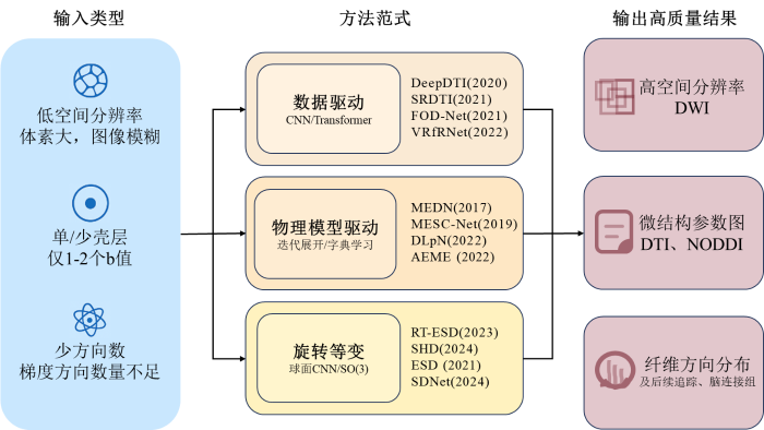
图1
dMRI超分辨率任务与方法体系总览图
Fig. 1
Overview map of the dMRI super-resolution task and method system
2 dMRI超分辨率重建模型
2.1 基础扩散指标的超分辨率重建方法
基础扩散指标以DTI模型为代表,它是最基础的dMRI建模方法,其定量指标的准确性高度依赖空间分辨率和采样方向数,当分辨率偏低或方向数不足时,张量估计会出现系统偏差,并表现出明显的部分容积效应. 本节介绍基础扩散指标的两种并列方法,2.1.1节关注稀疏采样下的参数直接估计,从有限的方向数中直接恢复高质量指标,2.1.2节介绍分辨率增强的重建方法,通过更高的分辨率缓解部分容积效应.
2.1.1 稀疏采样下的参数直接估计
Tian等提出的DeepDTI是早期的代表性研究[9],针对角度采样不足与扫描时间过长的问题,使用10层三维CNN,从仅6个方向的低维扩散信号重建高角度DTI张量参数,实现了由6向到30向的高精度映射. 此类模型输出的DWI仍需使用传统拟合流程得到临床需要的参数指标. Li等提出SuperDTI模型则跨越了传统张量拟合[10],直接让模型从少量DWI输入生成FA、MD等定量图像. 该模型以U-Net为核心架构,通过多尺度特征聚合实现了超快速的DTI参数重建与纤维束可视化,减弱了传统拟合对噪声与异常值的敏感性,展示了端到端学习DWI至参数映射的可行性. Sabidussi等提出的dtiRIM(DTI Recurrent Inference Machine)框架将q空间采样几何与张量模型嵌入循环推理机的物理约束中[11],使训练完成后的网络无需重新训练即可泛化至任意未知的采样协议.
近年来,生成模型在多个领域展现出强大潜力. 基于联合扩散模型的框架Diff-DTI(Diffusion DTI)[12]通过同时建模DWI与DTI参数图的联合概率分布,实现了在推理阶段利用极少方向DWI的引导生成,大幅降低扫描需求. Martin等提出的DiffDL(Diffusion Deep Learning)框架[13],将条件扩散概率模型引入DTI,在生成高质量参数图的同时,具备不确定性建模与多样本生成能力,且联合输出了DKI相关指标. 不确定性建模能力源于扩散模型独特的概率生成机制. 扩散模型通过一个渐进加噪的前向过程和一个逐步去噪的反向过程进行训练. 在推理阶段,模型从纯噪声开始多次迭代生成样本,生成过程中的多样性蕴含了不确定性. 该过程的核心公式为:
其中,
2.1.2 分辨率增强的重建方法
Tian等提出的另一模型SRDTI(Super-Resolution for Diffusion Tensor MRI)则重点提升空间分辨率[14],采用三维CNN与残差学习机制,并通过方向优化策略保持张量空间一致性,使分辨率由2 mm提升至 1.25 mm,在细纤维区域显著改善了FA与MD的保真度. DeepDTI与SRDTI分别代表了DTI超分辨率的两种互补范式:DeepDTI通过在角度域挖掘信号冗余以实现采样压缩与扫描加速,而SRDTI则聚焦于空间域重建以增强结构分辨率与微结构可视化能力. 尽管两者在网络结构与物理约束上具有相似性,但在输入形式、重建目标与评价指标上形成了区别.
图像质量迁移(Image Quality Transfer, IQT)是自然图像领域常用的超分辨率策略. Ma等将其引入dMRI[15],提出7 T至3 T跨场强映射框架,以改进的自编码器为核心,用7 T高分辨图像作为结构先验,在无需成对扫描的前提下实现跨设备、跨分辨率的知识迁移. 新生儿数据以运动伪影为特征,Karimi等[16]将从高质量新生儿数据中获取的精确扩散参数图作为解剖先验,与真实胎儿数据特征结合合成训练数据,解决了胎儿DWI缺乏可靠真实标签的瓶颈,使标准CNN能够直接从质量较差的胎儿DWI中稳定重建参数图. 随后,Karimi与Gholipour引入Transformer架构[17]设计的双阶段网络,先学习邻域体素间的信号相关性,再关注原始扩散信号和一阶段估计结果,学习张量本身在空间上的规律性先验.
近期,面向临床的一站式集成框架成为重要发展方向. Altmann等提出了结合k空间深度学习重建与图像域超分辨率的端到端流程[20],将先进的超分辨率技术封装为可直接应用于原始采集数据的黑箱解决方案. 临床研究表明,仅使用单次采集平均的DWI在主观图像质量与诊断信心上均显著优于传统四次平均的重建结果,且未引入额外伪影.
总体来看,基础扩散指标超分辨率重建方法正逐步从早期的卷积网络映射、端到端参数回归,发展到融合空间先验、q空间几何约束及生成式模型的多范式框架,且已有向验证临床可用性、实现工作流加速的务实转变. 数据驱动方法依赖大规模数据学习映射,实现快速重建但可解释性弱;几何约束方法嵌入q空间采样几何,提升协议泛化能力但计算复杂;生成式模型具备不确定性量化与高保真生成能力,更适合临床可信决策,但对计算资源要求较高. 当前最优方法趋向于融合生成式框架与物理先验,在保证精度的同时兼顾可解释性与泛化性.
2.2 针对高阶微结构指标提升的重建方法
DTI等模型虽能提供对组织结构的描述,但其参数与特定的细胞微观结构之间缺乏直接的生物学对应关系. 为了获取更具生物学特异性的微结构指标,出现了NODDI、球面平均技术(Spherical Mean Technique,SMT)[4]、体素内不相干运动(Intravoxel Incoherent Motion,IVIM)[21]等生物物理模型. 这类复杂模型的参数估计,更加依赖于多壳层、高方向数的密集采样,这不仅显著增加了扫描时间,还放大了对运动伪影和噪声的敏感性. 因此,利用深度学习技术准确重建这些微结构参数,具有重要的临床和研究意义. 深度学习针对高阶微结构模型的参数估计更倾向于通过端到端的形式解决,这类方法虽未输出高分辨率的中间步骤,但其目标是从低信息量观测中恢复本需高分辨率采集才能稳定拟合的参数,本质上是学习一种从低分辨率观测空间到高分辨率参数空间的隐式映射. 本节将这类端到端方法从算法设计角度分为数据驱动(2.2.1节)、模型驱动(2.2.2节)和基于旋转等变网络(2.2.3节)三类。2.2.4节则聚焦于以空间分辨率提升为目标的增强方法。这些研究从不同角度补偿采样不足导致的信息缺失,共同推动高阶微结构指标的重建精度提升.
综合来看,高阶微结构指标重建方法依其先验来源可分为数据驱动、模型驱动与旋转等变三类. 数据驱动方法从大规模数据中学习统计映射,适应性强但依赖标注数据;模型驱动方法将生物物理模型嵌入网络,提升可解释性与数据效率,但受限于模型假设;旋转等变性方法通过球面卷积内建旋转对称性,显著提升角度估计一致性,尤其适用于复杂纤维区域. 三类方法在数据需求、物理可解释性与计算效率上存在权衡.
2.2.1 数据驱动的重建方法
数据驱动方法旨在从大规模数据集中,直接学习从低信息量的观测信号到高质量微结构参数图之间的映射. 其核心假设是,存在一个由高质量数据分布所决定的、相对固定的统计关系,网络可以隐式地学习这种关系,从而为稀疏观测补充缺失的信息.
早期研究[22]将此过程简化为体素级的黑盒回归,利用多层感知机学习单个体素内q向量与目标参数间的映射,其核心目标虽非显式的空间或角度分辨率提升,但为后续结合结构先验的超分辨率方法奠定了基础. 这种方法学习的先验是特定采样模式与参数值之间的统计关联,但因其完全忽略了空间与角度结构,所利用的先验信息有限. 为融入空间先验,Gibbons等使用2D CNN同时估计NODDI和广义分数各向异性参数图[23],通过利用切片内的空间关系,补偿了因空间分辨率不足导致的体素内信号混叠. Nath等使用具有瓶颈层的多任务网络[24]进一步学习了不同微结构模型间的共同低维特征先验,经由多个任务头输出IVIM、SMT与NODDI等模型的13种微结构参数,验证了不同生物物理模型间存在的低维共享表示,为多任务联合超分辨率提供了新思路. 其新增信息来源于不同微结构模型间的泛化性特征,瓶颈层学习到的6维特征可视作一种微结构本质表示.
将q空间信号简单向量化处理会忽略梯度方向之间的内在角度关联性. 为了在角度维度进行信息补全,Chen等将每个q空间采样点视为图节点[25],并以节点间的空间角度距离作为边权构建图. 网络在图上进行消息传递的过程,学习了如何在q空间球面上,从稀疏离散的观测点恢复出可用于参数估计的连续角度信号表示. 这种设计对q空间的几何结构建模,使网络能进行角度维度的信息生成,实现角度超分辨率. 在欠采样72、48、36个方向的实验中,图CNN在峰值信噪比和计算效率上显著优于传统加速微结构成像凸优化(Accelerated Microstructure Imaging via Convex Optimization,AMICO). 其改进模型混合图Transformer(HGT)[26],同时捕捉体素间长程依赖的先验与用于建模q空间局部角度连续性先验,实现了空间与角度双重维度信息的高效融合与超分辨率重建,更全面地补偿了空间与角度两个维度的信息缺失.
数据驱动方法通过训练从数据中自动挖掘并学习复杂的先验信息,包括空间相关性、跨参数共享表示以及角度连续性等. 这些学习到的先验知识被编码在网络权重中,使得模型在面对新的稀疏观测时,能够依据这些先验生成合理信息,以逼近全采样下的估计结果. 尽管数据驱动方法在不断降低采样需求,但其性能高度依赖于大规模、高质量且分布均衡的训练数据集,泛化能力与稳定性不足.
2.2.2 模型驱动的重建方法
纯数据驱动的方法利用深度学习直接学习信号到参数的映射,缺乏可解释性. 为此,研究者们提出了模型驱动的重建方法,将生物物理模型本身的数学求解过程展开为可训练的网络架构,以物理先验提升数据效率. 这类方法以参数估计为主要目标,但其中一系列工作嵌入了空间角度先验,学习了低、高分辨率信息的可靠映射,实质性地推动了dMRI重建的发展,网络恢复的新增信息来源于对数据中固有结构性规律的挖掘与物理约束的遵循.
Ye等将AMICO算法展开为可训练网络,早期工作使用深度网络的微结构估计(Microstructure Estimation using a Deep Network,MEDN)及其改进版在迭代硬阈值机制(Iterative Hard-thresholding,IHT)中引入邻域加权层[27],通过学习局部空间平滑性先验,利用相邻体素的信息来补偿单个低分辨率体素内丢失的空间细节. 为进一步提升自适应优化能力,后续研究引入长短期记忆网络以优化路径中的历史信息先验,并构建空间-角域联合稀疏表示(MESC-Net),实现了对NODDI、SMT和系统平衡传播子(Ensemble Average Propagator,EAP)等多种模型的泛化[28]. 进一步的工作将联合字典分解为空间与角域两个独立的低维字典,以更少的参数实现了更具表达力的稀疏编码[29],建模了扩散信号在q空间球面上的连续性.
Faiyaz等认为IHT的8层迭代存在冗余[30],以一个字典学习网络直接估计各向同性体积分数作为先验,然后嵌入NODDI框架中估计NDI和ODI,将三参数拟合问题转化为两参数,体现了利用互补成像对比信息来弥补单一模态采样不足的思想. 针对IHT类方法的收敛不稳定问题,Zheng等提出外梯度增强自适应网络(Adaptive Network with Extragradient for dMRI-based Microstructure Estimation,AEME)[31],引入外梯度投影以在数学上保证收敛,并设计自适应迭代停止机制,根据验证损失动态调整网络深度,避免过度参数化. 后续的AEME+中加入噪声调谐[32],在迭代块中加入可控噪声,帮助网络逃离局部极小值.
总结来看,模型驱动方法将组织微结构在特定模型下具有的数学特性作为强先验嵌入网络,技术脉络是从基础的稀疏编码出发,逐步融入空间上下文、解耦的角度表示、稳定的优化器及稳定求解块. 其核心优势在于将生物物理约束与数据驱动学习相结合,为从低分辨率采样中恢复信息提供了理论依据. 然而,该类方法的性能上限受限于其所基于的生物物理模型本身的准确性. 在模型假设不完全成立的脑区,尤其是纤维交叉复杂或存在显著部分容积效应的区域,其估计精度可能会受到制约.
2.2.3 基于旋转等变网络的重建方法
传统的卷积神经网络由于其结构本质地具备平移等变性,在处理规则网格上的二维图像数据时表现出色. 而扩散MRI信号采样自球面,在梯度方向空间具有球面对称性,因此在卷积网络中,该信号的理想特征表示应对球面上的三维旋转(SO(3))保持等变性. 然而,传统卷积网络并不具备该性质,使得网络需要额外从数据中学习基础几何规律,且难以保证外推时的方向一致性.
为此,研究者提出了一系列依赖于球面卷积的球形CNN架构. 普通平面卷积一般表示为[33]:
其中,h表示卷积核的权重,I表示二维离散输入图像,
其中,
以上工作奠定了旋转等变球形CNN的基础,但均建立在等角网格采样的理想化假设之上,而dMRI信号在采样方向覆盖整个球面,并不在等角规则网格. 为适应常见随机均匀采样的dMRI信号,Sedlar等提出全谱域球形CNN[36],在S2和SO(3)的谐波系数空间中表示所有可训练参数,实现了完全在谱域中进行卷积与非线性变换. 整个过程遵循球面信号的谐波变换规律,使其在从稀疏角度采样恢复连续球面信号的角度超分辨率任务上具有优势. 可解释性是当前深度学习模型发展的重要趋势. Consagra等嵌入旋转等变球面U-Net架构[37],构建了基于序列反演的似然自由推断框架(LFI)[38],通过信号解混和混合密度网络构建参数的后验分布,将多纤维的微结构参数与不确定性量化统一输出. 该框架学习了强大的物理先验与纤维交叉角先验,实现了在q空间壳层维度上的信息增强,并提供了可靠性度量.
旋转等变方法通过球面卷积将旋转对称性编码为网络的刚性归纳偏置,为角度超分辨率提供了可靠的信息生成原则,从理论保证上消除了参数及方向估计的系统性偏差. 网络无需学习旋转对称性,可将全部容量用于学习信号与微结构间更复杂的映射关系,与经典的球面反卷积理论一脉相承,提升了数据效率与物理可解释性. 当然,其性能优势紧密依赖于嵌入的物理先验,在高度偏离此先验的病变或非白质场景下可能受限. 值得注意的是,本节模型的优势在更为复杂的纤维方向分布重建任务中得到了更充分的体现,成为该领域较为主流的技术路径之一.
2.2.4 分辨率提升的重建方法
空间分辨率的提升模型通过输入一个覆盖较大物理范围的三维图像块,网络学习挖掘其中与中心目标区域相关的解剖和微结构规律,从而补偿单个低分辨率体素丢失的细节,实现分辨率提升,典型提升倍数为2倍. 新增的高分辨率空间信息主要来自于对空间上下文先验的学习与利用.
多模态版本MSR-q-DL[41]考虑到临床常规中往往同步采集T1和T2加权解剖图像,在SR-q-DL框架中引入了注意力模块. 该模块从高分辨率解剖图像中计算出相关性图谱,用以加权调整低分辨率扩散信号的稀疏表示. 它将高分辨率解剖信息视为空间引导先验,用于指示领域信息对于重建目标体素的重要程度. MSR-q-DL新增的信息源于对解剖结构与微结构相互关系的学习,实现了跨模态的知识引导与信息增强. 此类方法直接提升空间分辨率,在一定程度上保持了模型驱动的可解释内核,验证了多模态引导的可行性与有效性. 但同样面临数据驱动和模型驱动相同的挑战,多模态方法的增益则依赖于辅助模态与目标微结构间的解剖相关性.
2.3 针对提升fODF分辨率的重建方法
纤维方向分布函数是解析体素内复杂纤维结构,进而支撑纤维追踪与脑连接组分析的关键工具. 其传统计算流程始于原始DWI信号,先通过球谐函数拟合得到信号的紧凑参数化表示,继而基于不同的球面反卷积模型求解出fODF. 对于单壳数据,目前最先进的传统重建方法为单壳层三组织约束球面反卷积(Single-shell 3-Tissue Constrained Spherical Deconvolution,SS3T-CSD)[42]. 对于多壳数据,常用的传统重建方法为多壳层多组织约束球面反卷积(Multi-shell Multi-tissue Constrained Spherical Deconvolution,MSMT-CSD)[43].
为突破高质量fODF对昂贵多壳高角度采集协议的依赖,深度学习提供了两种主要技术路径(图2). 一类模型旨在绕过传统的反卷积算法,端到端地建立从低质量DWI信号到高质量fODF的映射(2.3.1和2.3.2节). 第二类则呈现出增强策略,模型以由SS3T-CSD初步估计的fODF为输入,通过深度学习实现角度超分辨率增强,以逼近高质量采集所得的fODF(2.3.3节).
图2
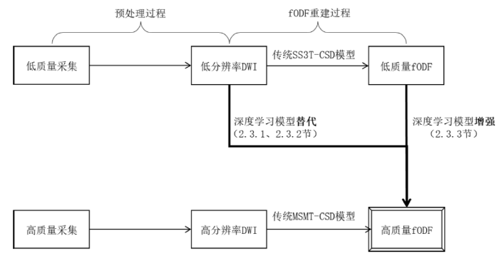
图2
提升fODF分辨率重建方法的分类图
Fig. 2
Classification schematic of reconstruction methods for enhancing fODF resolution
2.3.1 基于深度学习的端到端fODF重建
Nath等则专注于壳层维度的压缩与恢复[46],残差深度学习神经网络(Residual Deep Learning Neural Network,ResDNN)以组织学真值作为监督信号,从活体可行序列中挖掘了未被利用的微结构信息,证明了单壳协议中确实潜藏着高阶信息. 然而医学场景难以获取大量真值,残差卷积神经网络(Residual Convolutional Neural Network,ResCNN)转向数据驱动并联合输出组织分数[47]. ResCNN取3×3×3邻域并仅预测中心体素,建模范围与信息利用效率受到了局限. 为突破此瓶颈,Jha等提出体积型感兴趣区fODF重建网络(Volumetric ROI fODF Reconstruction Network,VrfRNet)[48],将基本处理单元从体素邻域扩展为非重叠的三维图像块,实现了由体素级向体积级的范式转变. VRfRNet采用生成对抗框架,并引入了粗粒度重建、多尺度特征增强、特征级和Softmax级注意力四个模块,由粗到精适应更大空间尺度的建模需求.
为融合物理模型先验与深度学习的表达能力,Bartlett等提出了球面反卷积网络(Spherical Deconvolution Network,SDNet)[49]. 该网络将球面反卷积模型嵌入网络架构,通过交替的DWI一致性模块与深度正则化模块,实现对原始信号的物理约束与特征学习的结合. 同时,SDNet引入分类损失,以高层语义约束提升对纤维数量与方向的辨识力. 以上模型一般适用于固定的壳层和方向数,针对多中心协议壳组合不一、重训成本高的问题,又有Yao等提出统一动态头球面CNN[50],以1 000 s/mm2、2 000 s/mm2、3 000 s/mm2三壳任意组合的90个方向DWI为输入,动态生成首层卷积核,单次训练即可覆盖七种壳缺失情形,大幅延展了模型的适用场景. 针对可解释性,Consagra等使用神经场来参数化潜在fODF的随机级数表示[51],隐式地模拟了常被忽略的空间相关结构,并推导了后验预测分布的解析,可用于量化任何空间位置的fODF估计的不确定性.
端到端方法通过学习从稀疏DWI信号到高质量fODF的直接映射,绕开了传统反卷积的计算流程,融合了空间领域信息与信号衰减规律. 这类方法的缺陷是大多依赖大规模标注数据,且通常未显式嵌入球面几何先验,在纤维方向一致性与小角度分辨上可能存在局限. 新增的高分辨率角向信息主要源于网络从训练数据中挖掘的统计先验与局部空间依赖关系.
2.3.2 基于旋转等变网络的fODF重建
前期模型验证了深度学习端到端估计fODF的可行性,但大多仍沿用传统空间卷积,未充分考虑dMRI信号固有的球面几何特性. 与微结构指标研究类似(2.2.3节),Sedlar等将旋转等变性引入fODF估计领域[52],针对普通三维CNN处理球面信号时存在的计算低效与几何失配问题,构建了谱域球形U-Net,采用轴向对称核大幅降低了参数量与计算复杂度.
Elaldi等认为缺乏旋转等变性会导致小角度交叉纤维分辨不佳[37],且依赖MSMT-CSD伪真值会限制模型性能. 他们提出旋转等变的球面图卷积网络(ESD),将SO(3)等变作为核心归纳偏置,并采用自监督训练策略,在小角度交叉纤维恢复上取得显著优势. 随后,旋转平移等变球面反卷积(RT-ESD)模型将归纳偏置从SO(3)扩展至E(3)×SO(3)[53],实现对空间刚性变换与体素内旋转的联合等变,使模型利用空间信息取得更优的性能. 近年,空间-半球反卷积(SHD)模型重构球形图拉普拉斯为半球形式[54],并以密集矩阵乘法替代稀疏实现,系统性消解了RT-ESD在内存与计算上的可扩展性瓶颈,让其支持大规模训练与临床部署.
旋转等变网络通过引入球面卷积结构,将对称性内建为网络归纳偏置,确保了方向估计的旋转等变性. 其发展脉络体现了从理想等角采样建模到适应临床随机采样、从单一旋转等变到联合空间等变的演进. 新增的角分辨率信息本质上来源于q空间信号在球面上的连续性与对称性,网络无需学习基础旋转对称规律,提升了数据效率与物理可解释性,但计算复杂度较高.
2.3.3 基于fODF的超分辨率增强
用单个深度学习模型直接替代传统反卷积计算是极具吸引力的解决方式,但模型往往需要同时学习信号衰减的物理意义和纤维解剖的生物学意义,有较重的负担和泛化性风险. 使用SS3T-CSD算法得到的fODF作为模型的输入,则能适应不同的临床协议,使输入相对标准化. 此外,网络不需要理解原始DWI信号,只需在fODF域学习,任务更专注. 新增的角向细节信息来源于对初步fODF中方向分布模式与空间连续性的深入学习,本质上是对已有方向信息的锐化与补全,其性能上限在一定程度上受限于初步估计的质量. Zeng等提出的纤维方向分布网络(Fiber Orientation Distribution Network,FOD-Net)是直接在fODF空间进行学习的代表性模型[57]. 该方法基于体素级卷积神经网络,学习成对低、高分辨率球谐系数之间复杂的非线性映射,显著提升了fODF重建的角度精度.
在此基础上,研究者将注意力机制引入此类模型,进一步增强网络的方向敏感性和空间建模能力. Da Silva等的FOD-Swin-Net基于Swin UNETR框架[58],通过移位窗口自注意力机制捕获更长程的空间依赖关系,在训练速度与角相关系数上均较FOD-Net有显著提升. Li等提出的邻域体素注意力机制网络(Neighboring Voxel Attention Mechanism Network,NVAM-Net)[59],针对FOD-Net在体素间空间连续性建模不足的问题,设计了邻域体素注意力机制. 该注意力模块由体素注意力与表面注意力两部分组成,前者聚焦于目标体素的关键特征,后者建模目标体素与邻域体素在空间上的方向依赖,从不同尺度提取周边信息,精准重建纤维走向. 这两项研究体现了注意力机制在基于fODF超分辨率增强方法的重要潜力与发展趋势.
fODF重建方法呈现出从端到端黑箱映射向旋转等变建模演进,同步专注后处理的增强策略拓展的脉络. 当前最新方法如UFO-3与FOD-Swin-Net等分别在纤维方向一致性与空间连续性上表现突出,旋转等变模型计算成本较高,而增强策略更易与临床工作流集成. 总体而言,端到端方法推理快但依赖数据,旋转等变方法精度高但部署复杂,增强策略轻量但受限于输入质量,临床落地需在精度、效率与兼容性间权衡.
3 重建方法评估、公开数据集与量化总结
3.1 重建方法评估指标
dMRI超分辨率任务不仅包括传统意义上的空间分辨率提升,也涵盖从低角度采样中恢复高角度分辨率的效果,涉及从原始信号图像保真度、微结构参数准确性,到下游纤维重建与追踪可靠性等多个层面. 具体而言,本文将常用评估指标系统划分为图像保真度指标、微结构标量误差指标、纤维方向重建评估指标、纤维追踪评估指标和重测信度指标五类(表2).
图像保真度指标主要用于评估重建结果在像素或体素层面与真实高分辨率数据的接近程度,适用于各类空间或参数域的重建任务[60]. 最常用的指标包括峰值信噪比(Peak Signal-to-Noise Ratio,PSNR):
其中,
其中,
表2评估指标共同构成了一个多层次的评估体系,用于全面衡量dMRI超分辨率重建方法在不同维度上的性能. 需要指出的是,dMRI超分辨率评估具有内在的多维性与挑战性. 多维性意味着不同层级的评估结果可能出现脱节. 图像层面的高保真度并不等同于微结构参数估计的准确性,因为算法可能引入对后续模型拟合有偏的系统性误差. 同样,在体素层面取得高角度相关性的fODF,在后续的纤维追踪中,可能因误差累积与路径积分偏差,导致纤维束的解剖合理性下降. 这种从信号到参数,再到连接组学应用的评估链不一致性,是方法可靠性验证的关键难点. 而挑战性,体现在当前领域缺乏一个统一的、以临床问题为导向的金标准评估框架. 如何超越技术指标的比较,将超分辨率的性能增益切实转化为对疾病诊断、手术规划或连接组学分析能力的提升,并建立相应的临床验证标准,是连接方法学研究与临床应用的桥梁,也是一个关键而富有挑战的研究方向.
表2 评价指标分类
Table 2
| 类别 | 指标 | 中文名称 | 含义 | 说明 | ||
|---|---|---|---|---|---|---|
| 图像保真度指标 | Peak Signal-to-Noise Ratio (PSNR)[60] | 峰值信噪比 | 衡量重建图像与参考图像之间的全局误差,值越高表示重建质量越好 | 基于均方误差计算,对误差敏感,但可能与视觉感知不完全一致,常用于评估DWI或参数图的重建质量 | ||
| Structural Similarity Index (SSIM)[60] | 结构相似性指数 | 从亮度、对比度、结构三方面综合评估图像之间的相似度,更符合人眼视觉感知 | 值域为[0, 1],值越接近1表示相似度越高,适用于评估空间超分辨率结果的结构保真度 | |||
| 微结构标量误差指标 | Mean Absolute Error (MAE)[14] | 平均绝对误差 | 计算预测标量值与真实值之间绝对误差的平均值 | 最基础的误差度量 | ||
| Mean Squared Error (MSE)[22] | 均方误差 | 计算预测值与真实值之间平方误差的平均值 | 对较大误差更敏感 | |||
| Normalized Mean Absolute Error (NMAE)[13] | 归一化平均绝对误差 | 对MAE进行归一化处理,便于比较不同量级的数据 | 具体归一化方式需在上下文中明确,通常除以真实值的范围或均值 | |||
| Normalized Mean Squared Error (NMSE)[10] | 归一化均方误差 | 对MSE进行归一化处理,便于比较不同量级的数据 | 具体归一化方式需在上下文中明确,通常除以真实值的范围或均值 | |||
| 纤维方向重建评估指标 | Angular Correlation Coefficient (ACC)[57] | 角相关系数 | 评估预测的纤维方向与参考真实方向在空间上的一致性 | 评估纤维方向估计整体准确性的核心指标 | ||
| Mean Angular Error (MAE)[57] | 平均角度误差 | 衡量预测纤维主峰方向与参考真实方向之间的平均角度差 | 此MAE专指角度误差,与上文“平均绝对误差”含义不同 | |||
| Peak Error (PE)[57] | 峰值误差 | 衡量预测纤维主峰幅度与参考真实幅度之间的峰值差 | 反映对纤维强度估计的准确性 | |||
| Proportion of Correct Peaks (PCP)[38] | 正确峰比例 | 统计正确识别出的纤维方向的比例 | 一种基于分类正确率的评估方式,计算时设有阈值 | |||
| Earth Mover‘s Distance (EMD)[61] | 推土机距离 | 衡量整个fODF分布与参考真实分布之间差异 | 评估分布层面相似性的核心指标,反映整体匹配度,综合考量角度和幅度 | |||
| 纤维追踪评估指标 | Valid Streamlines[62] | 有效流线比例 | 在所有生成的流线中,属于任一真实纤维束的流线比例 | 反映追踪结果的纯净度,值低说明假阳性高 | ||
| Bundle Overlap[62] | 纤维束重叠度 | 估计的纤维束与真实纤维束体素交集与真实集的比值 | 衡量灵敏度,即找回真实纤维束的能力 | |||
| Bundle Overreach[62] | 纤维束过度延伸度 | 估计的纤维束超出真实纤维束的部分与真实集的比值 | 衡量特异度,值高表示假阳性多 | |||
| Valid Bundles[62] | 有效纤维束数量 | 被正确识别出的、与真实解剖结构相符的纤维束数量 | 衡量算法重建特定神经通路的能力 | |||
| Dice Coefficient[10] | Dice系数 | 两个二元分割结果的空间重叠程度 | 衡量纤维束分割结果 | |||
| 重测信度指标 | Coefficient of Variation (CV)[57] | 变异系数 | 计算同一受试者经多次扫描的某指标的标准差与均值的比值 | 反映数据相对于其平均值的离散程度,值越低表示稳定性越好 | ||
| Weighted Mean Coefficient of Variation (wmCoV)[57] | 加权平均变异系数 | 针对连接组矩阵,对每个连接边的CV进行加权平均求变异系数,权重取决于连接强度 | 由于连接组数据通常具有高度偏态分布,wmCoV通过对强连接赋予更高权重,更能代表整体连接组的重测信度 | |||
| Intraclass Correlation Coefficient (ICC)[63] | 组内相关系数 | 量化重测信度的核心统计量,评估同一受试者多次扫描结果的一致性程度 | ICC > 0.75通常被认为信度优秀,是比简单相关性更严格的指标 | |||
3.2 数据集总结
表3 常用公开dMRI数据集
Table 3
| 全称 | 缩写 | 主要人群/研究重点 | 特点 | 数据量 | 主要采集参数(b值非0) |
|---|---|---|---|---|---|
| The Rotterdam Study[65] | RDS | 荷兰鹿特丹地区老年人慢性病的发病率、预后 | 大型长期随访队列 | MRI约8000人 | b = 1000 s/mm2,25 个方向 |
| The Rhineland Study[66] | RLS | 德国莱茵地区人群的深度表型研究 | 人群基线广、影像丰富 | 千例以上 | b范围270 ~ 6800 s/mm2, 112个方向 |
| Pediatric Imaging Neurocognition and Genetics[67] | PING | 儿科影像、认知发展与遗传关联 | 儿童、青少年群体 | 约1493名 | b = 1000 s/mm2, 约30个方向 |
| developing Human Connectome Project[68] | dHCP | 典型与非典型早期脑发育 | 新生儿、早期发育阶段 | 1173名 | b = 400 s/mm2,64个方向; b = 1000 s/mm2,88个方向; b = 2600 s/mm2,128个方向 |
| Baby Connectome Project[69] | BCP | 婴儿脑连接发育 | 婴幼儿期 | 500名 | b = 500 ~ 3000 s/mm2,总约144个方向 |
| Chinese Connectome Project[70] | CHCP | 中国人群脑连接组 | 亚洲人群样本 | 366名 | b = 1000 s/mm2,93个方向; b = 2000 s/mm2,92个方向 |
| Amsterdam Open MRI Collection[71] | AOMIC | 健康年轻成人,情感与社会认知的神经机制 | 模态丰富,附带行为与心理学量表数据 | 约1370名 | b = 1000 s/mm2,32个方向 |
| ISMRM 2015 Tractometer Challenge Dataset[72] | / | 纤维束追踪算法的标准评估与对比 | 追踪算法验证与性能比较的常用基准 | 1名 | b = 1000 s/mm2, 90个方向; b = 2000 s/mm2, 90个方向; b = 3000 s/mm2,90个方向 |
3.3 重建性能的量化总结
研究表明,深度学习驱动的dMRI超分辨率重建已在多个维度实现了明确的量化性能提升. 超分辨率重建的关键量化,包括超分辨率倍数、角向维度信息提升、壳层信息获取等性能.
在q空间角度维度的信息补偿是更为重要的方向. 多数方法能够从6~30个极稀疏的方向采样中,重建出与60~90个方向采集质量相当的扩散信号或衍生参数,实现约3~5倍的方向数有效提升. 部分连续建模框架更进一步,支持从任意稀疏采样中查询全方向信号[19].
针对高阶微结构模型和fODF的重建,许多方法聚焦于壳层信息的生成. 其典型范式是从b=1 000 s/mm2的单壳或b=1 000或2 000 s/mm2的双壳数据出发,恢复出本需三壳及以上密集采样才能稳定估计的微结构参数或fODF[57],从而在壳层维度实现显著的信息增强,实现壳层维度的分辨率提升.
综上,当前dMRI超分辨率重建在空间上普遍实现2~4倍提升,角度上实现3~5倍方向数增强,壳层上则从单壳或双壳少方向恢复多壳多方向信息为主,为在临床可接受的扫描时间内获取高信息量的扩散影像提供了可行的技术路径.
4 讨论
尽管近年来深度学习在dMRI超分辨率重建中取得显著进展,其迈向临床应用仍面临来自模型原理、临床泛化及计算部署的多重挑战. 除了3.1节提到的因评价指标多维性产生的挑战外,还有以下三点.
4.1 模型的反演非唯一性与评价指标
生物物理模型的非唯一性是一项长期存在的核心难题. 尤其在多纤维交叉和复杂组织结构下,多参数组合可能导致同一观测信号,使模型学习到有偏映射. 2.2.2节讨论的模型驱动方法体现了这一点,其性能上限受限于所基于的生物物理模型本身的准确性,在模型假设不完全成立的脑区[73]面临了性能天花板. 尽管参数正则化、联合物理建模和球面几何先验在一定程度上缩小了解空间,但难以完全解决模型反演固有的多解性. 以生成式模型推断表达参数后验分布,或引入序列反演框架,可能是未来减少多解性风险的方式.
4.2 临床转化中的泛化性与集成性障碍
在泛化性方面,临床数据稀缺与跨协议、跨中心的域偏移问题突出. 正如2.2.1节所述,数据驱动方法高度依赖大规模、高质量的训练数据,面临泛化困境. 而3.2节所列举的现有常用公开数据集,在样本多样性、病理覆盖及采集协议统一性方面仍存在局限. 尽管无监督学习、模型驱动架构等策略降低了对标注数据的依赖,但多中心在采集参数以及硬件条件上的差异,使模型在真实临床场景中出现明显的性能衰退. 尤其是在以模拟降采样构建训练数据对的常见做法下,系统性差异进一步限制了模型适应能力. 当前模型在跨中心、跨设备、跨人群时可能出现的波动,目前仍缺乏标准化的性能验证流程. 未来可探索构建协议无关的q空间表示、编码采集协议参数以及通过自监督预训练在大规模临床数据中提升泛化能力.
在集成性方面,目前,临床神经影像分析高度依赖于FSL、Mrtrix等集成化软件,深度学习模型与之存在集成障碍. 模型与现有临床影像系统的对接也涉及了数据接口、处理流程与交互界面的整合. 因此,如何将超分辨率模型无缝封装,输出与主流软件兼容的标准格式数据,或进一步开发为插件、提供用户友好的交互界面乃至一键式流程,成为决定其能否被临床工作者采纳的关键. 缺乏便捷的集成方案,会显著增加技术推广与学习成本,阻碍其在常规分析中的普及. 未来研究需重视与现有工作流的整合,是当前方法从实验研究迈向临床应用必须面对的关键挑战.
4.3 计算效率与临床部署兼容性
这些计算特性直接推高了硬件部署门槛,并对多设备适配构成挑战. 临床环境设备配置多样,从配备高性能GPU的研究工作站到算力有限的扫描终端,模型需具备良好的跨平台适配能力. 因此,未来的研究在追求重建质量突破的同时,必须同等重视模型轻量化、推理加速与硬件感知优化. 可能的解决路径包括设计更高效的等变操作符、探索稀疏注意力机制或开发专为边缘设备优化的网络架构,以期在保持模型性能优势的同时,显著降低其计算与存储成本,提升临床系统集成与落地可行性.
综合来看,未来的dMRI超分辨率重建方法需在物理一致性、数据适应性、临床可集成性和计算效率之间取得新的平衡,以满足临床应用的严苛需求.
5 总结与展望
本文系统回顾了脑部dMRI超分辨率重建的主要进展. 从早期应用于基础扩散指标的角度与空间分辨率增强,到针对高级生物物理模型和fODF的精细重建,深度学习在dMRI数据处理的各个层级均展现出强大的能力. 技术路径清晰地呈现了从数据驱动的黑箱映射,发展到嵌入解剖和物理先验的模型驱动,再到尊重数据内在特性的球形CNN的演进趋势.
展望未来,dMRI超分辨率重建的发展将从单一任务驱动逐步迈向以物理一致性、可解释性与生成式推断为核心的综合框架. 随着模型的推陈出新,未来的模型将更强调在统一架构中整合信号衰减机理与先验信息,并探索直接输出纤维追踪等多任务映射,实现对复杂纤维交叉与多协议数据的稳定重建. 同时,自监督预训练、协议无关表示和跨中心学习将推动更具普适性的基础模型出现,使模型能够在不同扫描仪和人群中保持一致性能.
此外,随着生成式模型和不确定性量化技术的引入[74],超分辨率估计将从确定性输出走向后验分布的概率表达,提升微结构指标估计的可靠性. 不仅如此,不确定性量化本身提供比单点估计更丰富的信息维度,也许能产生对临床决策的支持价值. 在疾病进展监测中,通过比较不同时间点后验分布的变化,可更稳健地区分真实的生物学演变与估计噪声,实现对微妙病理改变的早期预警. 而在手术规划中,纤维束方向的不确定性可直接关联至手术风险等级,辅助制定个性化方案. 发展不确定性建模,有望使超分辨率技术从提供更清晰的图像,进阶为可支持动态、审计临床决策的智能工具.
最终,可解释性、稳健性与临床一致性将成为模型真正进入临床应用的关键,使其在脑网络分析、疾病表型识别、治疗规划与随访监测等后续应用中发挥更具实践价值的作用.
利益冲突
无
参考文献
Research progress on tractography of superficial white matter based on diffusion magnetic resonance imaging
[J].
基于扩散磁共振的大脑浅表白质纤维束研究进展
[J].
DOI:10.11938/cjmr20243126
[本文引用: 1]

近年来,基于扩散磁共振成像的大脑浅表白质纤维束成像技术取得了显著进展.浅表白质纤维束是连接皮层和皮层下神经结构的重要通路,在构建完整人类连接组和神经病理学研究中具有重要意义.本文首先总结了浅表白质纤维束成像技术的发展,其次着重讨论了不同方法在浅表白质纤维束分割中的优缺点,之后探讨了浅表白质纤维束图谱的构建流程,最后总结并对浅表白质纤维束成像技术与分割方法的研究方向进行了展望.
Semi-supervised learning with graph learning-convolutional networks
[C]//
Spherical CNNs
[C]//
Artificial intelligence for diffusion MRI-based tissue microstructure estimation in the human brain: an overview
[J].
DOI:10.3389/fneur.2023.1168833
URL
[本文引用: 2]
Artificial intelligence (AI) has made significant advances in the field of diffusion magnetic resonance imaging (dMRI) and other neuroimaging modalities. These techniques have been applied to various areas such as image reconstruction, denoising, detecting and removing artifacts, segmentation, tissue microstructure modeling, brain connectivity analysis, and diagnosis support. State-of-the-art AI algorithms have the potential to leverage optimization techniques in dMRI to advance sensitivity and inference through biophysical models. While the use of AI in brain microstructures has the potential to revolutionize the way we study the brain and understand brain disorders, we need to be aware of the pitfalls and emerging best practices that can further advance this field. Additionally, since dMRI scans rely on sampling of the q-space geometry, it leaves room for creativity in data engineering in such a way that it maximizes the prior inference. Utilization of the inherent geometry has been shown to improve general inference quality and might be more reliable in identifying pathological differences. We acknowledge and classify AI-based approaches for dMRI using these unifying characteristics. This article also highlighted and reviewed general practices and pitfalls involving tissue microstructure estimation through data-driven techniques and provided directions for building on them.
MR diffusion tensor spectroscopy and imaging
[J].
DOI:10.1016/S0006-3495(94)80775-1
PMID:8130344
[本文引用: 1]
This paper describes a new NMR imaging modality--MR diffusion tensor imaging. It consists of estimating an effective diffusion tensor, Deff, within a voxel, and then displaying useful quantities derived from it. We show how the phenomenon of anisotropic diffusion of water (or metabolites) in anisotropic tissues, measured noninvasively by these NMR methods, is exploited to determine fiber tract orientation and mean particle displacements. Once Deff is estimated from a series of NMR pulsed-gradient, spin-echo experiments, a tissue's three orthotropic axes can be determined. They coincide with the eigenvectors of Deff, while the effective diffusivities along these orthotropic directions are the eigenvalues of Deff. Diffusion ellipsoids, constructed in each voxel from Deff, depict both these orthotropic axes and the mean diffusion distances in these directions. Moreover, the three scalar invariants of Deff, which are independent of the tissue's orientation in the laboratory frame of reference, reveal useful information about molecular mobility reflective of local microstructure and anatomy. Inherently tensors (like Deff) describing transport processes in anisotropic media contain new information within a macroscopic voxel that scalars (such as the apparent diffusivity, proton density, T1, and T2) do not.
MRI quantification of non-Gaussian water diffusion by kurtosis analysis
[J].
DOI:10.1002/nbm.1518
PMID:20632416
[本文引用: 1]
Quantification of non-Gaussianity for water diffusion in brain by means of diffusional kurtosis imaging (DKI) is reviewed. Diffusional non-Gaussianity is a consequence of tissue structure that creates diffusion barriers and compartments. The degree of non-Gaussianity is conveniently quantified by the diffusional kurtosis and derivative metrics, such as the mean, axial, and radial kurtoses. DKI is a diffusion-weighted MRI technique that allows the diffusional kurtosis to be estimated with clinical scanners using standard diffusion-weighted pulse sequences and relatively modest acquisition times. DKI is an extension of the widely used diffusion tensor imaging method, but requires the use of at least 3 b-values and 15 diffusion directions. This review discusses the underlying theory of DKI as well as practical considerations related to data acquisition and post-processing. It is argued that the diffusional kurtosis is sensitive to diffusional heterogeneity and suggested that DKI may be useful for investigating ischemic stroke and neuropathologies, such as Alzheimer's disease and schizophrenia.Copyright © 2010 John Wiley & Sons, Ltd.
NODDI: practical in vivo neurite orientation dispersion and density imaging of the human brain
[J].
DOI:10.1016/j.neuroimage.2012.03.072
PMID:22484410
[本文引用: 1]
This paper introduces neurite orientation dispersion and density imaging (NODDI), a practical diffusion MRI technique for estimating the microstructural complexity of dendrites and axons in vivo on clinical MRI scanners. Such indices of neurites relate more directly to and provide more specific markers of brain tissue microstructure than standard indices from diffusion tensor imaging, such as fractional anisotropy (FA). Mapping these indices over the whole brain on clinical scanners presents new opportunities for understanding brain development and disorders. The proposed technique enables such mapping by combining a three-compartment tissue model with a two-shell high-angular-resolution diffusion imaging (HARDI) protocol optimized for clinical feasibility. An index of orientation dispersion is defined to characterize angular variation of neurites. We evaluate the method both in simulation and on a live human brain using a clinical 3T scanner. Results demonstrate that NODDI provides sensible neurite density and orientation dispersion estimates, thereby disentangling two key contributing factors to FA and enabling the analysis of each factor individually. We additionally show that while orientation dispersion can be estimated with just a single HARDI shell, neurite density requires at least two shells and can be estimated more accurately with the optimized two-shell protocol than with alternative two-shell protocols. The optimized protocol takes about 30 min to acquire, making it feasible for inclusion in a typical clinical setting. We further show that sampling fewer orientations in each shell can reduce the acquisition time to just 10 min with minimal impact on the accuracy of the estimates. This demonstrates the feasibility of NODDI even for the most time-sensitive clinical applications, such as neonatal and dementia imaging.Copyright © 2012 Elsevier Inc. All rights reserved.
Robust determination of the fibre orientation distribution in diffusion MRI: non-negativity constrained super-resolved spherical deconvolution
[J].
DOI:10.1016/j.neuroimage.2007.02.016
PMID:17379540
[本文引用: 1]
Diffusion-weighted (DW) MR images contain information about the orientation of brain white matter fibres that potentially can be used to study human brain connectivity in vivo using tractography techniques. Currently, the diffusion tensor model is widely used to extract fibre directions from DW-MRI data, but fails in regions containing multiple fibre orientations. The spherical deconvolution technique has recently been proposed to address this limitation. It provides an estimate of the fibre orientation distribution (FOD) by assuming the DW signal measured from any fibre bundle is adequately described by a single response function. However, the deconvolution is ill-conditioned and susceptible to noise contamination. This tends to introduce artefactual negative regions in the FOD, which are clearly physically impossible. In this study, the introduction of a constraint on such negative regions is proposed to improve the conditioning of the spherical deconvolution. This approach is shown to provide FOD estimates that are robust to noise whilst preserving angular resolution. The approach also permits the use of super-resolution, whereby more FOD parameters are estimated than were actually measured, improving the angular resolution of the results. The method provides much better defined fibre orientation estimates, and allows orientations to be resolved that are separated by smaller angles than previously possible. This should allow tractography algorithms to be designed that are able to track reliably through crossing fibre regions.
DeepDTI: High-fidelity six-direction diffusion tensor imaging using deep learning
[J].DOI:10.1016/j.neuroimage.2020.117017 URL [本文引用: 1]
SuperDTI: Ultrafast DTI and fiber tractography with deep learning
[J].
DOI:10.1002/mrm.28937
PMID:34309073
[本文引用: 3]
To develop a deep learning-based reconstruction framework for ultrafast and robust diffusion tensor imaging and fiber tractography.SuperDTI was developed to learn the nonlinear relationship between DWIs and the corresponding diffusion tensor parameter maps. It bypasses the tensor fitting procedure, which is highly susceptible to noises and motions in DWIs. The network was trained and tested using data sets from the Human Connectome Project and patients with ischemic stroke. Results from SuperDTI were compared against widely used methods for tensor parameter estimation and fiber tracking.Using training and testing data acquired using the same protocol and scanner, SuperDTI was shown to generate fractional anisotropy and mean diffusivity maps, as well as fiber tractography, from as few as six raw DWIs, with a quantification error of less than 5% in all white-matter and gray-matter regions of interest. It was robust to noises and motions in the testing data. Furthermore, the network trained using healthy volunteer data showed no apparent reduction in lesion detectability when directly applied to stroke patient data.Our results demonstrate the feasibility of superfast DTI and fiber tractography using deep learning with as few as six DWIs directly, bypassing tensor fitting. Such a significant reduction in scan time may allow the inclusion of DTI into the clinical routine for many potential applications.© 2021 International Society for Magnetic Resonance in Medicine.
dtiRIM: A generalisable deep learning method for diffusion tensor imaging
[J].DOI:10.1016/j.neuroimage.2023.119900 URL [本文引用: 1]
Diff-DTI: Fast diffusion tensor imaging using a feature-enhanced joint diffusion model
[J].DOI:10.1109/JBHI.2024.3523532 URL [本文引用: 1]
Conditional generative diffusion deep learning for accelerated diffusion tensor and kurtosis imaging
[J].DOI:10.1016/j.mri.2024.110309 URL [本文引用: 3]
Image quality transfer with auto-encoding applied to dMRI super-resolution
[C]//
Deep learning-based parameter estimation in fetal diffusion-weighted MRI
[J].DOI:10.1016/j.neuroimage.2021.118482 URL [本文引用: 1]
Diffusion tensor estimation with transformer neural networks
[J].DOI:10.1016/j.artmed.2022.102330 URL [本文引用: 1]
Geometric deep learning for diffusion mri signal reconstruction with continuous samplings (discus)
[J].
DOI:10.1162/imag_a_00344
URL
[本文引用: 2]
Functional responses in the Nucleus Accumbens (NAcc) to risk- and reward-related cues can predict real-life risk-taking behavior. Since NAcc activity depends on neurotransmission from connected brain regions, projections to the NAcc may also predict risk preference. To quantify risk preference, we employed latent variables previously derived in a comprehensive, independent study examining the psychometric structure of risk preference, which yielded a general risk preference factor as well as several specific factors, including a factor capturing impulsivity. Informed by previous work, we preregistered a set of hypotheses concerning the association between different risk preference factors and fractional anisotropy (or FA, which is sensitive to fiber coherence) for projections to the NAcc from Medial PreFrontal Cortex (MPFC), Anterior Insula, Amygdala, and an inferior tract from the Ventral Tegmental Area (iVTA). We tested our hypotheses in a community sample of 125 healthy human adults. As predicted, bilateral iVTA-NAcc tract FA showed a negative correlation with a psychometric factor that captured impulsivity, generalizing findings from prior research. Also as predicted, FA of the bilateral Amygdala-NAcc tract was positively associated with the impulsivity factor. Contrary to predictions, however, we observed no robust associations between the general risk preference factor and FA for projections from bilateral MPFC, right Anterior Insula, or bilateral Amygdala to the NAcc. Notably, exploratory unilateral analyses revealed an association between the general risk preference factor and left MPFC-NAcc tract FA. Taken together, these findings suggest that impulse control as a facet of risk preference maps onto specific neurobiological targets, while more general facets of risk preference may be supported by structural properties of lateral fronto-striatal projections. Although the exact associated functional mechanisms remain to be fully clarified, conNAcctomic approaches like the one presented here could pave the way for further research into the physiological foundations of risk preference and related constructs.
Deep learning accelerated brain diffusion-weighted MRI with super resolution processing
[J].
DOI:10.1016/j.acra.2024.02.049
PMID:38521612
[本文引用: 1]
To investigate the clinical feasibility and image quality of accelerated brain diffusion-weighted imaging (DWI) with deep learning image reconstruction and super resolution.85 consecutive patients with clinically indicated MRI at a 3 T scanner were prospectively included. Conventional diffusion-weighted data (c-DWI) with four averages were obtained. Reconstructions of one and two averages, as well as deep learning diffusion-weighted imaging (DL-DWI), were accomplished. Three experienced readers evaluated the acquired data using a 5-point Likert scale regarding overall image quality, overall contrast, diagnostic confidence, occurrence of artefacts and evaluation of the central region, basal ganglia, brainstem, and cerebellum. To assess interrater agreement, Fleiss' kappa (ϰ) was determined. Signal intensity (SI) levels for basal ganglia and the central region were estimated via automated segmentation, and SI values of detected pathologies were measured.Intracranial pathologies were identified in 35 patients. DL-DWI was significantly superior for all defined parameters, independently from applied averages (p-value <0.001). Optimum image quality was achieved with DL-DWI by utilizing a single average (p-value <0.001), demonstrating very good (80.9%) to excellent image quality (14.5%) in nearly all cases, compared to 12.5% with very good and 0% with excellent image quality for c-MRI (p-value <0.001). Comparable results could be shown for diagnostic confidence. Inter-rater Fleiss' Kappa demonstrated moderate to substantial agreement for virtually all defined parameters, with good accordance, particularly for the assessment of pathologies (p = 0.74). Regarding SI values, no significant difference was found.Ultra-fast diffusion-weighted imaging with super resolution is feasible, resulting in highly accelerated brain imaging while increasing diagnostic image quality.Copyright © 2024 The Association of University Radiologists. Published by Elsevier Inc. All rights reserved.
What can we see with IVIM MRI?
[J]
DOI:S1053-8119(17)31086-8
PMID:29277647
[本文引用: 1]
Intravoxel Incoherent Motion (IVIM) refers to translational movements which within a given voxel and during the measurement time present a distribution of speeds in orientation and/or amplitude. The IVIM concept has been used to estimate perfusion in tissues as blood flow in randomly oriented capillaries mimics a pseudo-diffusion process. IVIM-based perfusion MRI, which does not require contrast agents, has gained momentum recently, especially in the field oncology. In this introductory review the basic concepts, models, technical requirements and limitations inherent to IVIM-based perfusion MRI are outlined, as well as new, non-perfusion applications of IVIM MRI, such as virtual MR Elastography.Copyright © 2018 The Author. Published by Elsevier Inc. All rights reserved.
Q-space deep learning: twelve-fold shorter and model-free diffusion MRI scans
[J].DOI:10.1109/TMI.2016.2551324 URL [本文引用: 2]
Simultaneous NODDI and GFA parameter map generation from subsampled q-space imaging using deep learning
[J].
DOI:10.1002/mrm.27568
PMID:30426558
[本文引用: 1]
To develop a robust multidimensional deep-learning based method to simultaneously generate accurate neurite orientation dispersion and density imaging (NODDI) and generalized fractional anisotropy (GFA) parameter maps from undersampled q-space datasets for use in stroke imaging.Traditional diffusion spectrum imaging (DSI) capable of producing accurate NODDI and GFA parameter maps requires hundreds of q-space samples which renders the scan time clinically untenable. A convolutional neural network (CNN) was trained to generated NODDI and GFA parameter maps simultaneously from 10× undersampled q-space data. A total of 48 DSI scans from 15 stroke patients and 14 normal subjects were acquired for training, validating, and testing this method. The proposed network was compared to previously proposed voxel-wise machine learning based approaches for q-space imaging. Network-generated images were used to predict stroke functional outcome measures.The proposed network achieves significant performance advantages compared to previously proposed machine learning approaches, showing significant improvements across image quality metrics. Generating these parameter maps using CNNs also comes with the computational benefits of only needing to generate and train a single network instead of multiple networks for each parameter type. Post-stroke outcome prediction metrics do not appreciably change when using images generated from this proposed technique. Over three test participants, the predicted stroke functional outcome scores were within 1-6% of the clinical evaluations.Estimates of NODDI and GFA parameters estimated simultaneously with a deep learning network from highly undersampled q-space data were improved compared to other state-of-the-art methods providing a 10-fold reduction scan time compared to conventional methods.© 2018 International Society for Magnetic Resonance in Medicine.
DW-MRI microstructure model of models captured via single-shell bottleneck deep learning
[C]//
Estimating tissue microstructure with undersampled diffusion data via graph convolutional neural networks
[C]//
Hybrid graph transformer for tissue microstructure estimation with undersampled diffusion MRI data
[C]//
Tissue microstructure estimation using a deep network inspired by a dictionary-based framework
[J].
DOI:S1361-8415(17)30132-9
PMID:28910696
[本文引用: 1]
Diffusion magnetic resonance imaging (dMRI) captures the anisotropic pattern of water displacement in the neuronal tissue and allows noninvasive investigation of the complex tissue microstructure. A number of biophysical models have been proposed to relate the tissue organization with the observed diffusion signals, so that the tissue microstructure can be inferred. The Neurite Orientation Dispersion and Density Imaging (NODDI) model has been a popular choice and has been widely used for many neuroscientific studies. It models the diffusion signal with three compartments that are characterized by distinct diffusion properties, and the parameters in the model describe tissue microstructure. In NODDI, these parameters are estimated in a maximum likelihood framework, where the nonlinear model fitting is computationally intensive. Therefore, efforts have been made to develop efficient and accurate algorithms for NODDI microstructure estimation, which is still an open problem. In this work, we propose a deep network based approach that performs end-to-end estimation of NODDI microstructure, which is named Microstructure Estimation using a Deep Network (MEDN). MEDN comprises two cascaded stages and is motivated by the AMICO algorithm, where the NODDI microstructure estimation is formulated in a dictionary-based framework. The first stage computes the coefficients of the dictionary. It resembles the solution to a sparse reconstruction problem, where the iterative process in conventional estimation approaches is unfolded and truncated, and the weights are learned instead of predetermined by the dictionary. In the second stage, microstructure properties are computed from the output of the first stage, which resembles the weighted sum of normalized dictionary coefficients in AMICO, and the weights are also learned. Because spatial consistency of diffusion signals can be used to reduce the effect of noise, we also propose MEDN+, which is an extended version of MEDN. MEDN+ allows incorporation of neighborhood information by inserting a stage with learned weights before the MEDN structure, where the diffusion signals in the neighborhood of a voxel are processed. The weights in MEDN or MEDN+ are jointly learned from training samples that are acquired with diffusion gradients densely sampling the q-space. We performed MEDN and MEDN+ on brain dMRI scans, where two shells each with 30 gradient directions were used, and measured their accuracy with respect to the gold standard. Results demonstrate that the proposed networks outperform the competing methods.Copyright © 2017 Elsevier B.V. All rights reserved.
A deep network for tissue microstructure estimation using modified LSTM units
[J].
DOI:S1361-8415(18)30557-7
PMID:31022640
[本文引用: 1]
Diffusion magnetic resonance imaging (dMRI) offers a unique tool for noninvasively assessing tissue microstructure. However, accurate estimation of tissue microstructure described by complicated signal models can be challenging when a reduced number of diffusion gradients are used. Deep learning based microstructure estimation has recently been developed and achieved promising results. In particular, optimization-based learning, where deep network structures are constructed by unfolding the iterative processes performed for solving optimization problems, has demonstrated great potential in accurate microstructure estimation with a reduced number of diffusion gradients. In this work, using the optimization-based learning strategy, we propose a deep network structure that is motivated by the use of historical information in iterative optimization for tissue microstructure estimation, and such incorporation of historical information has not been previously explored in the design of deep networks for microstructure estimation. We assume that (1) diffusion signals can be sparsely represented by a dictionary and its coefficients jointly in the spatial and angular domain, and (2) tissue microstructure can be computed from the sparse representation. Following these assumptions, our network comprises two cascaded stages. The first stage takes image patches as input and computes the spatial-angular sparse representation of the input with learned weights. Specifically, the network structure in the first stage is constructed by unfolding an iterative process for solving sparse reconstruction problems, where historical information is incorporated. The components in this network can be shown to correspond to modified long short-term memory (LSTM) units. In the second stage, fully connected layers are added to compute the mapping from the sparse representation to tissue microstructure. The weights in the two stages are learned jointly by minimizing the mean squared error of microstructure estimation. Experiments were performed on dMRI scans with a reduced number of diffusion gradients. For demonstration, we evaluated the estimation of tissue microstructure described by three signal models: the neurite orientation dispersion and density imaging (NODDI) model, the spherical mean technique (SMT) model, and the ensemble average propagator (EAP) model. The results indicate that the proposed approach outperforms competing methods.Copyright © 2019 Elsevier B.V. All rights reserved.
An improved deep network for tissue microstructure estimation with uncertainty quantification
[J].DOI:10.1016/j.media.2020.101650 URL [本文引用: 1]
Single-shell NODDI using dictionary-learner-estimated isotropic volume fraction
[J].DOI:10.1002/nbm.v35.2 URL [本文引用: 1]
An adaptive network with extragradient for diffusion MRI-Based microstructure estimation
[C]//
A microstructure estimation Transformer inspired by sparse representation for diffusion MRI
[J].DOI:10.1016/j.media.2023.102788 URL [本文引用: 1]
Gradient-based learning applied to document recognition
[J].DOI:10.1109/5.726791 URL [本文引用: 1]
Learning SO(3) equivariant representations with spherical CNNs
[C]//
Clebsch-gordan nets: a fully fourier space spherical convolutional neural network
[J].
A spherical convolutional neural network for white matter structure imaging via dMRI
[C]//
Equivariant spherical deconvolution: Learning sparse orientation distribution functions from spherical data
[C]//
A deep learning approach to multi-fiber parameter estimation and uncertainty quantification in diffusion MRI
[J].DOI:10.1016/j.media.2025.103537 URL [本文引用: 2]
Super-resolved q-space deep learning
[C]//
Super-Resolved q-Space deep learning with uncertainty quantification
[J].DOI:10.1016/j.media.2020.101885 URL [本文引用: 1]
Multimodal super-resolved q-space deep learning
[J].DOI:10.1016/j.media.2021.102085 URL [本文引用: 1]
Improved white matter response function estimation for 3-tissue constrained spherical deconvolution
[C]//
Multi-tissue constrained spherical deconvolution for improved analysis of multi-shell diffusion MRI data
[J].
DOI:S1053-8119(14)00644-2
PMID:25109526
[本文引用: 1]
Constrained spherical deconvolution (CSD) has become one of the most widely used methods to extract white matter (WM) fibre orientation information from diffusion-weighted MRI (DW-MRI) data, overcoming the crossing fibre limitations inherent in the diffusion tensor model. It is routinely used to obtain high quality fibre orientation distribution function (fODF) estimates and fibre tractograms and is increasingly used to obtain apparent fibre density (AFD) measures. Unfortunately, CSD typically only supports data acquired on a single shell in q-space. With multi-shell data becoming more and more prevalent, there is a growing need for CSD to fully support such data. Furthermore, CSD can only provide high quality fODF estimates in voxels containing WM only. In voxels containing other tissue types such as grey matter (GM) and cerebrospinal fluid (CSF), the WM response function may no longer be appropriate and spherical deconvolution produces unreliable, noisy fODF estimates. The aim of this study is to incorporate support for multi-shell data into the CSD approach as well as to exploit the unique b-value dependencies of the different tissue types to estimate a multi-tissue ODF. The resulting approach is dubbed multi-shell, multi-tissue CSD (MSMT-CSD) and is compared to the state-of-the-art single-shell, single-tissue CSD (SSST-CSD) approach. Using both simulations and real data, we show that MSMT-CSD can produce reliable WM/GM/CSF volume fraction maps, directly from the DW data, whereas SSST-CSD has a tendency to overestimate the WM volume in voxels containing GM and/or CSF. In addition, compared to SSST-CSD, MSMT-CSD can substantially increase the precision of the fODF fibre orientations and reduce the presence of spurious fODF peaks in voxels containing GM and/or CSF. Both effects translate into more reliable AFD measures and tractography results with MSMT-CSD compared to SSST-CSD.Copyright © 2014 Elsevier Inc. All rights reserved.
Direct estimation of fiber orientations using deep learning in diffusion imaging
[C]//
Fast learning of fiber orientation distribution function for MR tractography using convolutional neural network
[J].
DOI:10.1002/mp.13555
PMID:31009085
[本文引用: 1]
In diffusion-weighted magnetic resonance imaging (DW-MRI), the fiber orientation distribution function (fODF) is of great importance for solving complex fiber configurations to achieve reliable tractography throughout the brain, which ultimately facilitates the understanding of brain connectivity and exploration of neurological dysfunction. Recently, multi-shell multi-tissue constrained spherical deconvolution (MSMT-CSD) method has been explored for reconstructing full fODFs. To achieve a reliable fitting, similar to other model-based approaches, a large number of diffusion measurements is typically required for MSMT-CSD method. The prolonged acquisition is, however, not feasible in practical clinical routine and is prone to motion artifacts. To accelerate the acquisition, we proposed a method to reconstruct the fODF from downsampled diffusion-weighted images (DWIs) by leveraging the strong inference ability of the deep convolutional neural network (CNN).The method treats spherical harmonics (SH)-represented DWI signals and fODF coefficients as inputs and outputs, respectively. To compensate for the reduced gradient directions with reduced number of DWIs in acquisition in each voxel, its surrounding voxels are incorporated by the network for exploiting their spatial continuity. The resulting fODF coefficients are fitted with applying the CNN in a multi-target regression model. The network is composed of two convolutional layers and three fully connected layers. To obtain an initial evaluation of the method, we quantitatively measured its performance on a simulated dataset. Then, for in vivo tests, we employed data from 24 subjects from the Human Connectome Project (HCP) as training set and six subjects as test set. The performance of the proposed method was primarily compared to the super-resolved MSMT-CSD with the decreasing number of DWIs. The fODFs reconstructed by MSMT-CSD from all available 288 DWIs were used as training labels and the reference standard. The performance was quantitatively measured by the angular correlation coefficient (ACC) and the mean angular error (MAE).For the simulated dataset, the proposed method exhibited the potential advantage over the model reconstruction. For the in vivo dataset, it achieved superior results over the MSMT-CSD in all the investigated cases, with its advantage more obvious when a limited number of DWIs were used. As the number of DWIs was reduced from 95 to 25, the median ACC ranged from 0.96 to 0.91 for the CNN, but 0.93 to 0.77 for the MSMT-CSD (with perfect score of 1). The angular error in the typical regions of interest (ROIs) was also much lower, especially in multi-fiber regions. The average MAE for the CNN method in regions containing one, two, three fibers was, respectively, 1.09°, 2.75°, and 8.35° smaller than the MSMT-CSD method. The visual inception of the fODF further confirmed this superiority. Moreover, the tractography results validated the effectiveness of the learned fODF, in preserving known major branching fibers with only 25 DWIs.Experiments on HCP datasets demonstrated the feasibility of the proposed method in recovering fODFs from up to 11-fold reduced number of DWIs. The proposed method offers a new streamlined reconstruction procedure and exhibits promising potential in acquisition acceleration for the reconstruction of fODFs with good accuracy.© 2019 American Association of Physicists in Medicine.
Deep learning reveals untapped information for local white-matter fiber reconstruction in diffusion-weighted MRI
[J].
DOI:S0730-725X(19)30171-7
PMID:31323317
[本文引用: 1]
Diffusion-weighted magnetic resonance imaging (DW-MRI) is of critical importance for characterizing in-vivo white matter. Models relating microarchitecture to observed DW-MRI signals as a function of diffusion sensitization are the lens through which DW-MRI data are interpreted. Numerous modern approaches offer opportunities to assess more complex intra-voxel structures. Nevertheless, there remains a substantial gap between intra-voxel estimated structures and ground truth captured by 3-D histology.Herein, we propose a novel data-driven approach to model the non-linear mapping between observed DW-MRI signals and ground truth structures using a sequential deep neural network regression using residual block deep neural network (ResDNN). Training was performed on two 3-D histology datasets of squirrel monkey brains and validated on a third. A second validation was performed using scan-rescan datasets of 12 subjects from Human Connectome Project. The ResDNN was compared with multiple micro-structure reconstruction methods and super resolved-constrained spherical deconvolution (sCSD) in particular as baseline for both the validations.Angular correlation coefficient (ACC) is a correlation/similarity measure and can be interpreted as accuracy when compared with a ground truth. The median ACC of ResDNN is 0.82 and median ACC's of different variants of CSD are 0.75, 0.77, 0.79. The mean, median and std. of ResDNN & sCSD ACC across 12 subjects from HCP are 0.74, 0.88, 0.31 and 0.61, 0.71, 0.31 respectively.This work highlights the ability of deep learning to capture linkages between ex-vivo ground truth data with feasible MRI sequences. The data-driven approach is applicable to human in-vivo data and results in intriguingly high reproducibility of orientation structure.Copyright © 2019 Elsevier Inc. All rights reserved.
Deep learning estimation of multi-tissue constrained spherical deconvolution with limited single shell DW-MRI
[C]// Medical Imaging 2020:
VRfRNet: Volumetric ROI fODF reconstruction network for estimation of multi-tissue constrained spherical deconvolution with only single shell dMRI
[J].
DOI:10.1016/j.mri.2022.03.004
PMID:35341904
[本文引用: 1]
Diffusion MRI (dMRI) is one of the most popular techniques for studying the brain structure, mainly the white matter region. Among several sampling methods in dMRI, the high angular resolution diffusion imaging (HARDI) technique has attracted researchers due to its more accurate fiber orientation estimation. However, the current single-shell HARDI makes the intravoxel structure challenging to estimate accurately. While multi-shell acquisition can address this problem, it takes a longer scanning time, restricting its use in clinical applications. In addition, most existing dMRI scanners with low gradient-strengths often acquire single-shell up to b=1000s/mm because of signal-to-noise ratio issues and severe image artefacts. Hence, we propose a novel generative adversarial network, VRfRNet, for the reconstruction of multi-shell multi-tissue fiber orientation distribution function from single-shell HARDI volumes. Such a transformation learning is performed in the spherical harmonics (SH) space, as raw input HARDI volume is transformed to SH coefficients to soften gradient directions. The proposed VRfRNet consists of several modules, such as multi-context feature enrichment module, feature level attention, and softmax level attention. In addition, three loss functions have been used to optimize network learning, including L, adversarial, and total variation. The network is trained and tested using standard qualitative and quantitative performance metrics on the publicly available HCP data-set.Copyright © 2022. Published by Elsevier Inc.
Recovering high-quality fiber orientation distributions from a reduced number of diffusion-weighted images using a model-driven deep learning architecture
[J].
DOI:10.1002/mrm.30187
PMID:38852179
[本文引用: 1]
The aim of this study was to develop a model-based deep learning architecture to accurately reconstruct fiber orientation distributions (FODs) from a reduced number of diffusion-weighted images (DWIs), facilitating accurate analysis with reduced acquisition times.Our proposed architecture, Spherical Deconvolution Network (SDNet), performed FOD reconstruction by mapping 30 DWIs to fully sampled FODs, which have been fit to 288 DWIs. SDNet included DWI-consistency blocks within the network architecture, and a fixel-classification penalty within the loss function. SDNet was trained on a subset of the Human Connectome Project, and its performance compared with FOD-Net, and multishell multitissue constrained spherical deconvolution.SDNet achieved the strongest results with respect to angular correlation coefficient and sum of squared errors. When the impact of the fixel-classification penalty was increased, we observed an improvement in performance metrics reliant on segmenting the FODs into the correct number of fixels.Inclusion of DWI-consistency blocks improved reconstruction performance, and the fixel-classification penalty term offered increased control over the angular separation of fixels in the reconstructed FODs.© 2024 The Author(s). Magnetic Resonance in Medicine published by Wiley Periodicals LLC on behalf of International Society for Magnetic Resonance in Medicine.
A unified learning model for estimating fiber orientation distribution functions on heterogeneous multi-shell diffusion-weighted MRI
[C]//
Neural orientation distribution fields for estimation and uncertainty quantification in diffusion MRI
[J].DOI:10.1016/j.media.2024.103105 URL [本文引用: 1]
Diffusion MRI fiber orientation distribution function estimation using voxel-wise spherical U-net
[C]//
E(3) × so(3)-equivariant networks for spherical deconvolution in diffusion MRI
[C]//
Equivariant spatio-hemispherical networks for diffusion MRI deconvolution
[J].
UFO-3: unsupervised three-compartment learning for fiber orientation distribution function estimation
[C]//
FOD-Net: A deep learning method for fiber orientation distribution angular super resolution
[J].DOI:10.1016/j.media.2022.102431 URL [本文引用: 7]
FOD-Swin-Net:angular super resolution of fiber orientation distribution using a transformer-based deep model
[C]// 2024 IEEE International Symposium on Biomedical Imaging (ISBI),
NVAM-Net: deep learning networks for reconstructing high-quality fiber orientation distributions
[J].
DOI:10.1007/s00234-024-03341-y
PMID:38563964
[本文引用: 1]
Diffusion magnetic resonance imaging (dMRI) is a widely used non-invasive method for investigating brain anatomical structures. Conventional techniques for estimating fiber orientation distribution (FOD) from dMRI data often neglect voxel-level spatial relationships, leading to ambiguous associations between target voxels and their neighbors, which, in turn, adversely impacts FOD accuracy. This study aims to address this issue by introducing a novel neural network, the neighboring voxel attention mechanism network (NVAM-Net), designed to reconstruct high-quality FOD images.The NVAM-Net leverages a Transformer architecture and incorporates two innovative attention mechanisms: voxel attention and surface attention. These mechanisms are specifically designed to capture overlooked features among neighboring voxels. The processed features are subsequently passed through two fully connected layers, further enhancing FOD estimation accuracy by separately estimating spherical harmonics (SH) coefficients of varying orders.The experimental findings, based on the Human Connectome Project (HCP) dataset, reveal that the reconstructed super-resolution FOD images achieve results comparable to those obtained through more advanced dMRI acquisition protocols. These results underscore the NVAM-Net's robust performance in reconstructing multi-shell multi-tissue constrained spherical deconvolution (MSMT-CSD).In summary, this research underscores the NVAM-Net's advantages and practical feasibility in reconstructing high-quality FOD images. It provides a reliable reference point for clinical applications in the field of diffusion magnetic resonance imaging.© 2024. The Author(s), under exclusive licence to Springer-Verlag GmbH Germany, part of Springer Nature.
Research progress of denoising algorithms for diffusion tensor images
[J].
扩散张量图像去噪算法研究进展
[J].
DOI:10.11938/cjmr20243087
[本文引用: 3]
扩散张量成像是研究组织大脑微结构与白质纤维束分布的重要手段,然而受扩散加权信号衰减与长回波时间的影响,扩散张量图像存在严重的低信噪比问题.因此,有效的去噪技术在提高图像质量方面发挥着重要的作用.本文首先阐述了扩散张量成像的原理及噪声类型;其次论述了经典的扩散张量图像去噪算法,包括基于传统图像处理方法与基于深度学习方法,并着重探讨了扩散张量图像去噪的研究现状及不足;接着介绍了去噪评估标准及常用的公开数据集;然后讨论分析了文中提及的扩散张量图像去噪方法;最后总结并对该领域未来的研究方向进行了展望.
The earth mover's distance as a metric for image retrieval
[J].DOI:10.1023/A:1026543900054 [本文引用: 1]
Validate your white matter tractography algorithms with a reappraised ISMRM 2015 Tractography Challenge scoring system
[J].
DOI:10.1038/s41598-023-28560-w
PMID:36759653
[本文引用: 4]
Since 2015, research groups have sought to produce the ne plus ultra of tractography algorithms using the ISMRM 2015 Tractography Challenge as evaluation. In particular, since 2017, machine learning has made its entrance into the tractography world. The ISMRM 2015 Tractography Challenge is the most used phantom during tractography validation, although it contains limitations. Here, we offer a new scoring system for this phantom, where segmentation of the bundles is now based on manually defined regions of interest rather than on bundle recognition. Bundles are now more reliably segmented, offering more representative metrics for future users. New code is available online. Scores of the initial 96 submissions to the challenge are updated. Overall, conclusions from the 2015 challenge are confirmed with the new scoring, but individual tractogram scores have changed, and the data is much improved at the bundle- and streamline-level. This work also led to the production of a ground truth tractogram with less broken or looping streamlines and of an example of processed data, all available on the Tractometer website. This enhanced scoring system and new data should continue helping researchers develop and evaluate the next generation of tractography techniques.© 2023. The Author(s).
Intraclass correlation for reliability assessment: the introduction of a validated program in SAS (ICC6)
[J].
The WU-Minn human connectome project: an overview
[J].
DOI:10.1016/j.neuroimage.2013.05.041
PMID:23684880
[本文引用: 1]
The Human Connectome Project consortium led by Washington University, University of Minnesota, and Oxford University is undertaking a systematic effort to map macroscopic human brain circuits and their relationship to behavior in a large population of healthy adults. This overview article focuses on progress made during the first half of the 5-year project in refining the methods for data acquisition and analysis. Preliminary analyses based on a finalized set of acquisition and preprocessing protocols demonstrate the exceptionally high quality of the data from each modality. The first quarterly release of imaging and behavioral data via the ConnectomeDB database demonstrates the commitment to making HCP datasets freely accessible. Altogether, the progress to date provides grounds for optimism that the HCP datasets and associated methods and software will become increasingly valuable resources for characterizing human brain connectivity and function, their relationship to behavior, and their heritability and genetic underpinnings. Copyright © 2013 Elsevier Inc. All rights reserved.
Objectives, design and main findings until 2020 from the Rotterdam Study
[J].
DOI:10.1007/s10654-020-00640-5
PMID:32367290
[本文引用: 1]
The Rotterdam Study is an ongoing prospective cohort study that started in 1990 in the city of Rotterdam, The Netherlands. The study aims to unravel etiology, preclinical course, natural history and potential targets for intervention for chronic diseases in mid-life and late-life. The study focuses on cardiovascular, endocrine, hepatic, neurological, ophthalmic, psychiatric, dermatological, otolaryngological, locomotor, and respiratory diseases. As of 2008, 14,926 subjects aged 45 years or over comprise the Rotterdam Study cohort. Since 2016, the cohort is being expanded by persons aged 40 years and over. The findings of the Rotterdam Study have been presented in over 1700 research articles and reports. This article provides an update on the rationale and design of the study. It also presents a summary of the major findings from the preceding 3 years and outlines developments for the coming period.
IC-P-165: MRI in the Rhineland study: a novel protocol for population neuroimaging
[J].
The pediatric imaging, neurocognition, and genetics (PING) data repository
[J].
DOI:S1053-8119(15)00357-2
PMID:25937488
[本文引用: 1]
The main objective of the multi-site Pediatric Imaging, Neurocognition, and Genetics (PING) study was to create a large repository of standardized measurements of behavioral and imaging phenotypes accompanied by whole genome genotyping acquired from typically-developing children varying widely in age (3 to 20 years). This cross-sectional study produced sharable data from 1493 children, and these data have been described in several publications focusing on brain and cognitive development. Researchers may gain access to these data by applying for an account on the PING portal and filing a data use agreement. Here we describe the recruiting and screening of the children and give a brief overview of the assessments performed, the imaging methods applied, the genetic data produced, and the numbers of cases for whom different data types are available. We also cite sources of more detailed information about the methods and data. Finally we describe the procedures for accessing the data and for using the PING data exploration portal.Copyright © 2015 Elsevier Inc. All rights reserved.
The developing human connectome project neonatal data release
[J].
The UNC/UMN Baby Connectome Project (BCP): An overview of the study design and protocol development
[J].
DOI:S1053-8119(18)30259-3
PMID:29578031
[本文引用: 1]
The human brain undergoes extensive and dynamic growth during the first years of life. The UNC/UMN Baby Connectome Project (BCP), one of the Lifespan Connectome Projects funded by NIH, is an ongoing study jointly conducted by investigators at the University of North Carolina at Chapel Hill and the University of Minnesota. The primary objective of the BCP is to characterize brain and behavioral development in typically developing infants across the first 5 years of life. The ultimate goals are to chart emerging patterns of structural and functional connectivity during this period, map brain-behavior associations, and establish a foundation from which to further explore trajectories of health and disease. To accomplish these goals, we are combining state of the art MRI acquisition and analysis techniques, including high-resolution structural MRI (T1-and T2-weighted images), diffusion imaging (dMRI), and resting state functional connectivity MRI (rfMRI). While the overall design of the BCP largely is built on the protocol developed by the Lifespan Human Connectome Project (HCP), given the unique age range of the BCP cohort, additional optimization of imaging parameters and consideration of an age appropriate battery of behavioral assessments were needed. Here we provide the overall study protocol, including approaches for subject recruitment, strategies for imaging typically developing children 0-5 years of age without sedation, imaging protocol and optimization, a description of the battery of behavioral assessments, and QA/QC procedures. Combining HCP inspired neuroimaging data with well-established behavioral assessments during this time period will yield an invaluable resource for the scientific community.Copyright © 2018 Elsevier Inc. All rights reserved.
Increasing diversity in connectomics with the Chinese Human Connectome Project
[J].
The amsterdam open MRI collection, a set of multimodal MRI datasets for individual difference analyses
[J].
DOI:10.1038/s41597-021-00870-6
PMID:33741990
[本文引用: 1]
We present the Amsterdam Open MRI Collection (AOMIC): three datasets with multimodal (3 T) MRI data including structural (T1-weighted), diffusion-weighted, and (resting-state and task-based) functional BOLD MRI data, as well as detailed demographics and psychometric variables from a large set of healthy participants (N = 928, N = 226, and N = 216). Notably, task-based fMRI was collected during various robust paradigms (targeting naturalistic vision, emotion perception, working memory, face perception, cognitive conflict and control, and response inhibition) for which extensively annotated event-files are available. For each dataset and data modality, we provide the data in both raw and preprocessed form (both compliant with the Brain Imaging Data Structure), which were subjected to extensive (automated and manual) quality control. All data is publicly available from the OpenNeuro data sharing platform.
The challenge of mapping the human connectome based on diffusion tractography
[J].
DOI:10.1038/s41467-017-01285-x
[本文引用: 1]
Tractography based on non-invasive diffusion imaging is central to the study of human brain connectivity. To date, the approach has not been systematically validated in ground truth studies. Based on a simulated human brain data set with ground truth tracts, we organized an open international tractography challenge, which resulted in 96 distinct submissions from 20 research groups. Here, we report the encouraging finding that most state-of-the-art algorithms produce tractograms containing 90% of the ground truth bundles (to at least some extent). However, the same tractograms contain many more invalid than valid bundles, and half of these invalid bundles occur systematically across research groups. Taken together, our results demonstrate and confirm fundamental ambiguities inherent in tract reconstruction based on orientation information alone, which need to be considered when interpreting tractography and connectivity results. Our approach provides a novel framework for estimating reliability of tractography and encourages innovation to address its current limitations.
Improved constrained spherical deconvolution model for brain gray matter microstructure imaging
[J].
改进约束球面反卷积模型的脑灰质微结构成像
[J].
DOI:10.11938/cjmr20243117
[本文引用: 1]
在扩散磁共振成像中,传统多壳约束球面反卷积方法通常将白质微结构建模为各向异性,而灰质微结构建模为各向同性.然而,组织学和高分辨率扩散磁共振成像的研究表明,灰质中水分子的扩散过程具有明显的各向异性特征,因此传统约束球面反卷积方法必不能准确地描述灰质微结构.针对这一问题,本文提出了一种基于体细胞和神经突密度成像(Soma And Neurite Density Imaging,SANDI)模型的灰质多壳约束球面反卷积方法,旨在更准确地揭示灰质微结构特性.利用神经元数据对胞体部分信号进行仿真,模拟灰质特有的信号模式,使用SANDI模型生成了灰质的信号以及相应的响应函数,并将其应用于真实大脑数据的灰质进行微结构重建.结果表明,该方法在提取灰质区域各向异性特征方面表现出色,提高了纤维方向估计的准确度.
Validation of deep learning techniques for quality augmentation in diffusion MRI for clinical studies
[J].
