
波谱学杂志 ›› 2026, Vol. 43 ›› Issue (2): 223-240.doi: 10.11938/cjmr20253196cstr: 32225.14.cjmr20253196
• 综述评论 • 上一篇
谢心怡, 王远军*( )
)
收稿日期:2025-12-29
出版日期:2026-06-05
在线发表日期:2026-04-10
通讯作者:
王远军
E-mail:yjusst@126.com
基金资助:
XIE Xinyi, WANG Yuanjun*()
Received:2025-12-29
Published:2026-06-05
Online:2026-04-10
Contact:
WANG Yuanjun
E-mail:yjusst@126.com
摘要:
扩散磁共振成像(dMRI)被广泛用于研究脑白质微结构与纤维束走向,高角度、多壳层以及高空间分辨率的数据采集通常需要更长的扫描时间. 近年来,深度学习技术被广泛用于dMRI超分辨率重建,从稀疏采样条件下快速扫描采集的图像重建出高分辨率的成像信号,以便更精准地拟合脑微结构成像参数. 本文调研分析了深度学习技术在脑dMRI重建任务中的最新研究进展,按模型的重建指标不同,将重建方法划分为针对基础扩散指标重建、针对高阶微结构指标重建和针对纤维方向分布函数(fODF)重建三类,并详细展开三类方法的实现技术、评价指标及常用公开数据集,最后总结了dMRI超分辨率重建面临的主要挑战及研究动向.
中图分类号:
谢心怡, 王远军. 脑部扩散磁共振成像超分辨率重建研究进展[J]. 波谱学杂志, 2026, 43(2): 223-240.
XIE Xinyi, WANG Yuanjun. Research Progress on Super-resolution Reconstruction of Brain Diffusion Magnetic Resonance Images[J]. Chinese Journal of Magnetic Resonance, 2026, 43(2): 223-240.
表1
dMRI中的重建应用主要涉及的任务
| 分类 | 典型模型 | 典型采集参数 | 目标输出 | 架构发展 |
|---|---|---|---|---|
| 基础扩散指标重建 | DTI:估计主扩散方向及 各向异性特征 | b=1000 s/mm2, 6~30个方向 | 张量参数FA、MD、AD、RD等 | 主要是数据驱动,也有考虑旋转等变性 |
| 高阶微结构指标重建 | NODDI:区分神经突密度、取向分散和自由水 | b=700 s/mm2,90个方向 b=2000 s/mm2,60个方向 | 微结构指标NDI、ODI、fISO等 | 数据驱动、模型驱动、旋转等变性 |
| fODF分辨率提升重建 | fODF:解析交叉纤维等 复杂纤维构型 | b=3000 s/mm2, 60~90个方向 | fODF估计、下游任务有纤维追踪、脑连接组等 | 替代球面反卷积计算,或在单壳重建fODF上增强 |

图1
dMRI超分辨率任务与方法体系总览图
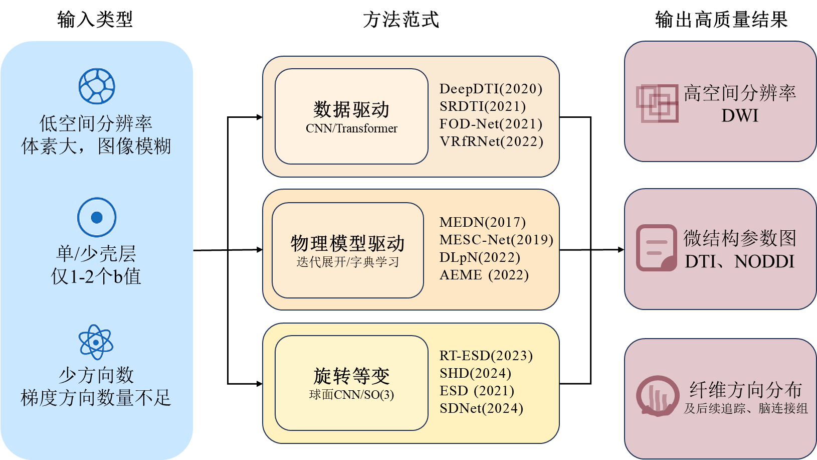

图2
提升fODF分辨率重建方法的分类图

表2
评价指标分类
| 类别 | 指标 | 中文名称 | 含义 | 说明 | ||
|---|---|---|---|---|---|---|
| 图像保真度指标 | Peak Signal-to-Noise Ratio (PSNR)[ | 峰值信噪比 | 衡量重建图像与参考图像之间的全局误差,值越高表示重建质量越好 | 基于均方误差计算,对误差敏感,但可能与视觉感知不完全一致,常用于评估DWI或参数图的重建质量 | ||
| Structural Similarity Index (SSIM)[ | 结构相似性指数 | 从亮度、对比度、结构三方面综合评估图像之间的相似度,更符合人眼视觉感知 | 值域为[0, 1],值越接近1表示相似度越高,适用于评估空间超分辨率结果的结构保真度 | |||
| 微结构标量误差指标 | Mean Absolute Error (MAE)[ | 平均绝对误差 | 计算预测标量值与真实值之间绝对误差的平均值 | 最基础的误差度量 | ||
| Mean Squared Error (MSE)[ | 均方误差 | 计算预测值与真实值之间平方误差的平均值 | 对较大误差更敏感 | |||
| Normalized Mean Absolute Error (NMAE)[ | 归一化平均绝对误差 | 对MAE进行归一化处理,便于比较不同量级的数据 | 具体归一化方式需在上下文中明确,通常除以真实值的范围或均值 | |||
| Normalized Mean Squared Error (NMSE)[ | 归一化均方误差 | 对MSE进行归一化处理,便于比较不同量级的数据 | 具体归一化方式需在上下文中明确,通常除以真实值的范围或均值 | |||
| 纤维方向重建评估指标 | Angular Correlation Coefficient (ACC)[ | 角相关系数 | 评估预测的纤维方向与参考真实方向在空间上的一致性 | 评估纤维方向估计整体准确性的核心指标 | ||
| Mean Angular Error (MAE)[ | 平均角度误差 | 衡量预测纤维主峰方向与参考真实方向之间的平均角度差 | 此MAE专指角度误差,与上文“平均绝对误差”含义不同 | |||
| Peak Error (PE)[ | 峰值误差 | 衡量预测纤维主峰幅度与参考真实幅度之间的峰值差 | 反映对纤维强度估计的准确性 | |||
| Proportion of Correct Peaks (PCP)[ | 正确峰比例 | 统计正确识别出的纤维方向的比例 | 一种基于分类正确率的评估方式,计算时设有阈值 | |||
| Earth Mover‘s Distance (EMD)[ | 推土机距离 | 衡量整个fODF分布与参考真实分布之间差异 | 评估分布层面相似性的核心指标,反映整体匹配度,综合考量角度和幅度 | |||
| 纤维追踪评估指标 | Valid Streamlines[ | 有效流线比例 | 在所有生成的流线中,属于任一真实纤维束的流线比例 | 反映追踪结果的纯净度,值低说明假阳性高 | ||
| Bundle Overlap[ | 纤维束重叠度 | 估计的纤维束与真实纤维束体素交集与真实集的比值 | 衡量灵敏度,即找回真实纤维束的能力 | |||
| Bundle Overreach[ | 纤维束过度延伸度 | 估计的纤维束超出真实纤维束的部分与真实集的比值 | 衡量特异度,值高表示假阳性多 | |||
| Valid Bundles[ | 有效纤维束数量 | 被正确识别出的、与真实解剖结构相符的纤维束数量 | 衡量算法重建特定神经通路的能力 | |||
| Dice Coefficient[ | Dice系数 | 两个二元分割结果的空间重叠程度 | 衡量纤维束分割结果 | |||
| 重测信度指标 | Coefficient of Variation (CV)[ | 变异系数 | 计算同一受试者经多次扫描的某指标的标准差与均值的比值 | 反映数据相对于其平均值的离散程度,值越低表示稳定性越好 | ||
| Weighted Mean Coefficient of Variation (wmCoV)[ | 加权平均变异系数 | 针对连接组矩阵,对每个连接边的CV进行加权平均求变异系数,权重取决于连接强度 | 由于连接组数据通常具有高度偏态分布,wmCoV通过对强连接赋予更高权重,更能代表整体连接组的重测信度 | |||
| Intraclass Correlation Coefficient (ICC)[ | 组内相关系数 | 量化重测信度的核心统计量,评估同一受试者多次扫描结果的一致性程度 | ICC > 0.75通常被认为信度优秀,是比简单相关性更严格的指标 | |||
表3
常用公开dMRI数据集
| 全称 | 缩写 | 主要人群/研究重点 | 特点 | 数据量 | 主要采集参数(b值非0) |
|---|---|---|---|---|---|
| The Rotterdam Study[ | RDS | 荷兰鹿特丹地区老年人慢性病的发病率、预后 | 大型长期随访队列 | MRI约8000人 | b = 1000 s/mm2,25 个方向 |
| The Rhineland Study[ | RLS | 德国莱茵地区人群的深度表型研究 | 人群基线广、影像丰富 | 千例以上 | b范围270 ~ 6800 s/mm2, 112个方向 |
| Pediatric Imaging Neurocognition and Genetics[ | PING | 儿科影像、认知发展与遗传关联 | 儿童、青少年群体 | 约1493名 | b = 1000 s/mm2, 约30个方向 |
| developing Human Connectome Project[ | dHCP | 典型与非典型早期脑发育 | 新生儿、早期发育阶段 | 1173名 | b = 400 s/mm2,64个方向; b = 1000 s/mm2,88个方向; b = 2600 s/mm2,128个方向 |
| Baby Connectome Project[ | BCP | 婴儿脑连接发育 | 婴幼儿期 | 500名 | b = 500 ~ 3000 s/mm2,总约144个方向 |
| Chinese Connectome Project[ | CHCP | 中国人群脑连接组 | 亚洲人群样本 | 366名 | b = 1000 s/mm2,93个方向; b = 2000 s/mm2,92个方向 |
| Amsterdam Open MRI Collection[ | AOMIC | 健康年轻成人,情感与社会认知的神经机制 | 模态丰富,附带行为与心理学量表数据 | 约1370名 | b = 1000 s/mm2,32个方向 |
| ISMRM 2015 Tractometer Challenge Dataset[ | / | 纤维束追踪算法的标准评估与对比 | 追踪算法验证与性能比较的常用基准 | 1名 | b = 1000 s/mm2, 90个方向; b = 2000 s/mm2, 90个方向; b = 3000 s/mm2,90个方向 |
| [1] | MENG J X, WANG Y J. Research progress on tractography of superficial white matter based on diffusion magnetic resonance imaging[J]. Chinese J Magn Reson, 2025, 42(2): 205-220. |
|
孟靖欣, 王远军. 基于扩散磁共振的大脑浅表白质纤维束研究进展[J]. 波谱学杂志, 2025, 42(2): 205-220.
doi: 10.11938/cjmr20243126 |
|
| [2] | JIANG B, ZHANG Z, LIN D, et al. Semi-supervised learning with graph learning-convolutional networks[C]// Proceedings of the IEEE/CVF conference on computer vision and pattern recognition, Long Beach, CA. Piscataway: IEEE, 2019: 11313-11320. |
| [3] | COHEN T S, GEIGER M, KÖHLER J, et al. Spherical CNNs[C]// International Conference on Learning Representations (ICLR), Vancouver, Canada: International Conference on Learning Representations, 2018: 1-15. |
| [4] |
FAIYAZ A, DOYLEY M M, SCHIFITTO G, et al. Artificial intelligence for diffusion MRI-based tissue microstructure estimation in the human brain: an overview[J]. Front Neurol, 2023, 14: 1168833.
doi: 10.3389/fneur.2023.1168833 |
| [5] |
BASSER P J, MATTIELLO J, LEBIHAN D. MR diffusion tensor spectroscopy and imaging[J]. Biophys J, 1994, 66(1): 259-267.
doi: 10.1016/S0006-3495(94)80775-1 pmid: 8130344 |
| [6] |
JENSEN J H, HELPERN J A. MRI quantification of non-Gaussian water diffusion by kurtosis analysis[J]. NMR Biomed, 2010, 23(7): 698-710.
doi: 10.1002/nbm.1518 pmid: 20632416 |
| [7] |
ZHANG H, SCHNEIDER T, WHEELER-KINGSHOTT C A, et al. NODDI: practical in vivo neurite orientation dispersion and density imaging of the human brain[J]. NeuroImage, 2012, 61(4): 1000-1016.
doi: 10.1016/j.neuroimage.2012.03.072 pmid: 22484410 |
| [8] |
TOURNIER J D, CALAMANTE F, CONNELLY A. Robust determination of the fibre orientation distribution in diffusion MRI: non-negativity constrained super-resolved spherical deconvolution[J]. NeuroImage, 2007, 35(4): 1459-1472.
doi: 10.1016/j.neuroimage.2007.02.016 pmid: 17379540 |
| [9] |
TIAN Q, BILGIC B, FAN Q, et al. DeepDTI: High-fidelity six-direction diffusion tensor imaging using deep learning[J]. NeuroImage, 2020, 219: 117017.
doi: 10.1016/j.neuroimage.2020.117017 |
| [10] |
LI H, LIANG Z, ZHANG C, et al. SuperDTI: Ultrafast DTI and fiber tractography with deep learning[J]. Magn Reson Med, 2021, 86(6): 3334-3347.
doi: 10.1002/mrm.28937 pmid: 34309073 |
| [11] |
SABIDUSSI E R, KLEIN S, JEURISSEN B, et al. dtiRIM: A generalisable deep learning method for diffusion tensor imaging[J]. NeuroImage, 2023, 269: 119900.
doi: 10.1016/j.neuroimage.2023.119900 |
| [12] |
ZHANG L, HE J, LI W, et al. Diff-DTI: Fast diffusion tensor imaging using a feature-enhanced joint diffusion model[J]. IEEE J Biomed Health Inform, 2026, 30(2): 1300-1313.
doi: 10.1109/JBHI.2024.3523532 |
| [13] |
MARTIN P, ALTBACH M, BILGIN A. Conditional generative diffusion deep learning for accelerated diffusion tensor and kurtosis imaging[J]. Magn Reson Imaging, 2025, 117: 110309.
doi: 10.1016/j.mri.2024.110309 |
| [14] | TIAN Q, LI Z, FAN Q, et al. SRDTI: Deep learning-based super-resolution for diffusion tensor MRI[PP/OL]. arXiv (2021-02-17) [2025-12-28]. https://arxiv.org/abs/2102.09069. |
| [15] | MA W, PENG L. Image quality transfer with auto-encoding applied to dMRI super-resolution[C]// 2021 4th International Conference on Advanced Electronic Materials, Computers and SoftwareEngineering AEMCSE, Changsha, China. Piscataway: IEEE, 2021: 828-831. |
| [16] |
KARIMI D, JAIMES C, MACHADO-RIVAS F, et al. Deep learning-based parameter estimation in fetal diffusion-weighted MRI[J]. NeuroImage, 2021, 243: 118482.
doi: 10.1016/j.neuroimage.2021.118482 |
| [17] |
KARIMI D, GHOLIPOUR A. Diffusion tensor estimation with transformer neural networks[J]. Artif Intell Med, 2022, 130: 102330.
doi: 10.1016/j.artmed.2022.102330 |
| [18] | FAIYAZ A, UDDIN M N, SCHIFITTO G. Angular upsampling in diffusion MRI using contextual hemihex sub-sampling in q-space[PP/OL]. arXiv (2022-11-01) [2025-12-28]. https://arxiv.org/abs/2211.00240. |
| [19] |
EWERT C, KUGLER D, STIRNBERG R, et al. Geometric deep learning for diffusion mri signal reconstruction with continuous samplings (discus)[J]. Imaging Neurosci, 2024, 2: 1-18.
doi: 10.1162/imag_a_00344 |
| [20] |
ALTMANN S, GRAUHAN N F, MERCADO M A A, et al. Deep learning accelerated brain diffusion-weighted MRI with super resolution processing[J]. Acad Radiol, 2024, 31(10): 4171-4182.
doi: 10.1016/j.acra.2024.02.049 pmid: 38521612 |
| [21] |
LE BIHAN D. What can we see with IVIM MRI?[J]NeuroImage, 2019, 187: 56-67.
doi: S1053-8119(17)31086-8 pmid: 29277647 |
| [22] |
GOLKOV V, DOSOVITSKIY A, SPERL J I, et al. Q-space deep learning: twelve-fold shorter and model-free diffusion MRI scans[J]. IEEE Trans Med Imaging, 2016, 35(5): 1344-1351.
doi: 10.1109/TMI.2016.2551324 |
| [23] |
GIBBONS E K, HODGSON K K, CHAUDHARI A S, et al. Simultaneous NODDI and GFA parameter map generation from subsampled q-space imaging using deep learning[J]. Magn Reson Med, 2019, 81(4): 2399-2411.
doi: 10.1002/mrm.27568 pmid: 30426558 |
| [24] | NATH V, RAMADASS K, SCHILLING K G, et al. DW-MRI microstructure model of models captured via single-shell bottleneck deep learning[C]// Computational Diffusion MRI: International MICCAI Workshop, Lima, Peru. Cham: Springer, 2021: 147-157. |
| [25] | CHEN G, HONG Y, ZHANG Y, et al. Estimating tissue microstructure with undersampled diffusion data via graph convolutional neural networks[C]// International Conference on Medical Image Computing and Computer-Assisted Intervention, Lima, Peru. Cham: Springer, 2020: 280-290. |
| [26] | CHEN G, JIANG H, LIU J, et al. Hybrid graph transformer for tissue microstructure estimation with undersampled diffusion MRI data[C]// International Conference on Medical Image Computing and Computer-Assisted Intervention, Singapore. Cham: Springer, 2022: 113-122. |
| [27] |
YE C. Tissue microstructure estimation using a deep network inspired by a dictionary-based framework[J]. Med Image Anal, 2017, 42: 288-299.
doi: S1361-8415(17)30132-9 pmid: 28910696 |
| [28] |
YE C, LI X, CHEN J. A deep network for tissue microstructure estimation using modified LSTM units[J]. Med Image Anal, 2019, 55: 49-64.
doi: S1361-8415(18)30557-7 pmid: 31022640 |
| [29] |
YE C, LI Y, ZENG X. An improved deep network for tissue microstructure estimation with uncertainty quantification[J]. Med Image Anal, 2020, 61: 101650.
doi: 10.1016/j.media.2020.101650 |
| [30] |
FAIYAZ A, DOYLEY M, SCHIFITTO G, et al. Single-shell NODDI using dictionary-learner-estimated isotropic volume fraction[J]. NMR Biomed, 2022, 35(2): e4628.
doi: 10.1002/nbm.v35.2 |
| [31] | ZHENG T, ZHENG W, SUN Y, et al. An adaptive network with extragradient for diffusion MRI-Based microstructure estimation[C]// International Conference on Medical Image Computing and Computer-Assisted Intervention, Singapore. Cham: Springer, 2022: 153-162. |
| [32] |
ZHENG T, YAN G, LI H, et al. A microstructure estimation Transformer inspired by sparse representation for diffusion MRI[J]. Med Image Anal, 2023, 86: 102788.
doi: 10.1016/j.media.2023.102788 |
| [33] |
LECUN Y, BOTTOU L, BENGIO Y, et al. Gradient-based learning applied to document recognition[J]. Proc IEEE, 2002, 86(11): 2278-2324.
doi: 10.1109/5.726791 |
| [34] | ESTEVES C, ALLEN-BLANCHETTE C, MAKADIA A, et al. Learning SO(3) equivariant representations with spherical CNNs[C]// European Conference on Computer Vision (ECCV), Munich, Germany. Cham: Springer, 2018: 52-68. |
| [35] | KONDOR R, LIN Z, TRIVEDI S. Clebsch-gordan nets: a fully fourier space spherical convolutional neural network[J]. Adv Neural Inf Process Syst, 2018: 31. |
| [36] | SEDLAR S, ALIMI A, PAPADOPOULO T, et al. A spherical convolutional neural network for white matter structure imaging via dMRI[C]// International Conference on Medical Image Computing and Computer-Assisted Intervention, Strasbourg, France. Cham: Springer, 2021: 529-539. |
| [37] | ELALDI A, DEY N, KIM H, et al. Equivariant spherical deconvolution: Learning sparse orientation distribution functions from spherical data[C]// International Conference on Information Processing in Medical Imaging, Bornholm, Denmark. Cham: Springer, 2021: 267-278. |
| [38] |
CONSAGRA W, NING L, RATHI Y. A deep learning approach to multi-fiber parameter estimation and uncertainty quantification in diffusion MRI[J]. Med Image Anal, 2025, 102: 103537.
doi: 10.1016/j.media.2025.103537 |
| [39] | YE C, QIN Y, LIU C, et al. Super-resolved q-space deep learning[C]// International Conference on Medical Image Computing and Computer-Assisted Intervention, Shenzhen, China. Cham: Springer, 2019: 582-589. |
| [40] |
QIN Y, LIU Z, LIU C, et al. Super-Resolved q-Space deep learning with uncertainty quantification[J]. Med Image Anal, 2021, 67: 101885.
doi: 10.1016/j.media.2020.101885 |
| [41] |
QIN Y, LI Y, ZHUO Z, et al. Multimodal super-resolved q-space deep learning[J]. Med Image Anal, 2021, 71: 102085.
doi: 10.1016/j.media.2021.102085 |
| [42] | DHOLLANDER T, MITO R, RAFFELT D, et al. Improved white matter response function estimation for 3-tissue constrained spherical deconvolution[C]// Proceedings of the International Society for Magnetic Resonance in Medicine (ISMRM) 2019. Montreal, Quebec, Canada: ISMRM, 2019: 555. |
| [43] |
JEURISSEN B, TOURNIER J D, DHOLLANDER T, et al. Multi-tissue constrained spherical deconvolution for improved analysis of multi-shell diffusion MRI data[J]. NeuroImage, 2014, 103: 411-426.
doi: S1053-8119(14)00644-2 pmid: 25109526 |
| [44] | KOPPERS S, MERHOF D. Direct estimation of fiber orientations using deep learning in diffusion imaging[C]// International Workshop on Machine Learning in Medical Imaging, Athens, Greece. Cham: Springer, 2016: 53-60. |
| [45] |
LIN Z, GONG T, WANG K, et al. Fast learning of fiber orientation distribution function for MR tractography using convolutional neural network[J]. Med Phys, 2019, 46(7): 3101-3116.
doi: 10.1002/mp.13555 pmid: 31009085 |
| [46] |
NATH V, SCHILLING K G, PARVATHANENI P, et al. Deep learning reveals untapped information for local white-matter fiber reconstruction in diffusion-weighted MRI[J]. Magn Reson Imaging, 2019, 62: 220-227.
doi: S0730-725X(19)30171-7 pmid: 31323317 |
| [47] | NATH V, PATHAK S K, SCHILLING K G, et al. Deep learning estimation of multi-tissue constrained spherical deconvolution with limited single shell DW-MRI[C]// Medical Imaging 2020: Image Processing, Bel Houston, Texas, USA. Bellingham: SPIE, 2020, 11313: 162-171. |
| [48] |
JHA R R, PATHAK S K, NATH V, et al. VRfRNet: Volumetric ROI fODF reconstruction network for estimation of multi-tissue constrained spherical deconvolution with only single shell dMRI[J]. Magn Reson Imaging, 2022, 90: 1-16.
doi: 10.1016/j.mri.2022.03.004 pmid: 35341904 |
| [49] |
BARTLETT J J, DAVEY C E, JOHNSTON L A, et al. Recovering high-quality fiber orientation distributions from a reduced number of diffusion-weighted images using a model-driven deep learning architecture[J]. Magn Reson Med, 2024, 92(5): 2193-2206.
doi: 10.1002/mrm.30187 pmid: 38852179 |
| [50] | YAO T, NEWLIN N, KANAKARAJ P, et al. A unified learning model for estimating fiber orientation distribution functions on heterogeneous multi-shell diffusion-weighted MRI[C]// International Workshop on Computational Diffusion MRI, Vancouver, BC, Canada. Cham: Springer, 2023: 13-22. |
| [51] |
CONSAGRA W, NING L, RATHI Y. Neural orientation distribution fields for estimation and uncertainty quantification in diffusion MRI[J]. Med Image Anal, 2024, 93: 103105.
doi: 10.1016/j.media.2024.103105 |
| [52] | SEDLAR S, PAPADOPOULO T, DERICHE R, et al. Diffusion MRI fiber orientation distribution function estimation using voxel-wise spherical U-net[C]// Computational Diffusion MRI: International MICCAI Workshop, Lima, Peru. Cham: Springer, 2021: 95-106. |
| [53] | ELALDI A, GERIG G, DEY N. E(3) × so(3)-equivariant networks for spherical deconvolution in diffusion MRI[C]// Medical Imaging with Deep Learning, Paris, France. Cambridge: PMLR, 2024: 301-319. |
| [54] | ELALDI A, GERIG G, DEY N. Equivariant spatio-hemispherical networks for diffusion MRI deconvolution[J]. Adv Neural Inf Process Syst, 2024, 37: 52095-52126. |
| [55] | GAO X, LIN R, FENG J, et al. UFO-3: unsupervised three-compartment learning for fiber orientation distribution function estimation[C]// International Conference on Medical Image Computing and Computer-Assisted Intervention, Daejeon, South Korea. Cham: Springer, 2025: 638-649. |
| [56] | SNOUSSI H, KARIMI D. Equivariant spherical CNNs for accurate fiber orientation distribution estimation in neonatal diffusion MRI with reduced acquisition time[PP/OL]. arXiv (2025-04-02) [2025-12-28]. https://arxiv.org/abs/2504.01925. |
| [57] |
ZENG R, LV J, WANG H, et al. FOD-Net: A deep learning method for fiber orientation distribution angular super resolution[J]. Med Image Anal, 2022, 79: 102431.
doi: 10.1016/j.media.2022.102431 |
| [58] | DA SILVA M O, SANTANA C P, DO CARMO D S, et al. FOD-Swin-Net:angular super resolution of fiber orientation distribution using a transformer-based deep model[C]// 2024 IEEE International Symposium on Biomedical Imaging (ISBI), Athens, Greece. Piscataway: IEEE, 2024: 1-5. |
| [59] |
LI J, AI L, YAO R. NVAM-Net: deep learning networks for reconstructing high-quality fiber orientation distributions[J]. Neuroradiology, 2024, 66(7): 1177-1187.
doi: 10.1007/s00234-024-03341-y pmid: 38563964 |
| [60] | YANG L M, WANG Y J. Research progress of denoising algorithms for diffusion tensor images[J]. Chinese J Magn Reson, 2024, 41(3): 341-361. |
|
杨黎明, 王远军. 扩散张量图像去噪算法研究进展[J]. 波谱学杂志, 2024, 41(3): 341-361.
doi: 10.11938/cjmr20243087 |
|
| [61] |
RUBNER Y, TOMASI C, GUIBAS L J. The earth mover's distance as a metric for image retrieval[J]. Int J Comput Vision, 2000, 40(2): 99-121.
doi: 10.1023/A:1026543900054 |
| [62] |
RENAULD E, THÉBERGE A, PETIT L, et al. Validate your white matter tractography algorithms with a reappraised ISMRM 2015 Tractography Challenge scoring system[J]. Sci Rep, 2023, 13(1): 2347.
doi: 10.1038/s41598-023-28560-w pmid: 36759653 |
| [63] | SENTHIL KUMAR V S, SHAHRAZ S. Intraclass correlation for reliability assessment: the introduction of a validated program in SAS (ICC6)[J]. Health Serv Outcome, 2024, 24(1): 1-13. |
| [64] |
VAN ESSEN D C, SMITH S M, BARCH D M, et al. The WU-Minn human connectome project: an overview[J]. NeuroImage, 2013, 80: 62-79.
doi: 10.1016/j.neuroimage.2013.05.041 pmid: 23684880 |
| [65] |
IKRAM M A, BRUSSELLE G, GHANBARI M, et al. Objectives, design and main findings until 2020 from the Rotterdam Study[J]. Eur J Epidemiol, 2020, 35(5): 483-517.
doi: 10.1007/s10654-020-00640-5 pmid: 32367290 |
| [66] | BRETELER M M B, STÖCKER T, PRACHT E, et al. IC-P-165: MRI in the Rhineland study: a novel protocol for population neuroimaging[J]. Alzheimer's & Dementia, 2014, 10: P92-P92. |
| [67] |
JERNIGAN T L, BROWN T T, HAGLER JR D J, et al. The pediatric imaging, neurocognition, and genetics (PING) data repository[J]. NeuroImage, 2016, 124: 1149-1154.
doi: S1053-8119(15)00357-2 pmid: 25937488 |
| [68] | EDWARDS A D, RUECKERT D, SMITH S M, et al. The developing human connectome project neonatal data release[J]. Front Neurol, 2022, 16: 886772. |
| [69] |
HOWELL B R, STYNER M A, GAO W, et al. The UNC/UMN Baby Connectome Project (BCP): An overview of the study design and protocol development[J]. NeuroImage, 2019, 185: 891-905.
doi: S1053-8119(18)30259-3 pmid: 29578031 |
| [70] | GE J, YANG G, HAN M, et al. Increasing diversity in connectomics with the Chinese Human Connectome Project[J]. Nat Neurosci, 2023, 26(1): 163-172. |
| [71] |
SNOEK L, VAN DER MIESEN M M, BEEMSTERBOER T, et al. The amsterdam open MRI collection, a set of multimodal MRI datasets for individual difference analyses[J]. Sci Data, 2021, 8(1): 85.
doi: 10.1038/s41597-021-00870-6 pmid: 33741990 |
| [72] |
MAIER-HEIN K H, NEHER P F, HOUDE J C, et al. The challenge of mapping the human connectome based on diffusion tractography[J]. Nat Commun, 2017, 8(1): 1349.
doi: 10.1038/s41467-017-01285-x |
| [73] | YANG J C, WANG Y J. Improved constrained spherical deconvolution model for brain gray matter microstructure imaging[J]. Chinese J Magn Reson, 2025, 42(1): 67-79. |
|
杨佳铖, 王远军. 改进约束球面反卷积模型的脑灰质微结构成像[J]. 波谱学杂志, 2025, 42(1): 67-79.
doi: 10.11938/cjmr20243117 |
|
| [74] | AJA-FERNÁNDEZ S, MARTÍN-MARTÍN C, PLANCHUELO-GÓMEZ Á, et al. Validation of deep learning techniques for quality augmentation in diffusion MRI for clinical studies[J]. NeuroImage, 2023, 39: 103483. |
| [1] | 李英豪, 王丽辉, 王苏成, 朱中旗, 黄长栋, 李仁峰, 曹开明, 胡海洋, 贾一鸣, 梁松涛, 杨光, 路青, 汪红志. 胰腺自动分割与区域定量及糖尿病评估研究[J]. 波谱学杂志, 2025, 42(4): 378-389. |
| [2] | 高照耀, 张展, 胡亮亮, 许光宇, 周胜, 胡雨欣, 林子捷, 周超. 基于虚拟线圈和GRAPPA增强网络的PMRI方法[J]. 波谱学杂志, 2025, 42(4): 390-401. |
| [3] | 张明玉, 肖洒, 石胜杰, 张学成, 周欣. 超极化129Xe MRI的多模态增强去噪扩散模型研究[J]. 波谱学杂志, 2025, 42(4): 364-377. |
| [4] | 孟靖欣, 王远军. 基于扩散磁共振的大脑浅表白质纤维束研究进展[J]. 波谱学杂志, 2025, 42(2): 205-220. |
| [5] | 曹飞, 徐芊芊, 陈浩, 祖洁, 李晓文, 田锦, 鲍磊. 基于交叉自监督和DWI的NIID智能诊断方法[J]. 波谱学杂志, 2025, 42(2): 154-163. |
| [6] | 杨佳铖, 王远军. 改进约束球面反卷积模型的脑灰质微结构成像[J]. 波谱学杂志, 2025, 42(1): 67-79. |
| [7] | 薛培阳, 耿辰, 李郁欣, 鲍奕仿, 鲁宇澄, 戴亚康. 基于3D ResNet50改进模型的TOF-MRA脑动脉瘤分类方法[J]. 波谱学杂志, 2025, 42(1): 56-66. |
| [8] | 宁欣宙, 黄臻, 陈西曲, 刘鑫杰, 陈罡, 张志, 鲍庆嘉, 刘朝阳. 用于超快时空编码MRI的Transformer超分辨率重建算法研究[J]. 波谱学杂志, 2024, 41(4): 454-468. |
| [9] | 杨黎明, 王远军. 扩散张量图像去噪算法研究进展[J]. 波谱学杂志, 2024, 41(3): 341-361. |
| [10] | 戴俊龙, 何聪, 武杰, 边云. 融合双解码和全局注意力上采样模块的胰腺囊性肿瘤分割网络[J]. 波谱学杂志, 2024, 41(2): 151-161. |
| [11] | 杨钰, 陈博, 吴柳滨, 林恩平, 黄玉清, 陈忠. Laplace NMR谱图重建——从经典正则化到深度学习[J]. 波谱学杂志, 2024, 41(2): 191-208. |
| [12] | 常博, 孙灏芸, 高清宇, 王丽嘉. 传统方法和深度学习用于不同模态心脏医学图像的分割研究进展[J]. 波谱学杂志, 2024, 41(2): 224-244. |
| [13] | 徐真顺, 袁小涵, 黄子珩, 邵成伟, 武杰, 边云. 基于深度学习的胰腺黏液性和浆液性囊性肿瘤的多源特征分类模型[J]. 波谱学杂志, 2024, 41(1): 19-29. |
| [14] | 赖嘉雯, 汪宇玲, 蔡晓宇, 周丽华. 基于CNN-SVM的多维度信息融合半月板撕裂分类方法[J]. 波谱学杂志, 2023, 40(4): 423-434. |
| [15] | 王慧, 王甜甜, 王丽嘉. 基于心脏磁共振电影图像的压缩激励残差U形网络左心肌分割[J]. 波谱学杂志, 2023, 40(4): 435-447. |
| 阅读次数 | ||||||
|
全文 |
|
|||||
|
摘要 |
|
|||||