
Chinese Journal of Magnetic Resonance ›› 2026, Vol. 43 ›› Issue (2): 175-185.doi: 10.11938/cjmr20253177cstr: 32225.14.cjmr20253177
• Articles • Previous Articles Next Articles
CAI Yue1,2,*( ), WANG Xuxia1,2, LIU Sijie1,2, GUO Haodong1,2, CHEN Xi1,2, CHENG Linlin1,3, KANG Yan1,2, LIN Fuchun1,2
Received:2025-07-23
Published:2026-06-05
Online:2025-09-08
Contact:
CAI Yue
E-mail:caiyue192@mails.ucas.ac.cn
CLC Number:
CAI Yue, WANG Xuxia, LIU Sijie, GUO Haodong, CHEN Xi, CHENG Linlin, KANG Yan, LIN Fuchun. Magnetic Resonance Imaging Study on the Microstructure Abnormalities of Striatal White Matter in Type 2 Diabetic Mellitus Rats[J]. Chinese Journal of Magnetic Resonance, 2026, 43(2): 175-185.
Add to citation manager EndNote|Reference Manager|ProCite|BibTeX|RefWorks
Table 1
Experiment reagents and chemicals
| 试剂名称 | 纯度 | 供应商 |
|---|---|---|
| 链脲佐菌素(STZ) | ≥99% | S0130, Sigma |
| 戊巴比妥钠 | ≥99% | 57-33-0, Sigma |
| 柠檬酸-柠檬酸钠缓冲液 | 0.1 mol/L, pH=4.5 | pH1716,飞净 |
| 0.9%生理盐水 | 0.9% (w/v) | 武汉滨湖双鹤药业有限公司 |
| 丙三醇 | ≥99% | 国药集团化学试剂有限公司 |
| 乙二醇 | ≥99.5% | 国药集团化学试剂有限公司 |
| 多聚甲醛 | ≥95% | 国药集团化学试剂有限公司 |
| 氯化钠 | ≥99.5% | 国药集团化学试剂有限公司 |
| 氯化钾 | ≥99.5% | 国药集团化学试剂有限公司 |
| 十二水合磷酸氢二钾 | ≥99% | 国药集团化学试剂有限公司 |
| 磷酸二氢钾 | ≥99.5% | 国药集团化学试剂有限公司 |
| 氢氧化钠 | ≥99.8% | 国药集团化学试剂有限公司 |
| 羊血清 | ≥99% | 武汉飞弈科技有限公司 |
| 异氟烷 | ≥99% | 深圳市瑞沃德生命科技股份有限公司 |
| 葡萄糖 | ≥99.5% | 国药集团化学试剂有限公司 |
| 蔗糖 | ≥99% | 国药集团化学试剂有限公司 |
| 髓鞘碱性蛋白(MBP)一抗 | ≥95% | Ab218011, sigma |
| 磷酸化神经丝(SMI-31)一抗 | 纯化型 | SMI-31P-100, Covance |
| 二抗羊抗小鼠 Alexa Fluor 594 | 免疫原亲和纯化型 | ab150116, abcam |
| 二抗羊抗兔 Alexa Fluor 488 | 免疫原亲和纯化型 | ab150077, abcam |
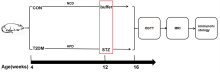
Fig. 1
The pipeline of whole experiment. CON: the saline control group rats; T2DM: the T2DM group rats; NCD: normal chow diet; HFD: high-fat diet; STZ: streptozotocin; OGTT; oral glucose tolerance test

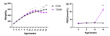
Fig. 2
The changes in body weight and fasting blood glucose level in rats. (a) Changes in body weight of rats in the two groups from 4 to 16 weeks of age; (b) Changes in fasting blood glucose in the two groups of rats aged 4 to 16 weeks. *p < 0.05; ***p < 0.001

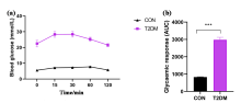
Fig. 3
Oral glucose tolerance test in rats. (a) The dynamic change curves of blood glucose in the OGTT test of the two groups of rats; (b) The areas under the OGTT change curves of the two groups of rats, with ***p < 0.001
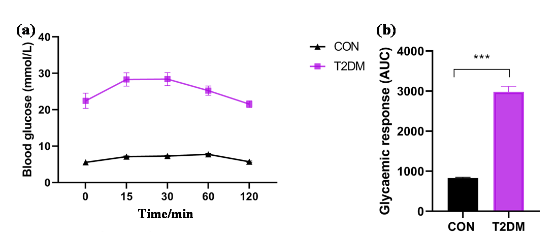

Fig. 4
Effect of T2DM on the microstructure of white matter in rats. CPU: caudate putamen (striatum), M1: primary motor cortex, S1FL: primary somatosensory cortex, forelimb area, S1HL: primary somatosensory cortex, hindlimb area. p<0.005 (FDR correction), voxel clusters of RARE =100, voxel clusters of DTI =50
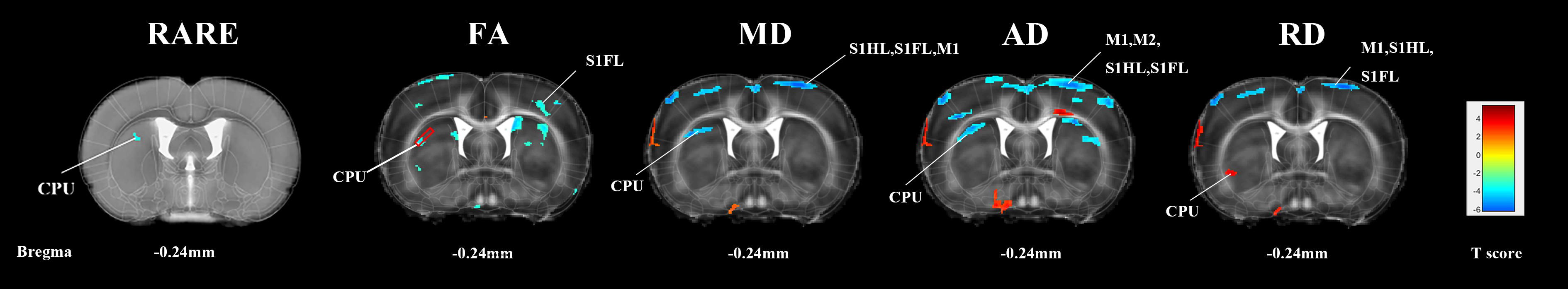
Fig. 5
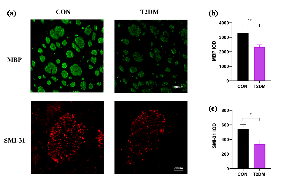
Immunohistological staining of the striatum in rats. (a) Immunohistochemical staining of MBP and SMI-31 in the striatum region of the two groups of rats. The scale of MBP immunoresponse staining was 200 μm, and the scale of SMI-31 immunoresponse staining was 20 μm; (b) The IOD value of MBP in the striatum region; (c) The IOD value of SMI-31 in the striatum region. * p < 0.05; ** p < 0.01
| [1] |
AHMAD E, LIM S, LAMPTEY R, et al. Type 2 diabetes[J]. Lancet, 2022, 400(10365): 1803-1820.
doi: 10.1016/S0140-6736(22)01655-5 pmid: 36332637 |
| [2] |
OGURTSOVA K, GUARIGUATA L, BARENGO N C, et al. IDF diabetes atlas: Global estimates of undiagnosed diabetes in adults for 2021[J]. Diabetes Res Clin Pract, 2022, 183: 109118.
doi: 10.1016/j.diabres.2021.109118 |
| [3] |
ZHENG Y, LEY S H, HU F B. Global aetiology and epidemiology of type 2 diabetes mellitus and its complications[J]. Nat Rev Endocrinol, 2018, 14(2): 88-98.
doi: 10.1038/nrendo.2017.151 pmid: 29219149 |
| [4] |
TOMIC D, SHAW J E, MAGLIANO D J. The burden and risks of emerging complications of diabetes mellitus[J]. Nat Rev Endocrinol, 2022, 18(9): 525-539.
doi: 10.1038/s41574-022-00690-7 |
| [5] |
HUANG M, GAO L, YANG L, et al. Abnormalities in the brain of streptozotocin-induced type 1 diabetic rats revealed by diffusion tensor imaging[J]. Neuroimage Clin, 2012, 1(1): 57-65.
doi: 10.1016/j.nicl.2012.09.004 |
| [6] |
ZHANG T, SHAW M, CHERBUIN N. Association between type 2 diabetes mellitus and brain atrophy: a meta-analysis[J]. Diabetes Metab J, 2022, 46(5): 781-802.
doi: 10.4093/dmj.2021.0189 pmid: 35255549 |
| [7] |
MA T, LI Z Y, YU Y, et al. Gray and white matter abnormality in patients with T2DM-related cognitive dysfunction: a systemic review and meta-analysis[J]. Nutr Diabetes, 2022, 12(1): 39.
doi: 10.1038/s41387-022-00214-2 pmid: 35970833 |
| [8] |
QIU W, YUE X, HUANG H, et al. Structural characteristics of amygdala subregions in type 2 diabetes mellitus[J]. Behav Brain Res, 2024, 466: 114992.
doi: 10.1016/j.bbr.2024.114992 |
| [9] |
TANG Q, LI S, YANG Z, et al. A narrative review of multimodal imaging of white matter lesions in type-2 diabetes mellitus[J]. Ann Palliat Med, 2021, 10(12): 12867-12876.
doi: 10.21037/apm-21-3299 pmid: 35016461 |
| [10] |
TOURNIER J D, MORI S, LEEMANS A. Diffusion tensor imaging and beyond[J]. Magn Reson Med, 2011, 65(6): 1532-1556.
doi: 10.1002/mrm.v65.6 |
| [11] |
ALOTAIBI A, TENCH C, STEVENSON R, et al. Investigating brain microstructural alterations in type 1 and type 2 diabetes using diffusion tensor imaging: a systematic review[J]. Brain Sci, 2021, 11(2): 140.
doi: 10.3390/brainsci11020140 |
| [12] |
XIONG Y, SUI Y, ZHANG S, et al. Brain microstructural alterations in type 2 diabetes: diffusion kurtosis imaging provides added value to diffusion tensor imaging[J]. Eur Radiol, 2019, 29(4): 1997-2008.
doi: 10.1007/s00330-018-5746-y pmid: 30338363 |
| [13] |
HUANG L, ZHANG Q, TANG T, et al. Abnormalities of brain white matter in type 2 diabetes mellitus: a meta-analysis of diffusion tensor imaging[J]. Front Aging Neurosci, 2021, 13: 693890.
doi: 10.3389/fnagi.2021.693890 |
| [14] |
TAN X, FANG P, AN J, et al. Micro-structural white matter abnormalities in type 2 diabetic patients: a DTI study using TBSS analysis[J]. Neuroradiology, 2016, 58(12): 1209-1216.
pmid: 27783100 |
| [15] |
WU C Y, HUANG S M, LIN Y H, et al. Reproducibility of diffusion tensor imaging-derived parameters: implications for the streptozotocin-induced type 1 diabetic rats[J]. Magn Reson Mater Phy, 2023, 36(4): 631-639.
doi: 10.1007/s10334-022-01048-w |
| [16] |
LI J, GUO Y, LI Q, et al. Presence of white matter lesions associated with diabetes-associated cognitive decline in male rat models of pre-type 2 diabetes[J]. Med Sci Monit, 2019, 25: 9679-9689.
doi: 10.12659/MSM.918557 |
| [17] |
LI M Z, ZHANG L, SHI Z Y, et al. Magnetic resonance imaging detects cerebral gray and white matter injury correlated with cognitive impairments in diabetic db/db mice[J]. Behav Brain Res, 2023, 451: 114510.
doi: 10.1016/j.bbr.2023.114510 |
| [18] | HU Y D, CAI Y, WANG X X, et al. Magnetic resonance imaging the brain structures involved in nicotine susceptibility in rats[J]. Chinese J Magn Reson, 2021, 38(3): 345-355. |
|
胡赢丹, 蔡悦, 王旭霞, 等. 尼古丁易感的脑结构特征的磁共振成像研究[J]. 波谱学杂志, 2021, 38(3): 345-355.
doi: 10.11938/cjmr20212890 |
|
| [19] | CHEN X, LIU S J, CAI Y, et al. Effects of seizure-inducing doses nicotine on hippocampal structure in adolescent female rats[J]. Chinese J Magn Reson, 2025, 42(4): 345-354. |
| 陈茜, 刘思婕, 蔡悦, 等. 致痫剂量尼古丁对青少年雌性大鼠海马结构的影响[J]. 波谱学杂志, 2025, 42(4): 345-354. | |
| [20] | HUANG W, CAO Z Y. STZ-induced progressive brain atrophy studied by magnetic resonance imaging and histochemical staining[J]. Chinese J Magn Reson, 2015, 32(3): 439-449. |
|
黄微, 曹子玉. STZ诱导大鼠1型糖尿病进行性脑萎缩的磁共振成像及组织化学研究[J]. 波谱学杂志, 2015, 32(3): 439-449.
doi: 10.11938/cjmr20150305 |
|
| [21] |
WANG X, LIN F, KANG Y, et al. Brain structural plasticity in rats subjected to early binocular enucleation characterized by high resolution anatomical magnetic resonance imaging and diffusion tensor imaging[J]. Magn Reson Lett, 2023, 3(1): 14-21.
doi: 10.1016/j.mrl.2022.10.001 pmid: 40919275 |
| [22] |
TANAKA S, HAYASHI T, TOYODA T, et al. High-fat diet impairs the effects of a single bout of endurance exercise on glucose transport and insulin sensitivity in rat skeletal muscle[J]. Metabolism, 2007, 56(12): 1719-1728.
doi: 10.1016/j.metabol.2007.07.017 |
| [23] |
SRINIVASAN K, VISWANAD B, ASRAT L, et al. Combination of high-fat diet-fed and low-dose streptozotocin-treated rat: a model for type 2 diabetes and pharmacological screening[J]. Pharmacol Res, 2005, 52(4): 313-320.
doi: 10.1016/j.phrs.2005.05.004 pmid: 15979893 |
| [24] |
KOWLURU R A. Retinopathy in a diet-induced type 2 diabetic rat model and role of epigenetic modifications[J]. Diabetes, 2020, 69(4): 689-698.
doi: 10.2337/db19-1009 pmid: 31949005 |
| [25] |
MORAN C, PHAN T G, CHEN J, et al. Brain atrophy in type 2 diabetes: regional distribution and influence on cognition[J]. Diabetes Care, 2013, 36(12): 4036-4042.
doi: 10.2337/dc13-0143 pmid: 23939539 |
| [26] |
YAU P L, JAVIER D C, RYAN C M, et al. Preliminary evidence for brain complications in obese adolescents with type 2 diabetes mellitus[J]. Diabetologia, 2010, 53(11): 2298-2306.
doi: 10.1007/s00125-010-1857-y pmid: 20668831 |
| [27] |
LI C, JIN R, LIU K, et al. White matter atrophy in type 2 diabetes mellitus patients with mild cognitive impairment[J]. Front Neurosci, 2020, 14: 602501.
doi: 10.3389/fnins.2020.602501 |
| [28] |
ZHOU Y, LI X L, XIE H L, et al. Voxel-based morphology analysis of STZ-induced type 1 diabetes mellitus rats with and without cognitive impairment[J]. Neurosci Lett, 2018, 684: 210-217.
doi: S0304-3940(18)30555-X pmid: 30125641 |
| [29] |
CHEN J, ZHANG J, LIU X, et al. Abnormal subcortical nuclei shapes in patients with type 2 diabetes mellitus[J]. Eur Radiol, 2017, 27(10): 4247-4256.
doi: 10.1007/s00330-017-4790-3 pmid: 28374074 |
| [30] |
SUN Q, CHEN G Q, WANG X B, et al. Alterations of white matter integrity and hippocampal functional connectivity in type 2 diabetes without mild cognitive impairment[J]. Front Neuroanat, 2018, 12: 00021.
doi: 10.3389/fnana.2018.00021 |
| [31] |
ABE Y, YAMAMOTO T, SOEDA T, et al. Diabetic striatal disease: clinical presentation, neuroimaging, and pathology[J]. Intern Med, 2009, 48(13): 1135-1141.
doi: 10.2169/internalmedicine.48.1996 |
| [32] |
LEI H, DOOLEY P, PEELING J, et al. Temporal profile of magnetic resonance imaging changes following forebrain ischemia in the gerbil[J]. Neurosci Lett, 1998, 257(2): 105-108.
pmid: 9865938 |
| [33] |
WANG H, WANG Z, GAO Y, et al. STZ-induced diabetes exacerbates neurons ferroptosis after ischemic stroke by upregulating LCN2 in neutrophils[J]. Exp Neurol, 2024, 377: 114797.
doi: 10.1016/j.expneurol.2024.114797 |
| [34] |
DE BRESSER J, KUIJF H J, ZAANEN K, et al. White matter hyperintensity shape and location feature analysis on brain MRI; proof of principle study in patients with diabetes[J]. Sci Rep, 2018, 8(1): 1893.
doi: 10.1038/s41598-018-20084-y pmid: 29382936 |
| [35] |
ZHANG J H, XU H Z, SHEN Q F, et al. Nepsilon-(carboxymethyl)-lysine, white matter, and cognitive function in diabetes patients[J]. Can J Neurol Sci, 2016, 43(4): 518-522.
doi: 10.1017/cjn.2015.398 |
| [36] |
YOON S, CHO H, KIM J, et al. Brain changes in overweight/obese and normal-weight adults with type 2 diabetes mellitus[J]. Diabetologia, 2017, 60(7): 1207-1217.
doi: 10.1007/s00125-017-4266-7 pmid: 28447116 |
| [37] |
ZHOU C, LI J, DONG M, et al. Altered white matter microstructures in type 2 diabetes mellitus: a coordinate-based meta-analysis of diffusion tensor imaging studies[J]. Front Endocrinol (Lausanne), 2021, 12: 658198.
doi: 10.3389/fendo.2021.658198 |
| [38] |
VAN DE VONDERVOORT I, AMIRI H, BRUCHHAGE M M K, et al. Converging evidence points towards a role of insulin signaling in regulating compulsive behavior[J]. Transl Psychiatry, 2019, 9(1): 225.
doi: 10.1038/s41398-019-0559-6 |
| [39] |
ZHANG J, CHEN S, SHI W, et al. Effects of Xiaoshuan enteric-coated capsule on white and gray matter injury evaluated by diffusion tensor imaging in ischemic stroke[J]. Cell Transplant, 2019, 28(6): 671-683.
doi: 10.1177/0963689718802755 |
| [40] |
GAO J, PAN P, LI J, et al. Analysis of white matter tract integrity using diffusion kurtosis imaging reveals the correlation of white matter microstructural abnormalities with cognitive impairment in type 2 diabetes mellitus[J]. Front Endocrinol (Lausanne), 2024, 15: 1327339.
doi: 10.3389/fendo.2024.1327339 |
| [41] |
PORTER A, LECKIE R, VERSTYNEN T. White matter pathways as both a target and mediator of health behaviors[J]. Ann N Y Acad Sci, 2018, 1428(1): 71-88.
doi: 10.1111/nyas.2018.1428.issue-1 |
| [42] |
SONG S K, SUN S W, JU W K, et al. Diffusion tensor imaging detects and differentiates axon and myelin degeneration in mouse optic nerve after retinal ischemia[J]. NeuroImage, 2003, 20(3): 1714-1722.
doi: 10.1016/j.neuroimage.2003.07.005 |
| [43] |
SOUSTELLE L, ANTAL M C, LAMY J, et al. Correlations of quantitative MRI metrics with myelin basic protein (MBP) staining in a murine model of demyelination[J]. NMR Biomed, 2019, 32(9): e4116.
doi: 10.1002/nbm.v32.9 |
| [44] | MUKHERJEE P, MILLER J H, SHIMONY J S, et al. Diffusion-tensor MR imaging of gray and white matter development during normal human brain maturation[J]. AJNR Am J Neuroradiol, 2002, 23(9): 1445-1456. |
| [45] | ZHU Y S, XIONG K L, ZHANG Y L, et al. Establishment of an acute diffuse axonal injury model and early diffusion tensor imaging manifestations[J]. Chin J Trauma, 2014, 30(5): 460-463. |
| 朱永山, 熊坤林, 张玉龙, 等. 急性弥漫性轴索损伤模型的建立与早期弥散张量成像表现[J]. 中华创伤杂志, 2014, 30(5): 460-463. | |
| [46] |
WANG S, WU E X, QIU D, et al. Longitudinal diffusion tensor magnetic resonance imaging study of radiation-induced white matter damage in a rat model[J]. Cancer Res, 2009, 69(3): 1190-1198.
doi: 10.1158/0008-5472.CAN-08-2661 pmid: 19155304 |
| [1] | FU Fenfang, LIN Guobing, LI Meifang. Advances in Magnetic Resonance Imaging for the Diagnosis and Prognosis of Mild Traumatic Brain Injury [J]. Chinese Journal of Magnetic Resonance, 2026, 43(2): 214-222. |
| [2] | NI Guangmao, LI Yuwei, HOU Wenxuan, LIU Caiyun, DONG Peng, ZHANG Yanhui. Research on the Influencing Factors of Acute Cerebral Infarction Recurrence Based on MR-DWI [J]. Chinese Journal of Magnetic Resonance, 2026, 43(1): 87-93. |
| [3] | CHEN Xi, LIU Sijie, CAI Yue, CHENG Linlin, WANG Xuxia, KANG Yan, LIN Fuchun, LEI Hao. Effects of Seizure-inducing Doses Nicotine on Hippocampal Structure in Adolescent Female Rats [J]. Chinese Journal of Magnetic Resonance, 2025, 42(4): 345-354. |
| [4] | LI Yinghao, WANG Lihui, WANG Sucheng, ZHU Zhongqi, HUANG Changdong, LI Renfeng, CAO Kaiming, HU Haiyang, JIA Yiming, LIANG Songtao, YANG Guang, LU Qing, WANG Hongzhi. Study on Pancreas Automatic Segmentation, Regional Quantification, and Diabetes Assessment [J]. Chinese Journal of Magnetic Resonance, 2025, 42(4): 378-389. |
| [5] | MA Yingxue, ZHAO Yanqiang, YANG Xiaodong, JIANG Bin, TAO Cheng. Opportunities and Challenges of High-field and Ultra-high-field Magnetic Resonance Imaging in China [J]. Chinese Journal of Magnetic Resonance, 2025, 42(3): 334-344. |
| [6] | SUI Meiju, ZHANG Lei, WANG Ruifang, LUO Yingying, LI Sha, QIU Maosong, XU Qiuyi, CHEN Daiqin, CHEN Shizhen, ZHOU Xin. MRI-traceable Nanoenzyme for Cascade Catalysis-enhanced Immunotherapy [J]. Chinese Journal of Magnetic Resonance, 2025, 42(3): 231-248. |
| [7] | CHEN Qun, YANG Zijian, CHENG Xinyi, JIA Siyi, DU Xiaoxia, WANG Mengxing. Application of Magnetic Resonance Imaging Technology in Pediatric Exercise Intervention Research [J]. Chinese Journal of Magnetic Resonance, 2025, 42(2): 195-204. |
| [8] | PANG Qifan, WANG Zhichao, WU Yupeng, LI Jianqi. The Impact of K-Space Filling Strategy on Fat Artifacts in APT Imaging Based on FLASH Sequence [J]. Chinese Journal of Magnetic Resonance, 2024, 41(4): 443-453. |
| [9] | XU Zhenshun, YUAN Xiaohan, HUANG Ziheng, SHAO Chengwei, WU Jie, BIAN Yun. Multi-source Feature Classification Model of Pancreatic Mucinous and Serous Cystic Neoplasms Based on Deep Learning [J]. Chinese Journal of Magnetic Resonance, 2024, 41(1): 19-29. |
| [10] | LIU Ying, LIN Ling, YUAN Binhua, ZHANG Haowei. Research Progress of MRI Gradient Waveform Generator [J]. Chinese Journal of Magnetic Resonance, 2024, 41(1): 99-115. |
| [11] | LI Pan,FANG Delei,ZHANG Junxia,MA Debei. Magnetic Resonance Compatibility Analysis Method of Surgical Robotic System Based on Image Quality Evaluation [J]. Chinese Journal of Magnetic Resonance, 2023, 40(1): 79-91. |
| [12] |
De-gang TANG,Hong-chuang LI,Xiao-ling LIU,Lei SHI,Hai-dong LI,Chao-hui YE,Xin ZHOU.
A Simulation Study on the Effect of the High Permittivity Materials Geometrical Structure on the Transmit Field |
| [13] | Zhen-yu WANG, Ying-shan WANG, Jin-ling MAO, Wei-wei MA, Qing LU, Jie SHI, Hong-zhi WANG. Magnetic Resonance Images Segmentation of Synovium Based on Dense-UNet++ [J]. Chinese Journal of Magnetic Resonance, 2022, 39(2): 208-219. |
| [14] | Yan MA, Cang-ju XING, Liang XIAO. Knee Joint Image Segmentation and Model Construction Based on Cascaded Network [J]. Chinese Journal of Magnetic Resonance, 2022, 39(2): 184-195. |
| [15] | Jun LUO, Sheng-ping LIU, Xing YANG, Jia-sheng WANG, Ye LI. Design of a 5 T Non-magnetic Magnetic Resonance Radio Frequency Power Amplifier [J]. Chinese Journal of Magnetic Resonance, 2022, 39(2): 163-173. |
| Viewed | ||||||
|
Full text |
|
|||||
|
Abstract |
|
|||||