
Chinese Journal of Magnetic Resonance ›› 2024, Vol. 41 ›› Issue (1): 19-29.doi: 10.11938/cjmr20233064cstr: 32225.14.cjmr20233064
• Articles • Previous Articles Next Articles
XU Zhenshun1,YUAN Xiaohan2,HUANG Ziheng1,SHAO Chengwei2,WU Jie1,#( ),BIAN Yun2,*()
Received:2023-04-19
Published:2024-03-05
Online:2023-06-08
Contact:
# Tel: 021-55271116, E-mail: CLC Number:
XU Zhenshun, YUAN Xiaohan, HUANG Ziheng, SHAO Chengwei, WU Jie, BIAN Yun. Multi-source Feature Classification Model of Pancreatic Mucinous and Serous Cystic Neoplasms Based on Deep Learning[J]. Chinese Journal of Magnetic Resonance, 2024, 41(1): 19-29.
Add to citation manager EndNote|Reference Manager|ProCite|BibTeX|RefWorks
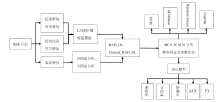
Fig. 1
Experimental workflow of multi-source feature model based on deep learning
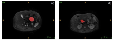
Fig. 2
Segmentation images of MCN and SCN. (a) Abdominal axial T2WI showed high MCN signal with regular boundary. (b) Abdominal axial T2WI showed high SCN signal with irregular boundary
Fig. 2
The process of ResNet50 extracting deep learning features, which includes four convolution blocks (Conv), average pooling layer (Av-pool) and fully connected layer (fc)
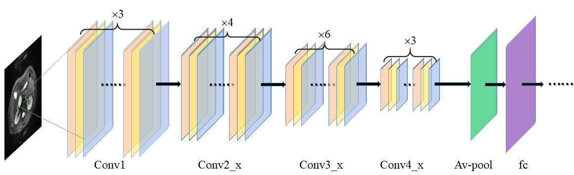

Table 1
Single factor logistic regression analysis of clinical features of MCN and SCN
| 特征名称 | p值 | OR值 | 95%CI | |
|---|---|---|---|---|
| 下限 | 上限 | |||
| 位置(胰头/胰体/胰尾) | <0.05 | 2.22 | 1.47 | 3.34 |
| 性别(男/女) | <0.05 | 6.30 | 1.82 | 21.82 |
| 年龄 | 0.14 | 0.98 | 0.96 | 1.01 |
| 肿瘤体积 | <0.05 | 1.00 | 1.00 | 1.00 |
Table 2
Multi-factor logistic regression analysis of clinical features of MCN and SCN
| 特征名称 | p值 | OR值 | 95%CI | |
|---|---|---|---|---|
| 下限 | 上限 | |||
| 位置(胰头/胰体/胰尾) | <0.05 | 2.12 | 1.39 | 3.24 |
| 性别(男/女) | <0.05 | 5.58 | 1.57 | 19.84 |
Fig. 3
(a) Mean Square Error (MSE) graphs corresponding to different λ values of radiomics features. The MSE is minimized by adjusting the λ value to determine the optimal λ value; (b) Convergence diagram of the optimal characteristic coefficient of radiomics features; (c) Weight of radiomics features screened by LASSO, Skewness.1 represents skewness, Busyness.1 represents complexity, MCC.2 represents morphological correlation coefficient, DependencyVariance.2 represents dependency difference, Idn.4 represents inverse difference moment, and Correlation.7 represents correlation; (d) Mean square error (MSE) graphs corresponding to different λ values of deep learning features; (e) Convergence diagram of the optimal characteristic coefficient of deep learning features; (f) Weight of deep learning features screened by LASSO
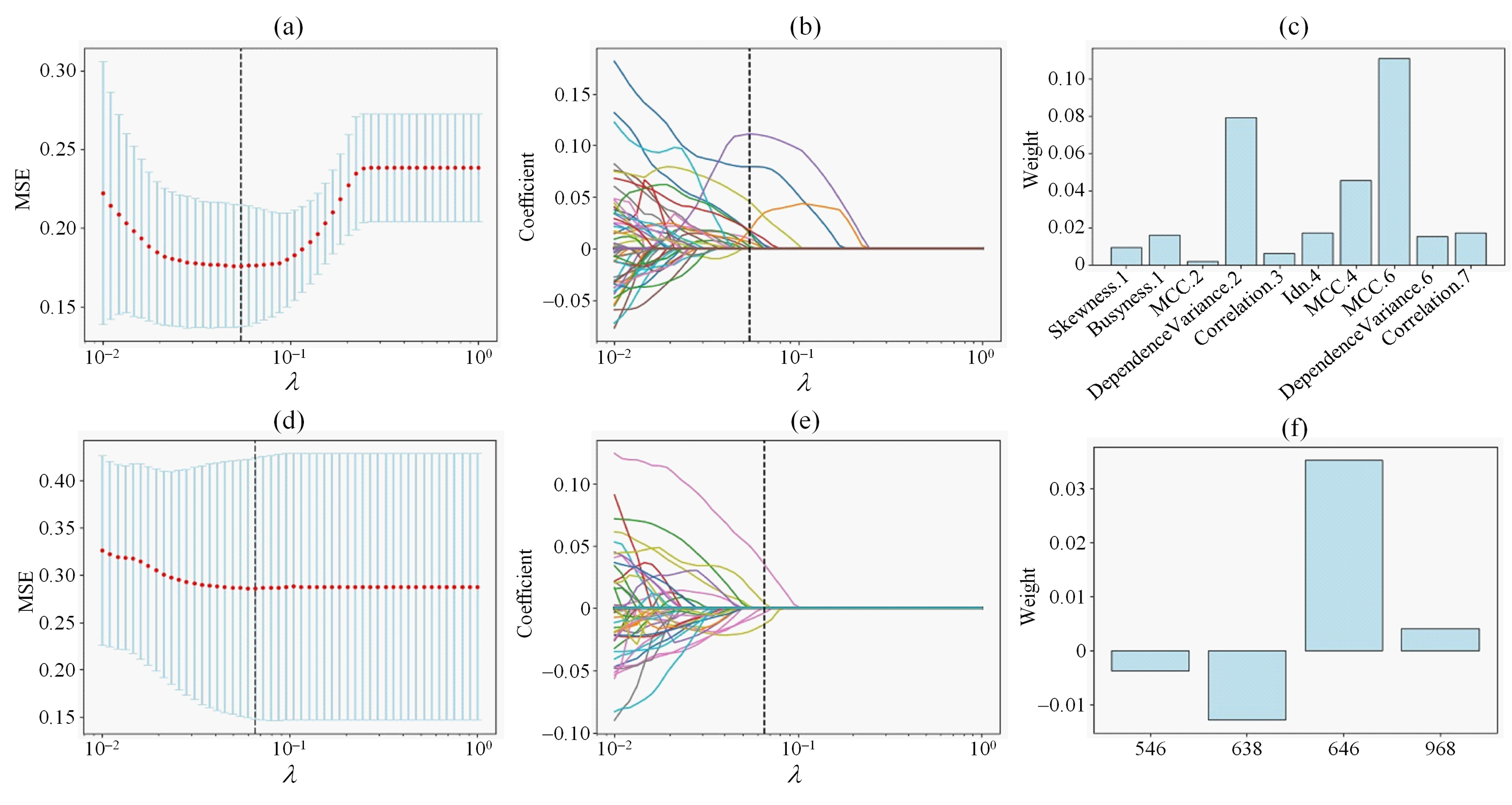
Table 3
Performance of the feature models in the four classifiers
| 特征模型 | 分类器 | 准确率 | 召回率 | 精确率 | AUC | F1 | |||
|---|---|---|---|---|---|---|---|---|---|
| RAD | SVM | 0.8077 | 0.5789 | 0.8462 | 0.7592 | 0.6875 | |||
| ADAboost | 0.8269 | 0.6842 | 0.8125 | 0.7967 | 0.7429 | ||||
| Random Forest | 0.7885 | 0.6742 | 0.7222 | 0.7663 | 0.6974 | ||||
| Logistic | 0.8269 | 0.6316 | 0.8571 | 0.7855 | 0.7273 | ||||
| DL | SVM | 0.6731 | 0.1176 | 0.5303 | 0.5595 | 0.1925 | |||
| ADAboost | 0.7692 | 0.9000 | 0.6429 | 0.8645 | 0.7500 | ||||
| Random Forest | 0.7115 | 0.7619 | 0.6154 | 0.8407 | 0.6809 | ||||
| Logistic | 0.7500 | 0.2857 | 0.5714 | 0.5770 | 0.3809 | ||||
| RAD_DL | SVM | 0.8462 | 0.6111 | 0.7908 | 0.8051 | 0.6894 | |||
| ADAboost | 0.8269 | 0.7727 | 0.8095 | 0.6882 | 0.7907 | ||||
| Random Forest | 0.8077 | 0.6364 | 0.8750 | 0.8051 | 0.7369 | ||||
| Logistic | 0.8462 | 0.9444 | 0.7083 | 0.8441 | 0.8095 | ||||
| Clinical_RAD_DL | SVM | 0.8846 | 0.8235 | 0.8235 | 0.8689 | 0.8235 | |||
| ADAboost | 0.8654 | 0.8823 | 0.7500 | 0.8697 | 0.8108 | ||||
| Random Forest | 0.7692 | 0.7059 | 0.6316 | 0.7529 | 0.6667 | ||||
| Logistic | 0.9231 | 0.8824 | 0.8820 | 0.9126 | 0.8822 | ||||
Fig. 4
ROC curve of four characteristic models. The abscissa is fpr (false positive rate) and the ordinate is tpr (true positive rate). The four classifiers are SVM, adaboost (ADAboost), randomforest (Random Forest) and logistic (Logistic). (a) ROC curve used by RAD feature model; (b) ROC curve of DL feature model; (c) ROC curve of RAD_DL feature model; (d) ROC curve of Clinical _RAD_DL feature model
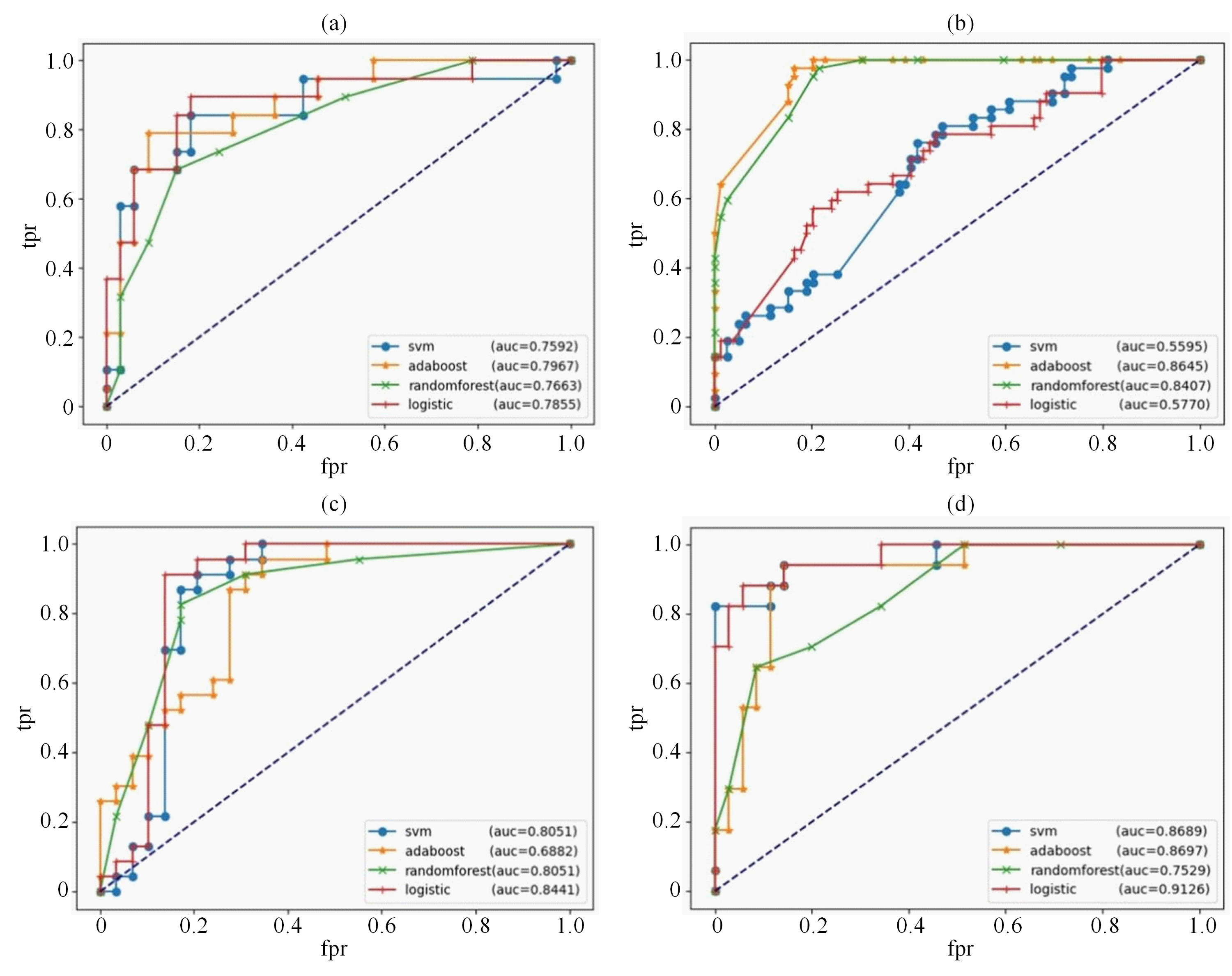
Table 4
Perform a Delong test to compare the performance differences between the Clinical_RAD_DL feature model and other models and obtain the p-value
| Clinical_RAD_DL与RAD-DL | Clinical_RAD_DL与DL | Clinical_RAD_DL与RAD | |
|---|---|---|---|
| MCN和SCN分类 | 0.069 | 0.029 | 0.051 |
Fig. 5
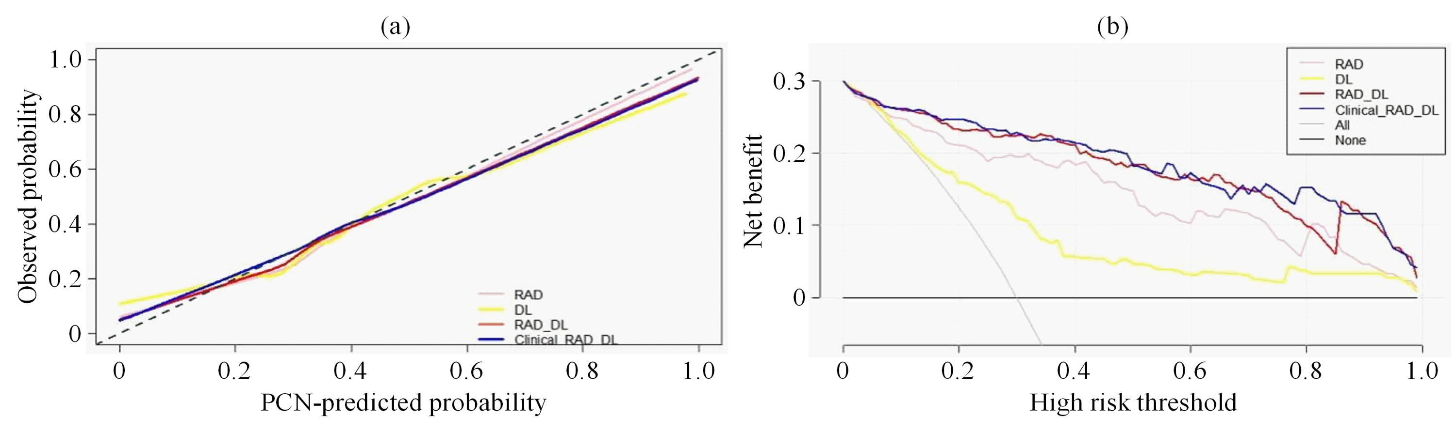
Calibration curves and decision curves. (a) Calibration curves of RAD, DL, RAD_DL, and Clinical-RAD_DL feature model. The abscissa represents the PCN-predicted probability of PCN classification model, and the ordinate represents the observed probability; (b) Decision curves used by RAD, DL, RAD_DL, and Clinical-RAD_DL feature model. The abscissa represents the high risk threshold, and the ordinate represents the net benefit of the model, the All curve represents the net benefit predicted as MCN, and the None curve represents the net benefit predicted as SCN
| [1] | LI Z S, JIN Z D, LI X. Chinese guidelines for the diagnosis and treatment of pancreas cystic neoplasm(2022)[J]. J Clin Hepatol, 2023, 39(2): 290-298. |
| 李兆申, 金震东, 李汛. 中国胰腺囊性肿瘤诊断指南(2022年)[J]. 临床肝胆病杂志, 2023, 39(2): 290-298. | |
| [2] |
BIAN Y, JIANG H, CAO K, et al. The relationship between microscopic tumor size and CT tumor size in pancreatic ductal adenocarcinoma[J]. Clin Imag, 2021, 76: 30-37.
doi: 10.1016/j.clinimag.2020.11.039 pmid: 33548890 |
| [3] |
JAYASREE C, ABHISHEK M, LIOR G, et al. CT radiomics to predict high-risk intraductal papillary mucinous neoplasms of the pancreas[J]. Med phys, 2018, 45(11): 5019-5029.
doi: 10.1002/mp.13159 pmid: 30176047 |
| [4] |
AVANZO M, WEI L S, STANCANELLO J, et al. Machine and deep learning methods for radiomics[J]. Med Phys, 2020, 47(5): 185-202.
doi: 10.1002/mp.13678 pmid: 32418336 |
| [5] | HAN B, XU J, WANG Y J, et al. Classification of BI-RADS 3-5 breast lesions based on MRI radiomics[J]. Chinese J Magn Reson, 2023, 40(1): 52-67. |
| 韩冰, 徐晶, 王远军, 等. 基于MRI影像组学的BI-RADS 3-5类乳腺病变三分类[J]. 波谱学杂志, 2023, 40(1): 52-67. | |
| [6] |
SHBOUL Z A, ALAM M, VIDYARATNE L, et al. Feature-guided deep radiomics for glioblastoma patient survival prediction[J]. Front Neurosci, 2019, 13: 966-982.
doi: 10.3389/fnins.2019.00966 |
| [7] |
LI J, LIU F, FANG X, et al. CT Radiomics features in differentiation of focal-type autoimmune pancreatitis from pancreatic ductal adenocarcinoma: a propensity score analysis[J]. Acad Radiol, 2021, 29(3): 358-366.
doi: 10.1016/j.acra.2021.04.014 pmid: 34108115 |
| [8] |
PREUSS K, THACH N, LIANG X, et al. Using quantitative imaging for personalized medicine in pancreatic cancer: a review of radiomics and deep learning applications[J]. Cancers (Basel), 2022, 14(7): 1654.
doi: 10.3390/cancers14071654 |
| [9] | NGUON L S, SEO K, LIM J H, et al. Deep learning-based differentiation between mucinous cystic neoplasm and serous cystic neoplasm in the pancreas using endoscopic ultrasonography[J]. Diagnostics (Basel), 2021, 11(6): 1052. |
| [10] | YANG Y F, QI Z X, NIE S D. Differentiation of benign and malignant breast lesions based on multimodal MRI and deep learning[J]. Chinese J Magn Reson, 2022, 39(4): 401-412. |
| 杨一风, 祁章璇, 聂生东. 基于多模态MRI与深度学习的乳腺病变良恶性鉴别[J]. 波谱学杂志, 2022, 39(4): 401-412. | |
| [11] | ZHANG Y F, XU S S, WU J, et al. Value of CT texture analysis in differentiating pancreatic serous cystadenoma from mucinous cystadenoma[J]. Journal of Southeast University, 2022, 41(3): 308-316. |
| 张怡帆, 徐姗姗, 吴锦, 等. CT纹理分析在鉴别胰腺浆液性囊腺瘤与黏液性囊腺瘤中的价值[J]. 东南大学学报, 2022, 41(3): 308-316. | |
| [12] |
PARK H J, SHIN K, YOU M W, et al. Deep learning-based detection of solid and cystic pancreatic neoplasms at contrast-enhanced CT[J]. Radiology, 2023, 306(1): 140-149.
doi: 10.1148/radiol.220171 |
| [13] |
ISLAM W, JONES M, FAIZ R, et al. Improving performance of breast lesion classification using a ResNet50 model optimized with a novel attention mechanism[J]. Tomography, 2022, 8(5): 2411-2425.
doi: 10.3390/tomography8050200 pmid: 36287799 |
| [14] | LU J, WU Y, XIONG Y, et al. Breast tumor computer-aided detection system based on magnetic resonance imaging using convolutional neural network[J]. Comput Model Eng Sci, 2022, 130(1): 365-377. |
| [15] | 中国人民解放军海军军医大学第一附属医院. 胰腺外分泌功能评价模型的训练方法、系统、电子设备及介质: 中国, CN2022107020513[P], 2022-08-09. |
| [16] |
LIANG W, TIAN W, WANG Y, et al. Classification prediction of pancreatic cystic neoplasms based on radiomics deep learning models[J]. BMC Cancer, 2022, 22(1): 1237-1246.
doi: 10.1186/s12885-022-10273-4 pmid: 36447168 |
| [17] | WANG D, HU Y, ZHAN C, et al. A nomogram based on radiomics signature and deep-learning signature for preoperative prediction of axillary lymph node metastasis in breast cancer[J]. Front Oncol, 2022, 12(20): 940-950. |
| [18] |
XIE T, WANG X, ZHANG Z, et al. CT-based radiomics analysis for preoperative diagnosis of pancreatic mucinous cystic neoplasm and atypical serous cystadenomas[J]. Front Oncol, 2021, 11(6): 621520.
doi: 10.3389/fonc.2021.621520 |
| [19] | SHAO C, FENG X, YU J, et al. A nomogram for predicting pancreatic mucinous cystic neoplasm and serous cystic neoplasm[J]. Abdom Radiol (NY), 2021, 46(8): 3963-3973. |
| [20] | CHEN S. Application of CT radiomics in differential diagnosis of pancreatic serous and mucinous cystic neoplasm[J]. Chinese Journal of CT and MRI, 2022, 20(10): 92-105. |
| 陈帅. 基于CT影像组学对胰腺浆液及黏液性囊性肿瘤鉴别诊断[J]. 中国CT和MRI杂志, 2022, 20(10): 92-105. |
| [1] | LI Na, XIAO Long, LI Sha, LIU Yudun, JIA Yushu, ZHANG Lei, CHEN Shizhen. 19F NMR Chemical Shift-encoded cRGD-targeted Dual-modal Probe for Multicolor Imaging [J]. Chinese Journal of Magnetic Resonance, 2026, 43(3): 279-290. |
| [2] | LIU Kewen, JIANG Yukang, CHEN Fang, CHEN Junfei, LU Yuan, CHEN Li, LIU Chaoyang. A Deep Learning-based Method for LF-NMR Relaxation Time Spectrum Inversion [J]. Chinese Journal of Magnetic Resonance, 2026, 43(3): 307-320. |
| [3] | SU Yilin, LIU Yuanyuan, CUI Zhuoxu, LIANG Dong. Physics Guided Multi-contrast Magnetic Resonance Reconstruction Diffusion Model [J]. Chinese Journal of Magnetic Resonance, 2026, 43(3): 321-338. |
| [4] | MA Suchao, WANG Yuanjun. Research Progress on Diffusion Magnetic Resonance Imaging Noise Reduction Methods Based on Deep Learning [J]. Chinese Journal of Magnetic Resonance, 2026, 43(3): 367-388. |
| [5] | CAI Yue, WANG Xuxia, LIU Sijie, GUO Haodong, CHEN Xi, CHENG Linlin, KANG Yan, LIN Fuchun. Magnetic Resonance Imaging Study on the Microstructure Abnormalities of Striatal White Matter in Type 2 Diabetic Mellitus Rats [J]. Chinese Journal of Magnetic Resonance, 2026, 43(2): 175-185. |
| [6] | XIE Xinyi, WANG Yuanjun. Research Progress on Super-resolution Reconstruction of Brain Diffusion Magnetic Resonance Images [J]. Chinese Journal of Magnetic Resonance, 2026, 43(2): 223-240. |
| [7] | FU Fenfang, LIN Guobing, LI Meifang. Advances in Magnetic Resonance Imaging for the Diagnosis and Prognosis of Mild Traumatic Brain Injury [J]. Chinese Journal of Magnetic Resonance, 2026, 43(2): 214-222. |
| [8] | NI Guangmao, LI Yuwei, HOU Wenxuan, LIU Caiyun, DONG Peng, ZHANG Yanhui. Research on the Influencing Factors of Acute Cerebral Infarction Recurrence Based on MR-DWI [J]. Chinese Journal of Magnetic Resonance, 2026, 43(1): 87-93. |
| [9] | CHEN Xi, LIU Sijie, CAI Yue, CHENG Linlin, WANG Xuxia, KANG Yan, LIN Fuchun, LEI Hao. Effects of Seizure-inducing Doses Nicotine on Hippocampal Structure in Adolescent Female Rats [J]. Chinese Journal of Magnetic Resonance, 2025, 42(4): 345-354. |
| [10] | LI Yinghao, WANG Lihui, WANG Sucheng, ZHU Zhongqi, HUANG Changdong, LI Renfeng, CAO Kaiming, HU Haiyang, JIA Yiming, LIANG Songtao, YANG Guang, LU Qing, WANG Hongzhi. Study on Pancreas Automatic Segmentation, Regional Quantification, and Diabetes Assessment [J]. Chinese Journal of Magnetic Resonance, 2025, 42(4): 378-389. |
| [11] | GAO Zhaoyao, ZHANG Zhan, HU Liangliang, XU Guangyu, ZHOU Sheng, HU Yuxin, LIN Zijie, ZHOU Chao. PMRI Image Reconstruction Method Based on Virtual Coils and GRAPPA-enhanced Network [J]. Chinese Journal of Magnetic Resonance, 2025, 42(4): 390-401. |
| [12] | ZHANG Mingyu, XIAO Sa, SHI Shengjie, ZHANG Xuecheng, ZHOU Xin. Research on a Multi-modal Enhanced Denoising Diffusion Model for Hyperpolarized 129Xe MRI [J]. Chinese Journal of Magnetic Resonance, 2025, 42(4): 364-377. |
| [13] | MA Yingxue, ZHAO Yanqiang, YANG Xiaodong, JIANG Bin, TAO Cheng. Opportunities and Challenges of High-field and Ultra-high-field Magnetic Resonance Imaging in China [J]. Chinese Journal of Magnetic Resonance, 2025, 42(3): 334-344. |
| [14] | SUI Meiju, ZHANG Lei, WANG Ruifang, LUO Yingying, LI Sha, QIU Maosong, XU Qiuyi, CHEN Daiqin, CHEN Shizhen, ZHOU Xin. MRI-traceable Nanoenzyme for Cascade Catalysis-enhanced Immunotherapy [J]. Chinese Journal of Magnetic Resonance, 2025, 42(3): 231-248. |
| [15] | CAO Fei, XU Qianqian, CHEN Hao, ZU Jie, LI Xiaowen, TIAN Jin, BAO Lei. An Intelligent Diagnosis Method for NIID Based on Cross Self-supervision and DWI [J]. Chinese Journal of Magnetic Resonance, 2025, 42(2): 154-163. |
| Viewed | ||||||
|
Full text |
|
|||||
|
Abstract |
|
|||||
