
Chinese Journal of Magnetic Resonance ›› 2025, Vol. 42 ›› Issue (4): 402-413.doi: 10.11938/cjmr20253156cstr: 32225.14.cjmr20253156
• Articles • Previous Articles Next Articles
WEN Yulin1,2, LI Gaiying1,2,*( ), WU Yupeng1,2, LI Jianqi1,2
Received:2025-04-02
Published:2025-12-05
Online:2025-05-12
Contact:
* Tel: 021-62233775, E-mail: ligaiying@phy.ecnu.edu.cn.
CLC Number:
WEN Yulin, LI Gaiying, WU Yupeng, LI Jianqi. Optimization of DW-MRS Acquisition Protocol: The Impact of Gating and Cycling Modes[J]. Chinese Journal of Magnetic Resonance, 2025, 42(4): 402-413.
Add to citation manager EndNote|Reference Manager|ProCite|BibTeX|RefWorks

Fig. 1
Sequence diagram of diffusion-weighted point resolved spectroscopy. RF denotes the radiofrequency pulse, while GX, GY, and GZ represent the gradients along the left-right, anterior-posterior, and head-foot directions, respectively. The blue color indicates the localization gradient, black represents the spoiling gradient, and green denotes the diffusion gradient. The echo time (TE) is defined as twice the sum of τ 1 and τ 2. ε denotes the duration of the applied diffusion gradient, while Δ represents the interval between the diffusion gradient pairs (i.e., the diffusion time), which is half the duration of TE
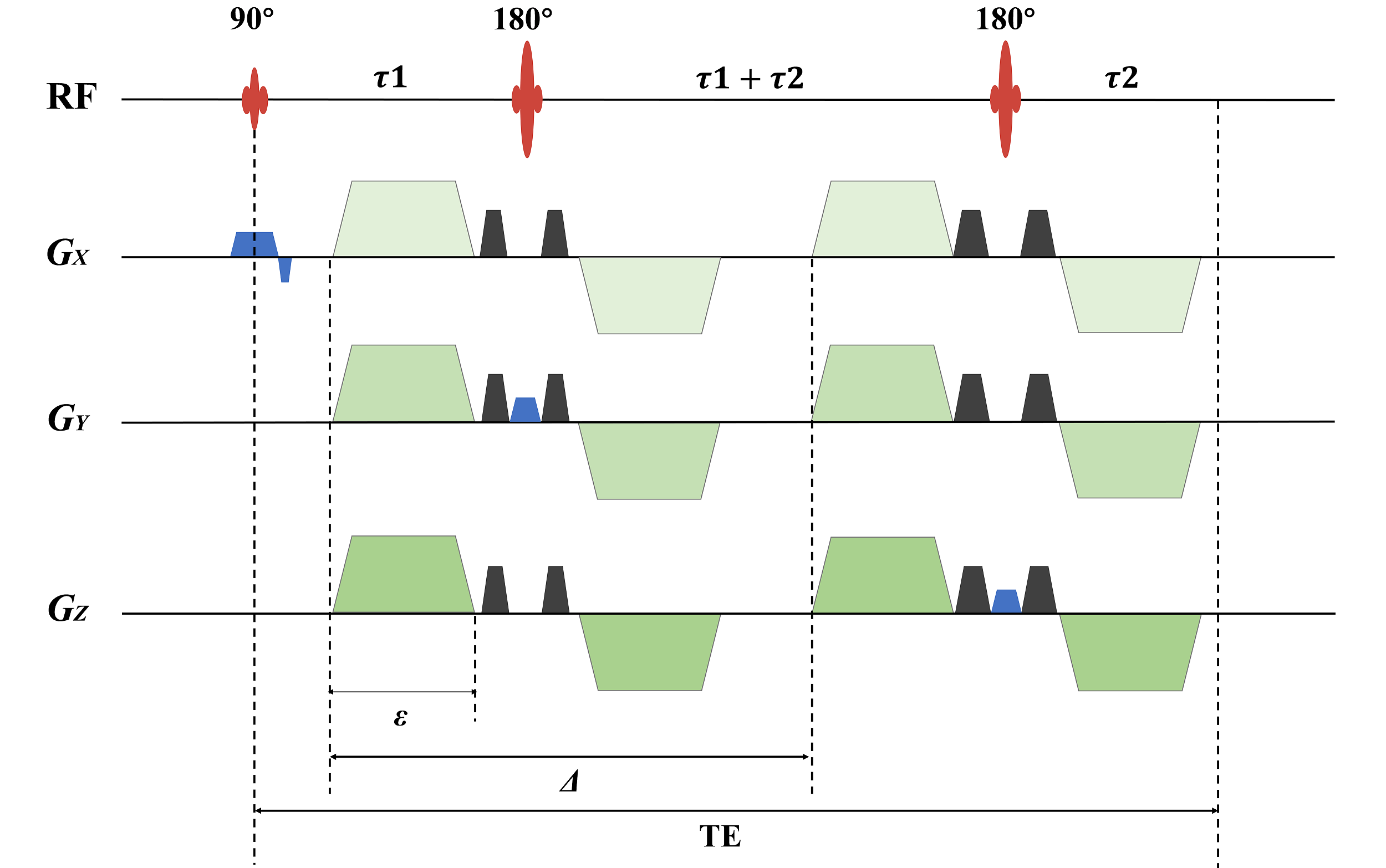
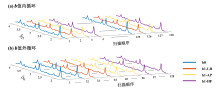
Fig. 2
Illustration of the two cycling modes. (a) Internal cycling of b-values: The b-value loop occurs within the accumulation loop, where data from different b-values are sequentially looped and collected before proceeding to the next accumulation. (b) External cycling of b-values: The b-value loop occurs outside the accumulation loop, with data for the same b-value first accumulated before moving on to the next b-value. The b0 denotes the signal when no diffusion gradient is applied. b1-LR, b1-AP, and b1-HF represent the signals when the diffusion gradient is applied in the left-right, anterior-posterior, and head-foot directions of the subject respectively. Blue, orange, yellow, and purple indicate b0, b1-LR, b1-AP, and b1-HF respectively
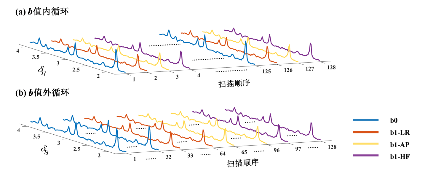
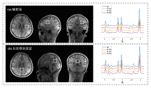
Fig. 3
Illustration of the volume of interest localization for diffusion-weighted magnetic resonance spectroscopy (left column) and the corresponding spectra (right column). (a) Corona radiata; (b) Posterior cingulate cortex. b0 denotes the spectrum acquired without diffusion gradient application, while b1-LR, b1-AP, and b1-HF represent spectra obtained with the diffusion gradient applied in the left-right, anterior-posterior, and head-foot directions, respectively

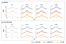
Fig. 4
Variations in signal intensity of the water peak in each transient spectrum acquired using different gating methods for a subject. (a) The volume of interest is located in the corona radiata region; (b) The volume of interest is located in the posterior cingulate cortex region. b0 represents the signal without diffusion gradient application, while b1-LR, b1-AP, and b1-HF denote the signals with the diffusion gradient applied in the left-right, anterior-posterior, and head-foot directions, respectively. Blue, orange, and yellow indicate the use of ECG gating, respiratory gating, and no gating, respectively
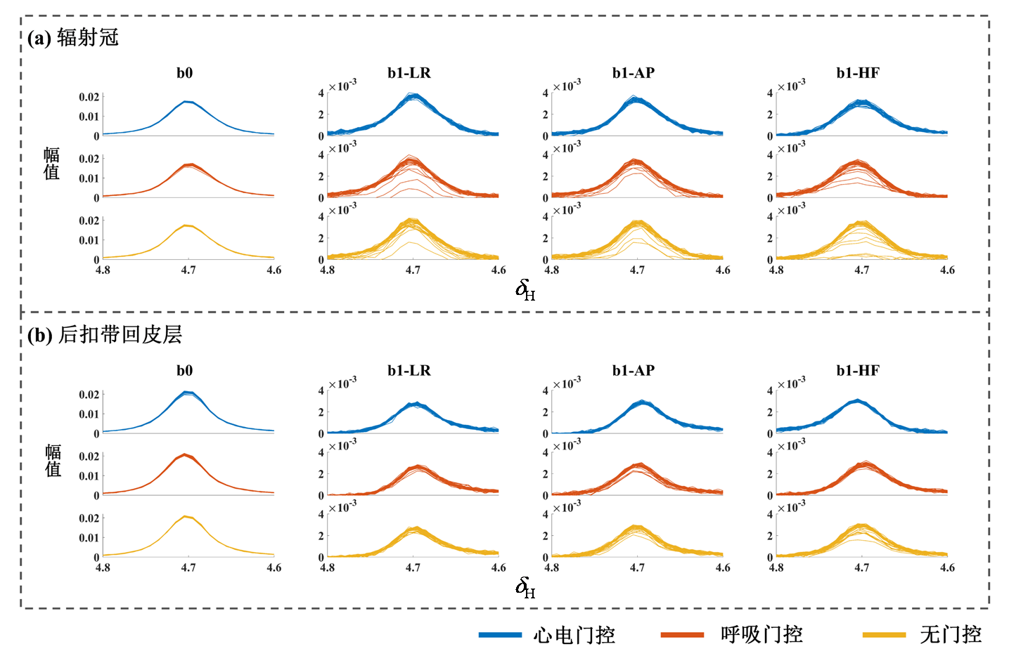
Table 1
Comparison of apparent diffusion coefficient (ADC) obtained under different gating conditions and results of linear mixed-effects model analysis in the corona radiata
| ADC/(μm2/ms) | P12 | P13 | P23 | ||||
|---|---|---|---|---|---|---|---|
| Cardiac gating | Respiratory gating | No gating | |||||
| tCho | LR | 0.144 ± 0.043 | 0.147 ± 0.017 | 0.164 ± 0.030 | 0.850 | 0.287 | 0.377 |
| AP | 0.116 ± 0.030 | 0.118 ± 0.026 | 0.135 ± 0.016 | 0.842 | 0.073 | 0.105 | |
| HF | 0.134 ± 0.013 | 0.152 ± 0.027 | 0.178 ± 0.036 | 0.193 | 0.005** | 0.077 | |
| mean | 0.131 ± 0.023 | 0.139 ± 0.018 | 0.159 ± 0.025 | 0.477 | 0.023* | 0.091 | |
| tCr | LR | 0.166 ± 0.045 | 0.191 ± 0.023 | 0.193 ± 0.034 | 0.151 | 0.204 | 0.858 |
| AP | 0.141 ± 0.026 | 0.167 ± 0.024 | 0.170 ± 0.020 | 0.072 | 0.049* | 0.839 | |
| HF | 0.162 ± 0.018 | 0.185 ± 0.015 | 0.195 ± 0.026 | 0.059 | 0.012* | 0.428 | |
| mean | 0.156 ± 0.025 | 0.181 ± 0.012 | 0.186 ± 0.024 | 0.044* | 0.028* | 0.817 | |
| tNAA | LR | 0.171 ± 0.041 | 0.186 ± 0.017 | 0.200 ± 0.038 | 0.432 | 0.125 | 0.427 |
| AP | 0.161 ± 0.020 | 0.179 ± 0.027 | 0.182 ± 0.023 | 0.076 | 0.046* | 0.794 | |
| HF | 0.153 ± 0.020 | 0.171 ± 0.016 | 0.193 ± 0.027 | 0.054 | <0.001*** | 0.016* | |
| mean | 0.162 ± 0.018 | 0.178 ± 0.013 | 0.192 ± 0.022 | 0.081 | 0.004** | 0.157 | |
Table 2
Comparison of apparent diffusion coefficient (ADC) obtained under different gating conditions and results of linear mixed-effects model results analysis in the posterior cingulate cortex
| ADC/(μm2/ms) | P12 | P13 | P23 | ||||
|---|---|---|---|---|---|---|---|
| Cardiac gating | Respiratory gating | No gating | |||||
| tCho | LR | 0.106 ± 0.024 | 0.112 ± 0.020 | 0.111 ± 0.023 | 0.674 | 0.721 | 0.949 |
| AP | 0.097 ± 0.027 | 0.099 ± 0.011 | 0.108 ± 0.018 | 0.876 | 0.346 | 0.428 | |
| HF | 0.092 ± 0.019 | 0.111 ± 0.023 | 0.111 ± 0.021 | 0.003** | 0.003** | 0.955 | |
| mean | 0.098 ± 0.019 | 0.107 ± 0.014 | 0.110 ± 0.014 | 0.303 | 0.178 | 0.734 | |
| tCr | LR | 0.130 ± 0.017 | 0.151 ± 0.015 | 0.139 ± 0.015 | 0.034* | 0.364 | 0.185 |
| AP | 0.114 ± 0.015 | 0.129 ± 0.013 | 0.131 ± 0.013 | 0.041* | 0.022* | 0.747 | |
| HF | 0.105 ± 0.009 | 0.119 ± 0.014 | 0.127 ± 0.015 | 0.055 | 0.004** | 0.215 | |
| mean | 0.116 ± 0.008 | 0.133 ± 0.012 | 0.132 ± 0.014 | 0.019* | 0.023* | 0.928 | |
| tNAA | LR | 0.144 ± 0.030 | 0.148 ± 0.009 | 0.142 ± 0.012 | 0.750 | 0.826 | 0.592 |
| AP | 0.120 ± 0.016 | 0.125 ± 0.008 | 0.128 ± 0.006 | 0.431 | 0.205 | 0.614 | |
| HF | 0.113 ± 0.013 | 0.124 ± 0.016 | 0.130 ± 0.017 | 0.219 | 0.073 | 0.529 | |
| mean | 0.126 ± 0.018 | 0.132 ± 0.008 | 0.133 ± 0.009 | 0.368 | 0.303 | 0.893 | |
Table 3
Comparison of the apparent diffusion coefficient (ADC) obtained using the internal and external b-value cycling methods and results of linear mixed-effects model analysis
| ADC/(μm2/ms) | P values | |||
|---|---|---|---|---|
| Internal b-value cycling | External b-value cycling | |||
| tCho | LR | 0.109 ± 0.014 | 0.109 ± 0.019 | 0.826 |
| AP | 0.112 ± 0.018 | 0.104 ± 0.024 | 0.158 | |
| HF | 0.097 ± 0.016 | 0.099 ± 0.021 | 0.614 | |
| mean | 0.106 ± 0.011 | 0.104 ± 0.018 | 0.601 | |
| tCr | LR | 0.133 ± 0.014 | 0.134 ± 0.019 | 0.680 |
| AP | 0.117 ± 0.016 | 0.120 ± 0.020 | 0.542 | |
| HF | 0.110 ± 0.011 | 0.109 ± 0.014 | 0.749 | |
| mean | 0.120 ± 0.010 | 0.121 ± 0.015 | 0.883 | |
| tNAA | LR | 0.144 ± 0.017 | 0.143 ± 0.021 | 0.806 |
| AP | 0.121 ± 0.008 | 0.122 ± 0.013 | 0.486 | |
| HF | 0.117 ± 0.014 | 0.116 ± 0.016 | 0.637 | |
| mean | 0.127 ± 0.011 | 0.127 ± 0.013 | 0.780 | |
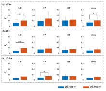
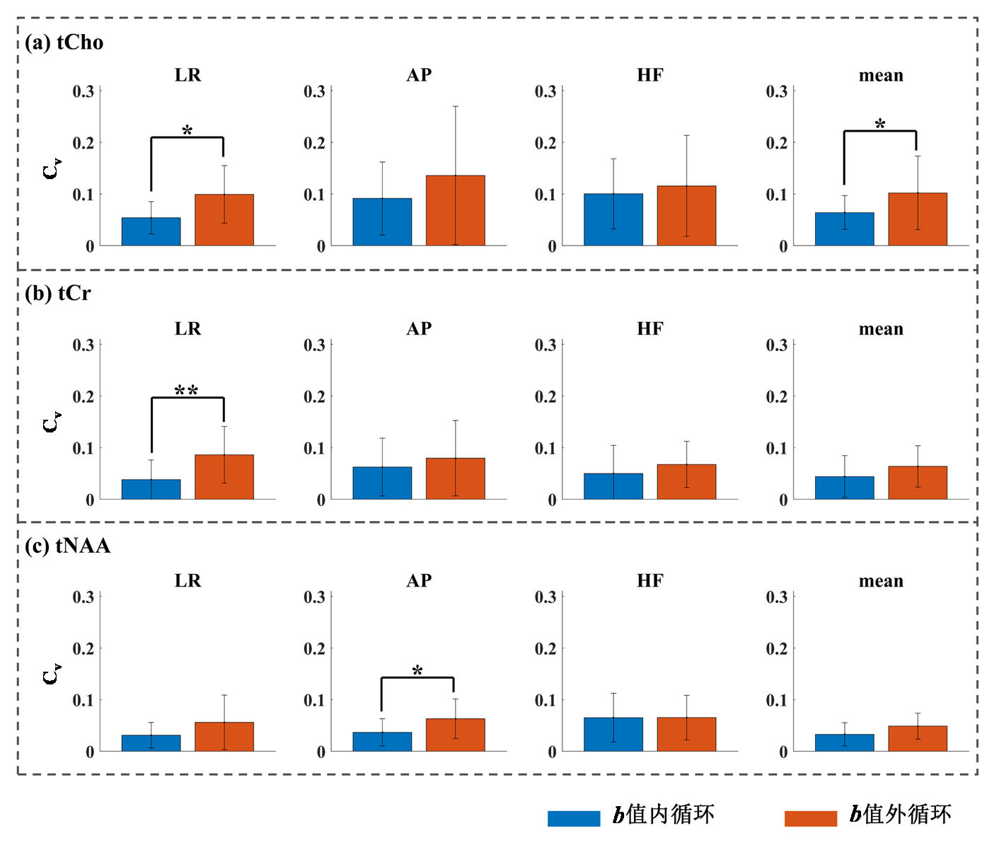
Fig. 5
The distribution of the coefficient of variation of the apparent diffusion coefficient (ADC) for three metabolites when using the internal and external b-value cycling modes. (a) tCho; (b) tCr; (c) tNAA. The LR, AP, and HF represent the coefficient of variation of the ADC when the diffusion gradient is applied in the left-right, anterior-posterior, and head-foot directions, respectively, while "mean" denotes the coefficient of variation of the averaged ADC across the three orthogonal directions. Blue indicates the internal b-value cycling mode; orange represents the external b-value cycling mode. * P < 0.05; ** P < 0.01
| [1] |
NEGENDANK W. Studies of human tumors by MRS: A review[J]. NMR Biomed, 1992, 5(5): 303-324.
pmid: 1333263 |
| [2] | LI R, CHANG X, ZHANG J, et al. Progress of magnetic resonance spectroscopy in the study of the effects of smoking on the brain[J]. Chinese J Magn Reson, 2023, 40(4): 471-480. |
|
李任, 常晓, 张捷, 等. 磁共振波谱技术在吸烟对大脑影响的研究进展[J]. 波谱学杂志, 2023, 40(4): 471-480.
doi: 10.11938/cjmr20233052 |
|
| [3] | ZHU X W, YANG X, WEI D X, et al. In vivo glutathione molecular MRS signal selection based on nuclear spin singlet states[J]. Chinese J Magn Reson, 2024, 41(4): 373-381. |
|
朱向炜, 杨雪, 魏达秀, 等. 基于核自旋单重态的活体谷胱甘肽分子MRS信号选择[J]. 波谱学杂志, 2024, 41(4): 373-381.
doi: 10.11938/cjmr20243105 |
|
| [4] |
TKÁC I, ÖZ G, ADRIANY G, et al. In vivo1H NMR spectroscopy of the human brain at high magnetic fields: Metabolite quantification at 4T vs. 7T[J]. Magn Reson Med, 2009, 62(4): 868-879.
doi: 10.1002/mrm.v62:4 |
| [5] |
NICOLAY K, BRAUN K P J, GRAAF R A D, et al. Diffusion NMR spectroscopy[J]. NMR Biomed, 2001, 14(2): 94-111.
pmid: 11320536 |
| [6] |
CAO P, WU E X. In vivo diffusion MRS investigation of non-water molecules in biological tissues[J]. NMR Biomed, 2017, 30(3): e3481.
doi: 10.1002/nbm.v30.3 |
| [7] |
LIGNEUL C, NAJAC C, DÖRING A, et al. Diffusion-weighted MR spectroscopy: Consensus, recommendations, and resources from acquisition to modeling[J]. Magn Reson Med, 2024, 91(3): 860-885.
doi: 10.1002/mrm.v91.3 |
| [8] |
PALOMBO M, SHEMESH N, RONEN I, et al. Insights into brain microstructure from in vivo DW-MRS[J]. NeuroImage, 2018, 182: 97-116.
doi: S1053-8119(17)30942-4 pmid: 29155183 |
| [9] | SPOTORNO N, NAJAC C, STRANDBERG O, et al. Diffusion weighted magnetic resonance spectroscopy revealed neuronal specific microstructural alterations in Alzheimer's disease[J]. Brain Commun, 2023, 6(1): fcae026. |
| [10] |
GENOVESE G, DIAZ-FERNANDEZ B, LEJEUNE F-X, et al. Longitudinal monitoring of microstructural alterations in cerebral ischemia with in vivo diffusion-weighted MR spectroscopy[J]. Radiology, 2022, 306(3): e220430.
doi: 10.1148/radiol.220430 |
| [11] |
RICIGLIANO V A G, TONIETTO M, PALLADINO R, et al. Thalamic energy dysfunction is associated with thalamo-cortical tract damage in multiple sclerosis: A diffusion spectroscopy study[J]. Mult Scler J, 2021, 27(4): 528-538.
doi: 10.1177/1352458520921362 |
| [12] |
BODINI B, BRANZOLI F, POIRION E, et al. Dysregulation of energy metabolism in multiple sclerosis measured in vivo with diffusion-weighted spectroscopy[J]. Mult Scler J, 2018, 24(3): 313-321.
doi: 10.1177/1352458517698249 |
| [13] |
HENRY-FEUGEAS M C, IDY-PERETTI I, BALEDENT O, et al. Cerebrospinal fluid flow waveforms-MR analysis in chronic adult hydrocephalus[J]. Invest Radiol, 2001, 36(3): 146-154.
doi: 10.1097/00004424-200103000-00003 |
| [14] |
WÅHLIN A, AMBARKI K, HAUKSSON J, et al. Phase contrast MRI quantification of pulsatile volumes of brain arteries, veins, and cerebrospinal fluids compartments: Repeatability and physiological interactions[J]. J Magn Reson Imaging, 2012, 35(5): 1055-1062.
doi: 10.1002/jmri.23527 pmid: 22170792 |
| [15] |
GENOVESE G, MARJAŃSKA M, AUERBACH E J, et al. In vivo diffusion-weighted MRS using semi-LASER in the human brain at 3 T: Methodological aspects and clinical feasibility[J]. NMR Biomed, 2021, 34(5): e4206.
doi: 10.1002/nbm.v34.5 |
| [16] |
DAOUK J, BOUZERAR R, BALEDENT O. Heart rate and respiration influence on macroscopic blood and CSF flows[J]. Acta Radiologica, 2017, 58(8): 977-982.
doi: 10.1177/0284185116676655 pmid: 28273732 |
| [17] |
KAN H E, TECHAWIBOONWONG A, VAN OSCH M J, et al. Differences in apparent diffusion coefficients of brain metabolites between grey and white matter in the human brain measured at 7 T[J]. Magn Reson Med, 2012, 67(5): 1203-1209.
doi: 10.1002/mrm.23129 pmid: 22083562 |
| [18] | ROBERT J O, PETER B K, JUNE S T. WET, a T1-and B1-insensitive water-suppression method for in vivo localized 1H NMR Spectroscopy[J]. J Magn Reson, 1994, 104: 1-10. |
| [19] |
OTA K, NAKAZATO Y, SEO K, et al. Clinical and magnetic resonance imaging features in acute ischemic stroke with early wallerian degeneration: a case-control study[J]. BMC Neurol, 2025, 25(1): 170.
doi: 10.1186/s12883-025-04179-4 |
| [20] |
SMITH S M, JOHANSEN-BERG H, JENKINSON M, et al. Acquisition and voxelwise analysis of multi-subject diffusion data with tract-based spatial statistics[J]. Nat Protoc, 2007, 2(3): 499-503.
doi: 10.1038/nprot.2007.45 pmid: 17406613 |
| [21] |
VILLAIN N, CHÉTELAT G, GRASSIOT B, et al. Regional dynamics of amyloid-β deposition in healthy elderly, mild cognitive impairment and Alzheimer's disease: a voxelwise PiB-PET longitudinal study[J]. Brain, 2012, 135: 2126-2139.
doi: 10.1093/brain/aws125 pmid: 22628162 |
| [22] |
GREICIUS M D, FLORES B H, MENON V, et al. Resting-state functional connectivity in major depression: Abnormally increased contributions from subgenual cingulate cortex and thalamus[J]. Biol Psychiatry, 2007, 62(5): 429-437.
doi: 10.1016/j.biopsych.2006.09.020 |
| [23] |
SIMPSON R, DEVENYI G A, JEZZARD P, et al. Advanced processing and simulation of MRS data using the FID appliance (FID-A)-An open source, MATLAB-based toolkit[J]. Magn Reson Med, 2017, 77(1): 23-33.
doi: 10.1002/mrm.26091 pmid: 26715192 |
| [24] |
NEAR J, HARRIS A D, JUCHEM C, et al. Preprocessing, analysis and quantification in single-voxel magnetic resonance spectroscopy: Experts' consensus recommendations[J]. NMR Biomed, 2021, 34(5): e4257.
doi: 10.1002/nbm.v34.5 |
| [25] |
LAUDADIO T, MASTRONARDI N, VANHAMME L, et al. Improved Lanczos algorithms for blackbox MRS data quantitation[J]. J Magn Reson, 2002, 157(2): 292-297.
pmid: 12323148 |
| [26] |
PROVENCHER S W. Automatic quantitation of localized in vivo 1H spectra with LCModel[J]. NMR Biomed, 2001, 14(4): 260-264.
doi: 10.1002/nbm.v14:4 |
| [27] |
HUI S C N, SALEH M G, ZÖLLNER H J, et al. MRSCloud: A cloud-based MRS tool for basis set simulation[J]. Magn Reson Med, 2022, 88(5): 1994-2004.
doi: 10.1002/mrm.29370 pmid: 35775808 |
| [28] |
BAESHEN A, WYSS P O, HENNING A, et al. Test-retest reliability of the brain metabolites GABA and Glx With JPRESS, PRESS, and MEGA-PRESS MRS sequences in vivo at 3T[J]. J Magn Reson Imaging, 2020, 51(4): 1181-1191.
doi: 10.1002/jmri.v51.4 |
| [29] |
MARGULIES D S, GHOSH S S, GOULAS A, et al. Situating the default-mode network along a principal gradient of macroscale cortical organization[J]. Proc Natl Acad Sci U S A, 2016, 113(44): 12574-12579.
doi: 10.1073/pnas.1608282113 |
| [30] |
LEECH R, SHARP D J. The role of the posterior cingulate cortex in cognition and disease[J]. Brain, 2013, 137(1): 12-32.
doi: 10.1093/brain/awt162 |
| [31] |
WOOD E T, ERCAN A E, BRANZOLI F, et al. Reproducibility and optimization of in vivo human diffusion-weighted MRS of the corpus callosum at 3 T and 7 T[J]. NMR Biomed, 2015, 28(8): 976-987.
doi: 10.1002/nbm.v28.8 |
| [32] |
TAL A. The future is 2D: Spectral-temporal fitting of dynamic MRS data provides exponential gains in precision over conventional approaches[J]. Magn Reson Med, 2023, 89(2): 499-507.
doi: 10.1002/mrm.29456 pmid: 36121336 |
| [33] |
CLARKE W T, LIGNEUL C, COTTAAR M, et al. Universal dynamic fitting of magnetic resonance spectroscopy[J]. Magn Reson Med, 2024, 91(6): 2229-2246.
doi: 10.1002/mrm.30001 pmid: 38265152 |
| [34] |
SIMICIC D, ZÖLLNER H J, DAVIES-JENKINS C W, et al. Model-based frequency-and-phase correction of 1H MRS data with 2D linear-combination modeling[J]. Magn Reson Med, 2024, 92(5): 2222-2236.
doi: 10.1002/mrm.v92.5 |
| [35] | ZHAN H L, FANG Q Y, LIU J W, et al. Noise reduction of nuclear magnetic resonance spectroscopy using lightweight deep neural network[J]. Acta Phys-Chim Sin, 2025, 41(2), 100017. |
| 詹昊霖, 房启元, 刘佳伟, 等. 基于轻量级深度神经网络的核磁共振波谱降噪[J]. 物理化学学报, 2025, 41(2): 90-97. | |
| [36] |
GENOVESE G, PALOMBO M, SANTIN M D, et al. Inflammation-driven glial alterations in the cuprizone mouse model probed with diffusion-weighted magnetic resonance spectroscopy at 11.7 T[J]. NMR Biomed, 2021, 34(4): e4480.
doi: 10.1002/nbm.4480 pmid: 33480101 |
| [37] |
DE MARCO R, RONEN I, BRANZOLI F, et al. Diffusion-weighted MR spectroscopy (DW-MRS) is sensitive to LPS-induced changes in human glial morphometry: A preliminary study[J]. Brain Behav Immun, 2022, 99: 256-265.
doi: 10.1016/j.bbi.2021.10.005 |
| [38] |
ERCAN E, MAGRO-CHECA C, VALABREGUE R, et al. Glial and axonal changes in systemic lupus erythematosus measured with diffusion of intracellular metabolites[J]. Brain, 2016, 139: 1447-1457.
doi: 10.1093/brain/aww031 pmid: 26969685 |
| [39] |
WOOD E T, RONEN I, TECHAWIBOONWONG A, et al. Investigating axonal damage in multiple sclerosis by diffusion tensor spectroscopy[J]. J Neurosci, 2012, 32(19): 6665-6669.
doi: 10.1523/JNEUROSCI.0044-12.2012 pmid: 22573688 |
| [1] | CHEN Xi, LIU Sijie, CAI Yue, CHENG Linlin, WANG Xuxia, KANG Yan, LIN Fuchun, LEI Hao. Effects of Seizure-inducing Doses Nicotine on Hippocampal Structure in Adolescent Female Rats [J]. Chinese Journal of Magnetic Resonance, 2025, 42(4): 345-354. |
| [2] | LI Lu, GAO Yong, ZHOU Yupan. Construction of Prostate Cancer Model Based on T2WI Combined with DWI Multi Parameter MRI Texture Feature Fusion [J]. Chinese Journal of Magnetic Resonance, 2025, 42(4): 355-363. |
| [3] | LI Yinghao, WANG Lihui, WANG Sucheng, ZHU Zhongqi, HUANG Changdong, LI Renfeng, CAO Kaiming, HU Haiyang, JIA Yiming, LIANG Songtao, YANG Guang, LU Qing, WANG Hongzhi. Study on Pancreas Automatic Segmentation, Regional Quantification, and Diabetes Assessment [J]. Chinese Journal of Magnetic Resonance, 2025, 42(4): 378-389. |
| [4] | YI Chunhai, LI Fang, YANG Xiaoyun. Spectroscopic Data of Fluopimomide and Interpretations [J]. Chinese Journal of Magnetic Resonance, 2025, 42(4): 429-436. |
| [5] | ZHANG Mingyu, XIAO Sa, SHI Shengjie, ZHANG Xuecheng, ZHOU Xin. Research on a Multi-modal Enhanced Denoising Diffusion Model for Hyperpolarized 129Xe MRI [J]. Chinese Journal of Magnetic Resonance, 2025, 42(4): 364-377. |
| [6] | ZHENG Jiaqi, WANG Yinong, YUAN Siwen, YIN Tianpeng. Structural Identification and Complete NMR Spectral Assignments of 4-Isopropoxy-1-(trifluoroacetyl)naphthalene [J]. Chinese Journal of Magnetic Resonance, 2025, 42(4): 437-444. |
| [7] | ZHANG Yuanyuan, WANG Pengcheng, LI Tao, HU Rui, YANG Yunhuang, LIU Maili. Development and Applications of HDX-NMR and HDX-MS in Protein Structure and Dynamics Research [J]. Chinese Journal of Magnetic Resonance, 2025, 42(4): 445-456. |
| [8] | TANG Shihao, YANG Jinyu, XU Yajie, WANG Ya, PENG Bowen, GAO Yuhao, YANG Xiaodong. A Design of Circularly Polarized Coil for Low-field Nuclear Magnetic Resonance Spectrometers [J]. Chinese Journal of Magnetic Resonance, 2025, 42(3): 308-320. |
| [9] | LI Keyan, CHENG Xin, CHEN Junfei, CAO Li, HUANG Zhen, LIU Chaoyang. Development of Low-noise Preamplifier for Low-field NMR [J]. Chinese Journal of Magnetic Resonance, 2025, 42(3): 321-333. |
| [10] | SUI Meiju, ZHANG Lei, WANG Ruifang, LUO Yingying, LI Sha, QIU Maosong, XU Qiuyi, CHEN Daiqin, CHEN Shizhen, ZHOU Xin. MRI-traceable Nanoenzyme for Cascade Catalysis-enhanced Immunotherapy [J]. Chinese Journal of Magnetic Resonance, 2025, 42(3): 231-248. |
| [11] | LIU Ying, YUAN Binhua, ZHANG Haowei. Design of a Portable Magnetic Resonance Multi-source RF Pulse Generator [J]. Chinese Journal of Magnetic Resonance, 2025, 42(3): 285-298. |
| [12] | KOU Xinhui, ZHANG Yubing. Study on the Enantiomeric Recognition of Chiral Ureas Containing Amino Acid Units [J]. Chinese Journal of Magnetic Resonance, 2025, 42(3): 221-230. |
| [13] | MA Yingxue, ZHAO Yanqiang, YANG Xiaodong, JIANG Bin, TAO Cheng. Opportunities and Challenges of High-field and Ultra-high-field Magnetic Resonance Imaging in China [J]. Chinese Journal of Magnetic Resonance, 2025, 42(3): 334-344. |
| [14] | JIANG Chaochao, YAO Shouquan, XU Juncheng, JIANG Yu. Design of the Broadband Magnetic Resonance Microcoil [J]. Chinese Journal of Magnetic Resonance, 2025, 42(3): 299-307. |
| [15] | SHU Wei. Diagnostic Efficacy Comparison of B-scan Ultrasonography and MRI in Fetal Skeletal Abnormalities [J]. Chinese Journal of Magnetic Resonance, 2025, 42(3): 265-274. |
| Viewed | ||||||
|
Full text |
|
|||||
|
Abstract |
|
|||||