
Chinese Journal of Magnetic Resonance ›› 2025, Vol. 42 ›› Issue (3): 265-274.doi: 10.11938/cjmr20253143cstr: 32225.14.cjmr20253143
• Articles • Previous Articles Next Articles
SHU Wei*( )
)
Received:2025-01-21
Published:2025-09-05
Online:2025-04-03
Contact:
* Tel: 15932589525, E-mail: meiwoj99917@163.com.CLC Number:
SHU Wei. Diagnostic Efficacy Comparison of B-scan Ultrasonography and MRI in Fetal Skeletal Abnormalities[J]. Chinese Journal of Magnetic Resonance, 2025, 42(3): 265-274.
Add to citation manager EndNote|Reference Manager|ProCite|BibTeX|RefWorks
Table 1
ROC curve analysis of diagnostic value of B-scan and MRI
| 检查方式 | AUC | 灵敏度 | 特异性 | 约登指数 | 标准误 | P值 | 95%CI |
|---|---|---|---|---|---|---|---|
| B超检查 | 0.792 | 81.65% | 77.83% | 0.595 | 0.12 | <0.001 | 0.705~0.808 |
| MRI检查 | 0.902 | 90.17% | 88.53% | 0.787 | 0.15 | <0.001 | 0.839~0.943 |
| 联合检查 | 0.925 | 90.39% | 89.04% | 0.794 | 0.10 | <0.001 | 0.894~0.944 |

Figure 1
ROC curve analysis of diagnostic value of B-scan and MRI

Table 2
Comparison of diagnostic image quality among different examination methods (n=58)
| 检查方式 | 优 | 良 | 差 | 优良 |
|---|---|---|---|---|
| B超检查 | 48(82.76) | 2(3.45) | 8(13.79) | 50(86.21) |
| MRI检查 | 52(89.66) | 4(6.89) | 2(3.45) | 56(96.55) |
| χ2 | 3.940 | |||
| P | 0.047 |
Table 3
Comparison of skeletal malformations detected by B-scan and MRI
| 骨骼畸形种类 | 实际发生例数 | B超检出例数 | MRI检出例数 |
|---|---|---|---|
| 足内翻 | 13(22.41) | 10(17.24) | 13(22.41) |
| 多(并)指、趾 | 9(15.52) | 7(12.07) | 9(15.52) |
| 手缺失 | 9(15.52) | 8(13.79) | 9(15.52) |
| 桡骨缺失 | 6(10.34) | 5(8.62) | 6(10.34) |
| 颅骨缺失 | 5(8.62) | 5(8.62) | 5(8.62) |
| 股骨不等长 | 3(5.19) | 2(3.45) | 2(3.45) |
| 尾端退化综合征 | 2(3.45) | 1(1.73) | 1(1.72) |
| 脊柱裂 | 7(12.06) | 5(8.62) | 6(10.34) |
| 骨不全 | 4(6.89) | 3(5.17) | 4(6.89) |
| 总计 | 58(100.00) | 46(79.31) | 55(94.84) |

Figure 2
Fetal MRI scans of myelomeningocele malformation and postnatal images. (a) Fetal sagittal position; (b) Fetal coronal position showing sacral T2 hyperintense cystic lesion; (c) Fetal sagittal position; (d) Fetal axial position showing communication between cystic component and intraspinal cerebrospinal fluid; (e) Postnatal image
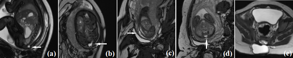
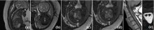
Figure 3
Fetal MRI Scans of Diastematomyelia and Postnatal Images. (a) Fetal sagittal view; (b) Fetal coronal view showing local dilatation of the spinal canal, spinal cord enlargement, and a T2-weighted hypointense lesion inside; (c) Fetal axial view; (d) Fetal axial view showing the spinal cord divided into left and right parts; (e) Postnatal image
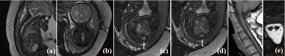

Figure 4
Fetal MRI Scans of Hemivertebra Malformation and Postnatal Images. (a) Fetal SWI coronal view; (b) Fetal coronal view; (c) Fetal coronal view; (d) Fetal coronal view showing hemivertebra malformation at the third lumbar vertebra (L3); (e) Postnatal CTVR reconstructed image
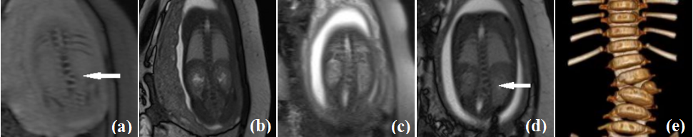
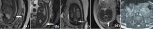
Figure 5
Fetal MRI Scans of Tethered Cord Malformation and Postnatal Images. (a) Fetal sagittal view; (b) Fetal sagittal view; (c) Fetal coronal view showing local dilatation of the spinal canal, enlargement of the terminal spinal cord, and a low position of the terminal spinal cord; (d) Fetal axial view showing abnormal signal in the spinal canal closely adjacent to the posterior wall of the spinal canal; (e) Postnatal fetal MRI image
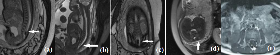
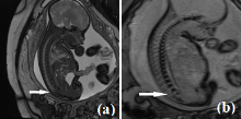
Figure 6
Fetal MRI Scans of Tethered Cord Malformation and Caudal Regression Syndrome Malformation. (a) Fetal sagittal view; (b) Fetal sagittal view showing absence of sacrococcygeal vertebrae and cauda equina
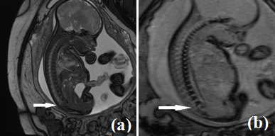
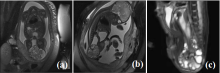
Figure 7
Fetal MRI Scans of Sacrococcygeal Teratoma Malformation and Postnatal Images. (a) Fetal coronal view showing a large externally protruding mass in the sacrococcygeal region; (b) Fetal sagittal view showing a large externally protruding mass in the sacrococcygeal region; (c) Postnatal fetal MRI image

| [1] |
BAI Y, SUN Y, LIU N, et al. Genetic analysis of 55 cases with fetal skeletal dysplasia[J]. Orphanet J Rare Dis, 2022, 17(1): 410.
doi: 10.1186/s13023-022-02559-4 pmid: 36352425 |
| [2] | WU W, ZENG R Y, QIU M F. Three-dimensional bone imaging diagnosis of fetal rib fusion costospinal junction deformity: a case report[J]. Chinese Journal of Ultrasound Medicine, 2020, 36(12): 1143. |
| 吴玮, 曾瑞盈, 邱木峰. 三维骨骼成像诊断胎儿肋骨融合肋脊连接畸形分析1例[J]. 中国超声医学杂志, 2020, 36(12): 1143. | |
| [3] | LIU H, ZHANG X Y, LIU L F, et al. Prenatal three-dimensional ultrasonography and chromosomal microarray analysis for screening fetal skeletal abnormalities and genetic etiology analysis[J]. Chinese Journal of Family Planning, 2023, 31(3): 646-649. |
| 刘皓, 张小燕, 刘利芳, 等. 产前三维超声与染色体微阵列分析技术筛查骨骼畸形异常胎儿及遗传学病因分析[J]. 中国计划生育学杂志, 2023, 31(3): 646-649. | |
| [4] | MA X L, ZHANG G F. Application of magnetic resonance imaging in fetal central nervous system[J]. Journal of Clinical Radiology, 2018, 37(6): 1056-1060. |
| 马晓亮, 张国福. 磁共振成像在胎儿中枢神经系统中的应用进展[J]. 临床放射学杂志, 2018, 37(6): 1056-1060. | |
| [5] | 徐梦莹. 超声与核磁共振对胎儿畸形产前诊断的比较性研究[D]. 长春: 吉林大学, 2023. |
| [6] | CAI X Y, CHEN X, SHAN R Q, et al. The value of magnetic sensitivity weighted imaging for fetal spinal anatomic and developmental malformation[J]. Chinese Journal of Radiology, 2018, 52(2): 119-124. |
| 蔡先云, 陈欣, 单瑞芹, 等. 磁敏感加权成像显示胎儿脊柱解剖及发育畸形的价值[J]. 中华放射学杂志, 2018, 52(2): 119-124. | |
| [7] | ZHOU Z, WU J L, CHEN W S, et al. Correlation analysis of prenatal ultrasound screening for fetal bone malformation and chromosomal microarray analysis[J]. Journal of Reproductive Medicine, 2020, 29(5): 593-597. |
| 周正, 吴洁丽, 陈文殊, 等. 产前超声筛查胎儿骨骼畸形与染色体微阵列分析特征的相关性分析[J]. 生殖医学杂志, 2020, 29(5): 593-597. | |
| [8] | CARTER J V, PAN J, RAI S N, et al. ROC-ing along: Evaluation and interpretation of receiver operating characteristic curves[J]. Surgery, 2016, 159(6): 1638-1645. |
| [9] |
LIU Y, WANG L, YANG Y K, et al. Prenatal diagnosis of fetal skeletal dysplasia using targeted next-generation sequencing: an analysis of 30 cases[J]. Diagn Pathol, 2019, 14(1): 76.
doi: 10.1186/s13000-019-0853-x pmid: 31299979 |
| [10] | HU L Q, YUAN H L. Ultrasound diagnosis and genetic analysis of congenital skeletal malformations in fetuses[J]. Chinese Journal of Family Planning, 2024, 32(2): 382-385. |
| 胡李琪, 袁洪亮. 先天性骨骼畸形胎儿的超声诊断和遗传学分析[J]. 中国计划生育学杂志, 2024, 32(2): 382-385. | |
| [11] | MASSELLI G, COZZI D, CECCANTIE S, et al. Fetal body MRI for fetal and perinatal management[J]. Clin Radiol, 2021, 76(9): 708. e1-708.e8. |
| [12] |
DULGHEROFF F F, PEIXOTO A B, PETRINI C G, et al. Fetal structural anomalies diagnosed during the first, second and third trimesters of pregnancy using ultrasonography: a retrospective cohort study[J]. Sao Paulo Med J, 2019, 137(5): 391-400.
doi: S1516-31802019000500391 pmid: 31939566 |
| [13] | DU C. Value of ultrasonography in the diagnosis of skeletal deformity of limbs of fetus in the second trimester[J]. Chinese Journal of Eugenics and Genetics, 2016, 24(11): 103-104+48. |
| 杜成. 中孕期胎儿肢体骨骼畸形的超声诊断价值[J]. 中国优生与遗传杂志, 2016, 24(11): 103-104+48. | |
| [14] | LUO D Q, CHEN X L, ZHU X, et al. Application of prenatal ultrasound and MRI in the diagnosis of fetal malformation[J]. Chinese Medical Imaging Technology, 2016, 32(4): 586-590. |
| 罗德清, 陈欣林, 朱霞, 等. 产前超声和MRI在诊断胎儿畸形中的应用[J]. 中国医学影像技术, 2016, 32(4): 586-590. | |
| [15] | LI S Y, WANG X H, WANG H P, et al. The clinical value of MRI in the diagnosis of fetal malformation[J]. Chinese Journal of Practical Neurological Diseases, 2014, 17(15): 35-36. |
| 李素英, 王新会, 王红泼, 等. MRI对胎儿畸形诊断的临床价值[J]. 中国实用神经疾病杂志, 2014, 17(15): 35-36. | |
| [16] | CAO Y. Comparative study on imaging quality of different magnetic resonance imaging sequences in prenatal fetal diagnosis[J]. Southwest Defense Medicine, 2017, 27(1): 62-66. |
| 曹伊. 磁共振不同成像序列在产前胎儿诊断成像质量的比较研究[J]. 西南国防医药, 2017, 27(1): 62-66. | |
| [17] | LIANG Y S, ZHANG X A, ZHAO X, et al. MRI diagnosis of fetal spinal cord malformation and sequence selection[J]. Chinese Medical Imaging Technology, 2020, 36(1): 111-115. |
| 梁艳山, 张小安, 赵鑫, 等. MRI诊断胎儿脊柱脊髓畸形及序列选择[J]. 中国医学影像技术, 2020, 36(1): 111-115. | |
| [18] | TANG B. Analysis of clinical application value of transabdominal ultrasonography and MRI in prenatal fetal malformation[J]. Tibetan Medicine, 2021, 42(1): 36-38. |
| 唐蓓. 经腹壁超声、MRI检查在产前胎儿畸形中的临床应用价值分析[J]. 西藏医药, 2021, 42(1): 36-38. | |
| [19] | ZHANG S, CHEN J L, HUANG Y Z, et al. Clinical diagnostic value of fourdimensional ultrasound combined with MRI in fetal malformation in second trimester and pregnancy outcomes[J]. Chinese Journal of CT and MRI, 2021, 19(8): 181-184. |
| 张帅, 陈建玲, 黄亚哲, 等. 四维超声联合MRI成像对中孕期胎儿畸形的临床诊断价值及其妊娠结局[J]. 中国CT和MRI杂志, 2021, 19(8): 181-184. | |
| [20] | LIU Z. Correlation between MRI diagnostic value of placenta implantation during pregnancy and pregnancy outcome[J]. Chinese Journal of Practical Medicine, 2017, 12(29): 83-85. |
| 刘姿. 妊娠期胎盘植入的MRI诊断价值与妊娠结局的相关性[J]. 中国实用医药, 2017, 12(29): 83-85. | |
| [21] | ZHEN J H, ZHOU W N, LIU Y N, et al. Application value of prenatal ultrasound and MRI in the diagnosis of fetal closed spina bifida[J]. Journal of Clinical Ultrasound Medicine, 2023, 25(6): 451-455. |
| 甄敬华, 周伟娜, 刘益宁, 等. 产前超声及MRI在胎儿闭合性脊柱裂诊断中的应用价值[J]. 临床超声医学杂志, 2023, 25(6): 451-455. |
| [1] | MA Yingxue, ZHAO Yanqiang, YANG Xiaodong, JIANG Bin, TAO Cheng. Opportunities and Challenges of High-field and Ultra-high-field Magnetic Resonance Imaging in China [J]. Chinese Journal of Magnetic Resonance, 2025, 42(3): 334-344. |
| [2] | SUI Meiju, ZHANG Lei, WANG Ruifang, LUO Yingying, LI Sha, QIU Maosong, XU Qiuyi, CHEN Daiqin, CHEN Shizhen, ZHOU Xin. MRI-traceable Nanoenzyme for Cascade Catalysis-enhanced Immunotherapy [J]. Chinese Journal of Magnetic Resonance, 2025, 42(3): 231-248. |
| [3] | MENG Jingxin, WANG Yuanjun. Research Progress on Tractography of Superficial White Matter Based on Diffusion Magnetic Resonance Imaging [J]. Chinese Journal of Magnetic Resonance, 2025, 42(2): 205-220. |
| [4] | CHEN Qun, YANG Zijian, CHENG Xinyi, JIA Siyi, DU Xiaoxia, WANG Mengxing. Application of Magnetic Resonance Imaging Technology in Pediatric Exercise Intervention Research [J]. Chinese Journal of Magnetic Resonance, 2025, 42(2): 195-204. |
| [5] | ZUO Bingyu, SHI Lili, SONG Jia, ZHAO Yang, LI Qian. Application of Estrogen and Tumor Markers Combined with DCE-MRI in Diagnosis and Clinical Staging of Cervical Cancer [J]. Chinese Journal of Magnetic Resonance, 2025, 42(2): 164-173. |
| [6] | YANG Jiacheng, WANG Yuanjun. Improved Constrained Spherical Deconvolution for Microstructural Imaging of Brain Gray Matter [J]. Chinese Journal of Magnetic Resonance, 2025, 42(1): 67-79. |
| [7] | PANG Qifan, WANG Zhichao, WU Yupeng, LI Jianqi. The Impact of K-Space Filling Strategy on Fat Artifacts in APT Imaging Based on FLASH Sequence [J]. Chinese Journal of Magnetic Resonance, 2024, 41(4): 443-453. |
| [8] | HUANG Min, ZHU Junlin, KAO Yuchen, ZHOU Dao, TANG Qiling. Multi-Coil MRI Image Reconstruction Based on ISTAVS-Net of Physical Model [J]. Chinese Journal of Magnetic Resonance, 2024, 41(4): 418-429. |
| [9] | XU Zhenshun, YUAN Xiaohan, HUANG Ziheng, SHAO Chengwei, WU Jie, BIAN Yun. Multi-source Feature Classification Model of Pancreatic Mucinous and Serous Cystic Neoplasms Based on Deep Learning [J]. Chinese Journal of Magnetic Resonance, 2024, 41(1): 19-29. |
| [10] | LIU Ying, LIN Ling, YUAN Binhua, ZHANG Haowei. Research Progress of MRI Gradient Waveform Generator [J]. Chinese Journal of Magnetic Resonance, 2024, 41(1): 99-115. |
| [11] | ZHOU Tianli, ZHANG Dian, WU Jizhi, JIA Huihui, CHANG Yan, SHENG Mao, YANG Xiaodong. Application of MRI-based Finite Element Modeling and Analysis in Periacetabular Osteotomy [J]. Chinese Journal of Magnetic Resonance, 2023, 40(4): 397-409. |
| [12] | CHEN Mengying, WU Yupeng, PANG Qifan, ZHONG Haodong, LI Gaiying, LI Jianqi. Simultaneously Neuromelanin-sensitive Imaging and Quantitative Susceptibility Mapping in the Whole Brain [J]. Chinese Journal of Magnetic Resonance, 2023, 40(4): 385-396. |
| [13] | REN Hongjin, MA Yan, XIAO Liang. Knee Joint Model Construction and Local Specific Absorption Rate Estimation Based on Generative Adversarial Networks [J]. Chinese Journal of Magnetic Resonance, 2023, 40(4): 410-422. |
| [14] | LAI Jiawen, WANG Yuling, CAI Xiaoyu, ZHOU Lihua. Multidimensional Information Fusion Method for Meniscal Tear Classification Based on CNN-SVM [J]. Chinese Journal of Magnetic Resonance, 2023, 40(4): 423-434. |
| [15] | Li Yijie, YANG Xinyu, YANG Xiaomei. Magnetic Resonance Image Reconstruction of Multi-scale Residual Unet Fused with Attention Mechanism [J]. Chinese Journal of Magnetic Resonance, 2023, 40(3): 307-319. |
| Viewed | ||||||
|
Full text |
|
|||||
|
Abstract |
|
|||||