
Chinese Journal of Magnetic Resonance ›› 2026, Vol. 43 ›› Issue (3): 367-388.doi: 10.11938/cjmr20263197cstr: 32225.14.cjmr20263197
• Review Articles • Previous Articles
MA Suchao, WANG Yuanjun*( )
)
Received:2026-01-06
Published:2026-09-05
Online:2026-08-11
Contact:
WANG Yuanjun *Tel: 13761603606, E-mail: yjusst@126.com.
CLC Number:
MA Suchao, WANG Yuanjun. Research Progress on Diffusion Magnetic Resonance Imaging Noise Reduction Methods Based on Deep Learning[J]. Chinese Journal of Magnetic Resonance, 2026, 43(3): 367-388.
Add to citation manager EndNote|Reference Manager|ProCite|BibTeX|RefWorks
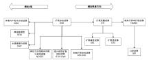
Fig. 1
The developmental history of DWI and its derivative models
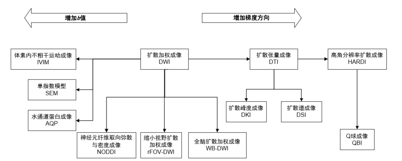

Table 1
Classification of noise mechanisms and artifacts in dMRI
| 类型 | 分类 | 来源 | 典型特征 | 降噪是否关注 |
|---|---|---|---|---|
| 随机噪声 | 热噪声 | 由接收链路与硬件产生的随机电子噪声 | 在k空间中近似独立同分布的复高斯噪声 | √ |
| 采集放大的噪声 | 数据采集策略放大已有噪声的方差与相关性 | 噪声方差增加,空间相关性增强,非平稳性显著 | √ | |
| 重建后的统计噪声 | 由复值高斯噪声经过模值运算和多通道合成后形成的统计分布 | 非高斯,非对称,存在偏置 | √ | |
| 空间相关与非平稳噪声 | 由于线圈灵敏度、并行成像与重建过程导致的不再满足空间独立同分布的噪声 | 具有空间相关性,噪声强度随位置变化 | √ | |
| 扩散特有噪声效应 | 扩散编码导致的信号衰减与噪声统计之间的耦合效应 | 高b值下SNR急剧下降;噪声偏置主导信号 | √ | |
| 结构性伪影 | 采集相关伪影 | 由设备不完美、模型失配以及其他偶然性因素导致的系统性误差 | 具有系统性、非随机性;通常表现为空间几何畸变或相位错误 | × |
| 重建与后处理伪影 | 由算法假设或数值处理引入的确定性误差 | 与重建/后处理参数强相关,具有明显的结构模式 | × |
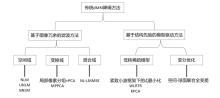
Fig. 2
Summary of traditional dMRI denoising methods
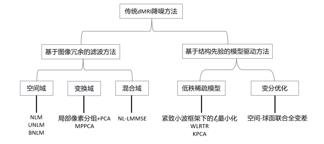
Fig. 3
Dual-channel input module in the DnCNN framework. Abbreviations: Conv, Convolution; BN, Batch Normalization; ReLU, Rectified Linear Unit; MSE, Mean Squared Error
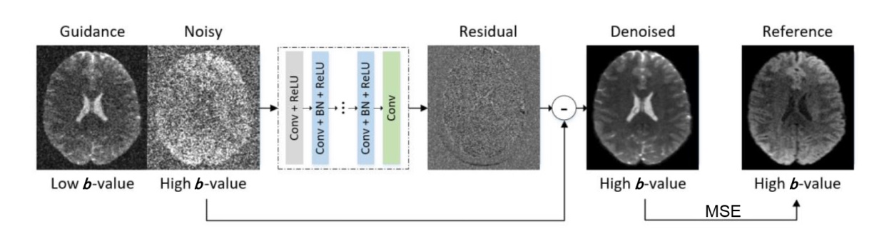
Fig. 4
Dual-branch network architecture integrating DWI and T1-weighted images[35]. Abbreviations: CSF, cerebrospinal fluid; GM, gray matter; WM, white matter; SD-LSTM, spatial-directional long short-term memory; FC, fully connected; AD, axial diffusivity; Vic, ICVF; Viso, isotropic volume fraction; OD, Orientation Dispersion Index
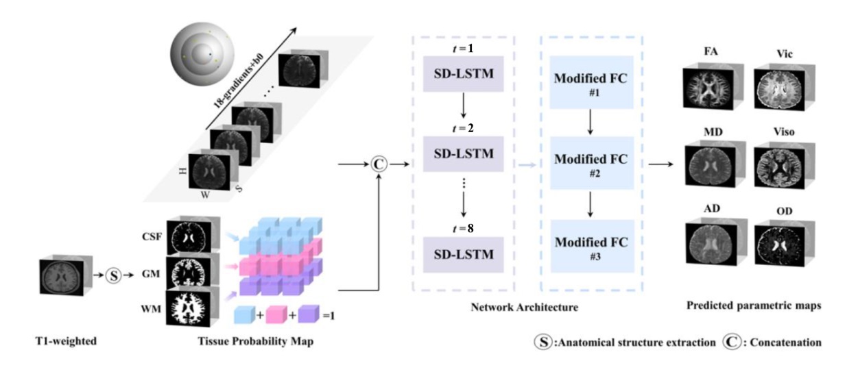
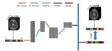
Fig. 5
Schematic diagram of the 1D-CNN denoising network architecture. The network takes a one-dimensional sequence composed of N highly noisy voxels at the same spatial location as input. It sequentially undergoes two layers of one-dimensional convolution (Conv) and Pooling operations for feature extraction. The first convolutional layer has 16 channels and a kernel size of 1; the second convolutional layer has 32 channels and a kernel size of 8. After feature Flatten, signal reconstruction is performed via a dense connected layer (Dense), with root mean squared error (RMSE) loss serving as the loss function for supervised training
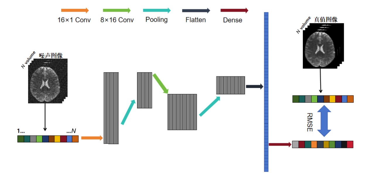
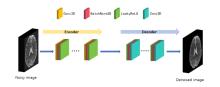
Fig. 6
Architecture diagram of the generative network. Abbreviations: Conv3D, 3D Convolution; BatchNorm3D, 3D Batch Normalization; LeakyReLU, Leaky Rectified Linear Unit
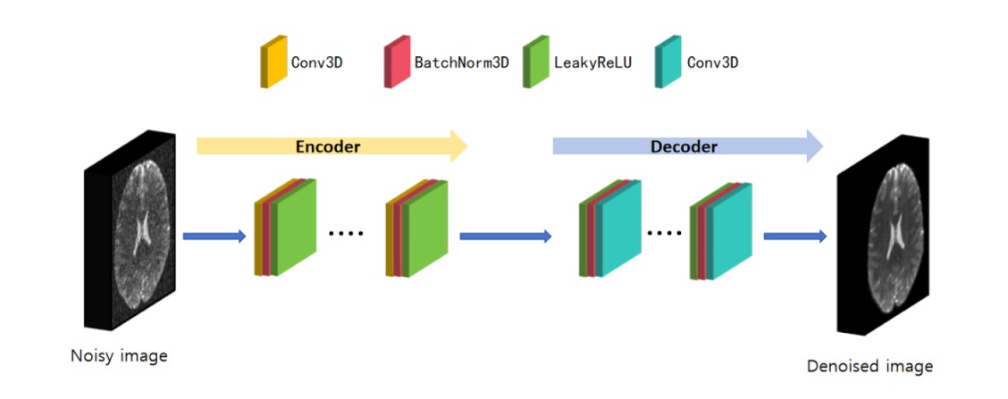
Fig. 7
Architecture diagram of the discriminator network. Abbreviations: Conv3D, 3D Convolution; LeakyReLU, Leaky Rectified Linear Unit; FC, Fully Connected
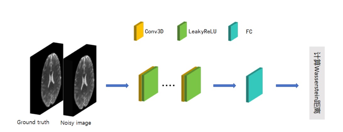
Table 2
Summary of deep learning-based denoising methods for dMRI
| 模型框架 | 对应方法 | 数据来源 | 优点 | 展望 |
|---|---|---|---|---|
| CNN | 复数域2DCNN[ | BrainWeb模拟脑数据库 | 分离实部和虚部处理,较好保留DWI完整信息,优于仅处理幅值图像的方法 | 目前主要用于2D切片,未来可扩展至3D重构,并结合多b值/多方向DWIs进一步提升降噪性能 |
| Guided DnCNN[ | 30名真实患者的前列腺MRI数据集 | 通过双输入引导和特征融合机制,利用低b值DWIs的解剖结构信息,提升高b值DWIs的降噪质量并减少过平滑 | 可进一步优化融合策略和注意力机制;结合k空间与图像域加速技术,有望实现更高效的加速重建 | |
| Residual DnCNN[ | 真实采集的带噪声DWIs与合成数据集 | 用残差学习预测噪声映射,兼顾降噪效率与细节保留,验证了方法在高维任务中的可行性 | 可探索更少平均次数输入以提升速度,并与运动校正模块结合,以适应临床动态场景 | |
| Enhanced SURE-based Method[ | fastMRI数据集和Siemens Healthineers数据集 | 仅需输入图像,无需额外噪声先验或扫描设定;利用相邻图像互补信息提升降噪效果 | 已在fastMRI和西门子7 T场强设备上验证,后续可推广到其他解剖部位,如腹部、膝部DWIs | |
| DeepDTI[ | Human Connectome Project(HCP)数据集 | 仅需1个b0图像和6个DWIs即可生成与90个DWIs接近质量的DTI,显著加快采集过程 | 方向敏感性可能受个体差异和扫描设备影响,需在更多临床人群和不同扫描协议下验证其泛化能力与诊断价值 | |
| SDnDTI[ | HCP数据集 | 通过子集划分进行损失约束,可在有限样本条件下有效利用7例DWIs进行自监督学习 | 对极高分辨率或超低信噪比DTI数据,可能需调整子集划分策略,并使用更多DWI方向或更先进的张量拟合方法 | |
| JD-CNN[ | 真实采集的低SNR数据和病例数据以及模拟数据集 | 同时输入所有b值的DWIs,利用跨b值结构相关性联合降噪,增强高b值DWIs的降噪能力 | 虽未假设噪声分布,但主要在高斯噪声下测试;未来需验证其在更复杂噪声模型下的表现 | |
| 1D-CNN[ | HCP数据集以及模拟数据 | 以重建图像作为监督目标进行训练和评估,可较好解决高b值DWI中SoS重建所引入的噪声 | 效果依赖高质量参考图像,限制了直接应用范围;后续需扩大数据规模并增强模型鲁棒性 | |
| SSECNN[ | 心脏DTI数据 | 利用不同扩散方向DWI的结构相似性构建自监督信号,并设计Sobel加权损失,有效抑制过平滑 | 可结合深度学习模型提取更具语义的切片相似性度量,进一步提升匹配精度和降噪效果 | |
| MCNN[ | 真实采集的健康受试者、多发性硬化症患者数据以及合成的训练数据 真实采集的健康受试者、多发性硬化症患者数据以及合成的训练数据 | 直接在幅度图像上完成训练和推理,输入与输出均为SoS图像;标准重建下对噪声峰值伪影更鲁棒 | 对重建顺序高度敏感;当重建流程变为“先切分后线圈组合”时性能明显下降,不推荐用于高加速场景 | |
| CCNN[ | 在复数图像上直接训练与推理,处理实部和虚部信息,较适用于平滑相位并能适应重建顺序变化 | 基于编码算子的CNN具有较大潜力,可为更强深度学习重建模型提供方向 | ||
| GNN | Node2Node[ | 离体猪心脏、在体人类心脏以及合成数据集 | 将不同方向DWIs视为图节点,并结合图卷积小波变换进行频谱分解与信息匹配,可同时滤除噪声和谐波干扰,提升结构感知能力 | 心脏成像易受运动影响,未来可结合运动校正;图信号处理计算开销较大,还需探索更高效实现以满足实时或准实时需求 |
| 扩散模型 | Di-Fusion[ | Stanford HARDI、Sherbrooke 3-Shell和PPMI数据集 | 基于J-invariance优化,仅用成对噪声DWIs训练,并通过迭代式精炼实现高保真重建,在复杂噪声下仍可保留DWIs结构细节 | 可进一步探索将dMRI物理模型,融入扩散模型或损失函数中,以增强物理可解释性 |
| DDM2[ | Sherbrooke 3-Shell、Stanford HARDI、PPMI以及真实采集数据 | 提出三阶段协同策略:先学习噪声分布,再桥接真实噪声与扩散过程,最后生成高质量降噪图像,实现由弱到强的降噪 | 推理耗时较长,难以满足实时需求;后续可通过推理加速、数据一致性约束及防止结构幻觉等策略进一步改进 |
Table 3
Comparison of SSIM metrics for different denoising methods at various noise levels
| b值/(s/mm²) | 噪声水平 | 噪声图像 | PCA | MPPCA | Patch2Self | 1D-CNN |
|---|---|---|---|---|---|---|
| b=0 | 2% | 0.82 | 0.93 | 0.95 | 0.93 | 0.94 |
| 4% | 0.62 | 0.88 | 0.90 | 0.89 | 0.91 | |
| 6% | 0.47 | 0.81 | 0.85 | 0.84 | 0.86 | |
| 8% | 0.37 | 0.77 | 0.80 | 0.78 | 0.77 | |
| 10% | 0.33 | 0.73 | 0.75 | 0.73 | 0.78 | |
| b=1000 | 2% | 0.48 | 0.73 | 0.76 | 0.77 | 0.80 |
| 4% | 0.25 | 0.55 | 0.64 | 0.66 | 0.69 | |
| 6% | 0.16 | 0.43 | 0.54 | 0.56 | 0.57 | |
| 8% | 0.12 | 0.34 | 0.44 | 0.45 | 0.46 | |
| 10% | 0.09 | 0.29 | 0.35 | 0.36 | 0.41 | |
| b=2000 | 2% | 0.36 | 0.58 | 0.63 | 0.66 | 0.68 |
| 4% | 0.17 | 0.38 | 0.47 | 0.51 | 0.56 | |
| 6% | 0.10 | 0.26 | 0.33 | 0.38 | 0.40 | |
| 8% | 0.08 | 0.21 | 0.27 | 0.28 | 0.31 | |
| 10% | 0.07 | 0.19 | 0.20 | 0.23 | 0.25 |
Table 4
Comparison of PSNR metrics for different denoising methods at various noise levels
| b值/(s/mm²) | 噪声水平 | 噪声图像 | PCA | MPPCA | Patch2Self | 1D-CNN |
|---|---|---|---|---|---|---|
| b=0 | 2% | 32.27 | 35.87 | 37.79 | 35.33 | 36.02 |
| 4% | 27.16 | 33.00 | 33.37 | 33.46 | 35.08 | |
| 6% | 23.72 | 29.49 | 29.67 | 30.19 | 31.77 | |
| 8% | 21.32 | 26.72 | 26.61 | 27.30 | 30.77 | |
| 10% | 19.40 | 24.29 | 24.06 | 24.73 | 27.58 | |
| b=1000 | 2% | 20.29 | 25.21 | 26.10 | 25.67 | 27.77 |
| 4% | 14.52 | 19.22 | 19.52 | 19.73 | 23.54 | |
| 6% | 10.90 | 14.58 | 14.72 | 15.08 | 20.46 | |
| 8% | 8.18 | 11.14 | 11.21 | 11.66 | 17.16 | |
| 10% | 5.97 | 8.48 | 8.42 | 8.91 | 15.96 | |
| b=2000 | 2% | 17.48 | 21.82 | 22.22 | 21.85 | 26.33 |
| 4% | 11.28 | 13.44 | 16.62 | 15.45 | 23.65 | |
| 6% | 7.32 | 9.70 | 12.80 | 10.63 | 20.51 | |
| 8% | 4.44 | 7.42 | 7.48 | 6.37 | 19.61 | |
| 10% | 2.19 | 5.95 | 3.58 | 4.21 | 17.92 |
Table 5
Comparison of RMSE metrics for different denoising methods at various noise levels
| b值/(s/mm²) | 噪声水平 | 噪声图像 | PCA | MPPCA | Patch2Self | 1D-CNN |
|---|---|---|---|---|---|---|
| b=0 | 2% | 0.024 | 0.016 | 0.013 | 0.018 | 0.015 |
| 4% | 0.042 | 0.022 | 0.020 | 0.025 | 0.017 | |
| 6% | 0.060 | 0.028 | 0.027 | 0.030 | 0.025 | |
| 8% | 0.077 | 0.039 | 0.035 | 0.036 | 0.031 | |
| 10% | 0.082 | 0.046 | 0.041 | 0.042 | 0.037 | |
| b=1000 | 2% | 0.089 | 0.055 | 0.040 | 0.046 | 0.039 |
| 4% | 0.141 | 0.084 | 0.073 | 0.071 | 0.067 | |
| 6% | 0.167 | 0.101 | 0.082 | 0.075 | 0.071 | |
| 8% | 0.179 | 0.118 | 0.107 | 0.090 | 0.085 | |
| 10% | 0.186 | 0.122 | 0.128 | 0.107 | 0.095 | |
| b=2000 | 2% | 0.107 | 0.076 | 0.055 | 0.054 | 0.049 |
| 4% | 0.146 | 0.123 | 0.084 | 0.086 | 0.072 | |
| 6% | 0.184 | 0.146 | 0.106 | 0.113 | 0.095 | |
| 8% | 0.205 | 0.153 | 0.143 | 0.166 | 0.111 | |
| 10% | 0.232 | 0.159 | 0.182 | 0.178 | 0.123 |
Fig. 8
Visual comparison of different denoising methods at a 6% noise level. From top to bottom, images under conditions of b=0, b=1 000 s/mm², and b=2 000 s/mm² are shown to reflect the performance differences of each method at varying diffusion sensitivities
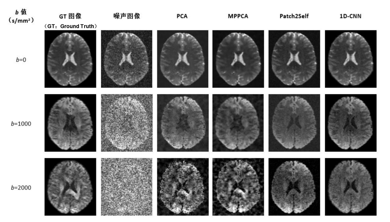
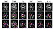
Fig. 9
Comparison of FA maps (Row 1), color-coded FA maps (Row 2), and local details (Row 3) across different denoising methods at a 4% noise level (b=1 000 s/mm²)
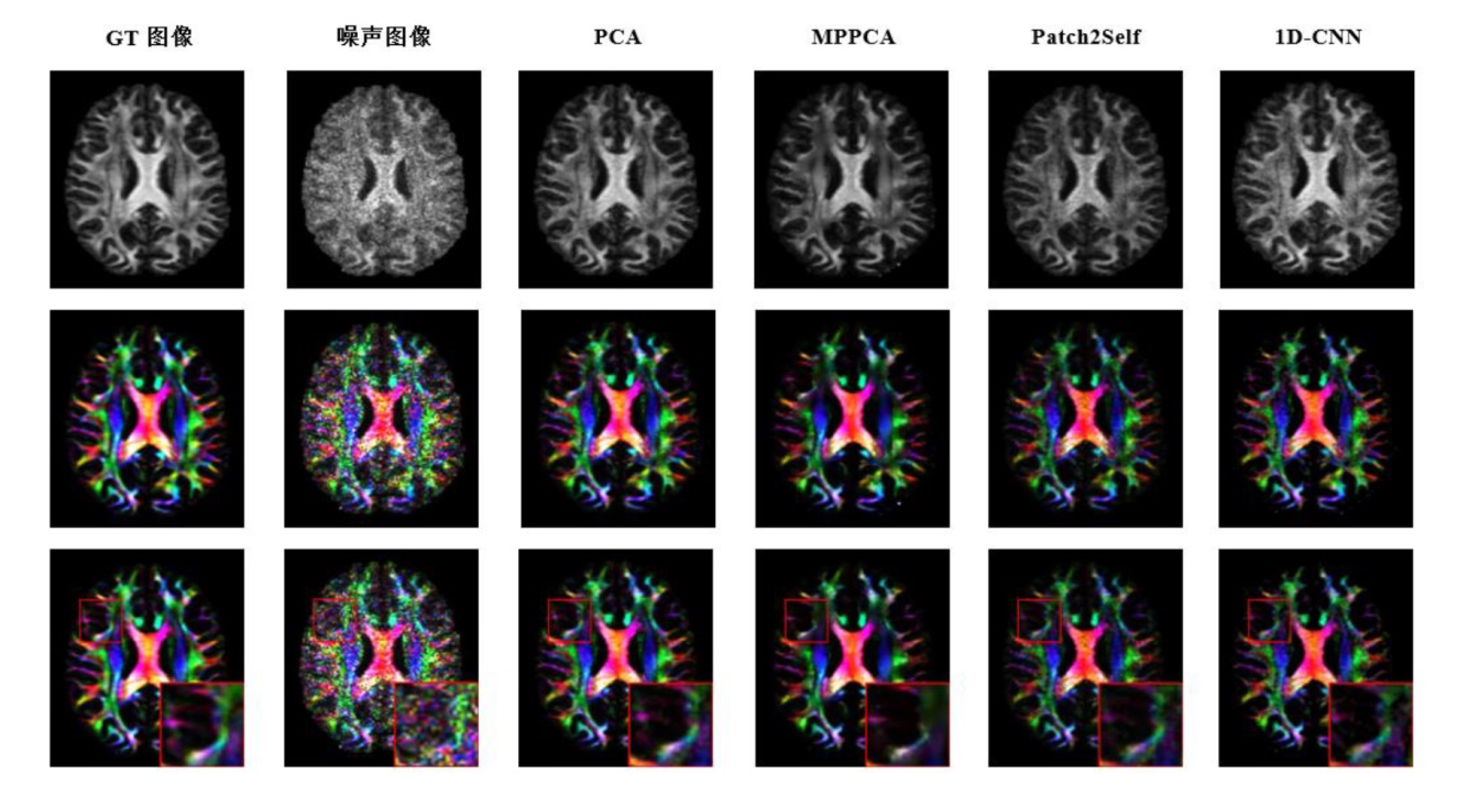
Table 6
Quantitative evaluation of denoising performance on DTI microstructural parameters. Values represent the voxel-wise MAPE of microstructural metrics (FA, MD, AD, RD) relative to the ground truth. Lower values indicate better preservation of microstructural details. Bold values indicate the best performance for each noise level
| 指标 | 噪声水平 | Noisy | PCA | MPPCA | Patch2Self | 1D-CNN |
|---|---|---|---|---|---|---|
| FAMAPE | 2% | 0.40 | 0.25 | 0.36 | 0.40 | 0.28 |
| 4% | 0.81 | 0.45 | 0.53 | 0.71 | 0.43 | |
| 6% | 1.30 | 0.55 | 0.60 | 0.84 | 0.51 | |
| 8% | 1.86 | 0.61 | 0.60 | 0.88 | 0.56 | |
| 10% | 2.40 | 0.63 | 0.62 | 0.89 | 0.58 | |
| MDMAPE | 2% | 0.16 | 0.24 | 0.25 | 0.26 | 0.30 |
| 4% | 0.51 | 0.52 | 0.51 | 0.54 | 0.34 | |
| 6% | 0.70 | 0.69 | 0.64 | 0.72 | 0.36 | |
| 8% | 0.86 | 0.79 | 0.73 | 0.82 | 0.51 | |
| 10% | 0.92 | 0.85 | 0.84 | 0.89 | 0.62 | |
| ADMAPE | 2% | 0.16 | 0.26 | 0.29 | 0.31 | 0.30 |
| 4% | 0.44 | 0.56 | 0.57 | 0.61 | 0.28 | |
| 6% | 0.61 | 0.72 | 0.71 | 0.77 | 0.24 | |
| 8% | 0.87 | 0.78 | 0.77 | 0.85 | 0.55 | |
| 10% | 1.08 | 0.87 | 0.86 | 0.91 | 0.66 | |
| RDMAPE | 2% | 0.16 | 0.21 | 0.19 | 0.20 | 0.27 |
| 4% | 0.45 | 0.31 | 0.30 | 0.45 | 0.21 | |
| 6% | 0.67 | 0.45 | 0.43 | 0.69 | 0.38 | |
| 8% | 0.85 | 0.61 | 0.57 | 0.83 | 0.52 | |
| 10% | 1.08 | 0.87 | 0.82 | 0.91 | 0.64 |
| [1] | LUNDELL H, STEELE C J. Cerebellar imaging with diffusion magnetic resonance imaging: approaches, challenges, and potential[J]. Curr Opin Behav Sci, 2024, 56(1): 101353. |
| [2] |
LIU X Y, WU Z K, WANG X C. An intrinsic anisotropic feature of DTI images derived by geometric properties on the Riemannian manifold[J]. Biomed Signal Proces, 2024, 87(1): 105478.
doi: 10.1016/j.bspc.2023.105478 |
| [3] |
LIAO Y, COELHO S, CHEN J, et al. Mapping tissue microstructure of brain white matter in vivo in health and disease using diffusion MRI[J]. Imaging Neurosci, 2024, 2(1): 1-17.
doi: 10.1162/imag_a_00344 |
| [4] |
LEBEL C, DEONI S. The development of brain white matter microstructure[J]. NeuroImage, 2018, 182(1): 207-218.
doi: 10.1016/j.neuroimage.2017.12.097 |
| [5] |
TAMNES C K, ROALF D R, GODDINGS A L, et al. Diffusion MRI of white matter microstructure development in childhood and adolescence: Methods, challenges and progress[J]. Dev Cogn Neurosci, 2018, 33(1): 161-175.
doi: 10.1016/j.dcn.2017.12.002 |
| [6] |
LEE H H, PAPAIOANNOU A, KIM S L, et al. A time-dependent diffusion MRI signature of axon caliber variations and beading[J]. Commun Biol, 2020, 3(1): 354-366.
doi: 10.1038/s42003-020-1050-x |
| [7] |
LAVDAS I, BEHAN K C, PAPADAKI A, et al. A phantom for diffusion-weighted MRI (DW-MRI)[J]. J Magn Reson Imaging, 2013, 38(1): 173-179.
doi: 10.1002/jmri.23950 pmid: 23576443 |
| [8] | YANG L M, WANG Y J. New method for diffusion-weighted images denoising based on patch-matching with higher-order singular value decomposition[J]. J Xray Sci Technol, 2025, 33(3): 526-539. |
| [9] | DENG L, WANG Y J. DTI brain template construction based on gaussian averaging[J]. Chinese J Magn Reson, 2022, 39(4): 413-427. |
|
邓岚, 王远军. 基于高斯平均的DTI脑模板构建方法[J]. 波谱学杂志, 2022, 39(4): 413-427.
doi: 10.11938/cjmr20212957 |
|
| [10] |
GUNDOGDU B, PITTMAN J M, CHATTERJEE A, et al. Directional and inter-acquisition variability in diffusion-weighted imaging and editing for restricted diffusion[J]. Magn Reson Med, 2022, 88(5): 2298-2310.
doi: 10.1002/mrm.29385 pmid: 35861268 |
| [11] |
ZHANG X, PENG J, XU M, et al. Denoise diffusion-weighted images using higher-order singular value decomposition[J]. NeuroImage, 2017, 156(1): 128-145.
doi: 10.1016/j.neuroimage.2017.04.017 |
| [12] |
MANJON J V, COUPE P, CONCHA L, et al. Diffusion weighted image denoising using overcomplete local PCA[J]. PloS ONE, 2013, 8(9): e73021.
doi: 10.1371/journal.pone.0073021 |
| [13] |
CHENG H, VINCI-BOOHER S, WANG J, et al. Denoising diffusion weighted imaging data using convolutional neural networks[J]. PloS ONE, 2022, 17(9): e0274396.
doi: 10.1371/journal.pone.0274396 |
| [14] | YANG L M, WANG Y J. Research progress of denoising algorithms for diffusion tensor images[J]. Chinese J Magn Reson, 2024, 41(3): 341-361. |
|
杨黎明, 王远军. 扩散张量图像降噪算法研究进展[J]. 波谱学杂志, 2024, 41(3): 341-361.
doi: 10.11938/cjmr20243087 |
|
| [15] |
LE BIHAN D. Looking into the functional architecture of the brain with diffusion MRI[J]. Nat Rev Neurosci, 2003, 4(6): 469-480.
doi: 10.1038/nrn1119 pmid: 12778119 |
| [16] | BUADES A, COLL B, MOREL J-M. A non-local algorithm for image denoising[C]// Proceedings of the 2005 IEEE Computer Society Conference on Computer Vision and Pattern Recognition (CVPR), San Diego, CA, USA. Piscataway: IEEE, 2005: 60-65. |
| [17] |
MANJON J V, CARBONELL-CABALLERO J, LULL J J, et al. MRI denoising using non-local means[J]. Med Image Anal, 2008, 12(4): 514-523.
doi: 10.1016/j.media.2008.02.004 pmid: 18381247 |
| [18] |
COUPE P, YGER P, PRIMA S, et al. An optimized blockwise nonlocal means denoising filter for 3-D magnetic resonance images[J]. IEEE T Med Imaging, 2008, 27(4): 425-441.
doi: 10.1109/TMI.2007.906087 pmid: 18390341 |
| [19] |
AJA-FERNANDEZ S, NIETHAMMER M, KUBICKI M, et al. Restoration of DWI data using a Rician LMMSE estimator[J]. IEEE T Med Imaging, 2008, 27(10): 1389-1403.
doi: 10.1109/TMI.2008.920609 |
| [20] | HENRIQUES R N. Advanced methods for diffusion MRI data analysis and their application to the healthy ageing brain[D]. Cambridge: University of Cambridge, 2017. |
| [21] |
TRISTAN-VEGA A, AJA-FERNANDEZ S. DWI filtering using joint information for DTI and HARDI[J]. Med Image Anal, 2010, 14(2): 205-218.
doi: 10.1016/j.media.2009.11.001 |
| [22] |
VERAART J, FIEREMANS E, NOVIKOV D S. Diffusion MRI noise mapping using random matrix theory[J]. Magn Reson Med, 2016, 76(5): 1582-1593.
doi: 10.1002/mrm.26059 pmid: 26599599 |
| [23] |
RAMOS-LLORDEN G, VEGAS-SANCHEZ-FERRERO G, LIAO C, et al. SNR-enhanced diffusion MRI with structure-preserving low-rank denoising in reproducing kernel Hilbert spaces[J]. Magn Reson Med, 2021, 86(3): 1614-1632.
doi: 10.1002/mrm.28752 |
| [24] |
MCGRAW T, VEMURI B, OZARSLAN E, et al. Variational denoising of diffusion weighted MRI[J]. Inverse Probl Imaging, 2009, 3(4): 625-648.
doi: 10.3934/ipi.2009.3.625 |
| [25] |
LAM F, BABACAN S D, HALDAR J P, et al. Denoising diffusion-weighted magnitude MR images using rank and edge constraints[J]. Magn Reson Med, 2014, 71(3): 1272-1284.
doi: 10.1002/mrm.24728 pmid: 23568755 |
| [26] |
JUREK J, MATERKA A, LUDWISIAK K, et al. Supervised denoising of diffusion-weighted magnetic resonance images using a convolutional neural network and transfer learning[J]. Biocybern Biomed Eng, 2023, 43(1): 206-232.
doi: 10.1016/j.bbe.2022.12.006 |
| [27] |
KAYE E A, AHERNE E A, DUZGOL C, et al. Accelerating prostate diffusion-weighted MRI using a guided denoising convolutional neural network: retrospective feasibility study[J]. Radiol Artif Intell, 2020, 2(5): e200007.
doi: 10.1148/ryai.2020200007 |
| [28] | PHIPPS K, BOOMEN M V D, EDER R, et al. Accelerated in vivo cardiac diffusion-tensor MRI using residual deep learning-based denoising in participants with obesity[J]. Radiol-Cardiothorac, 2021, 3(3): e200580. |
| [29] |
PFAFF L, DARWISH O, WAGNER F, et al. Enhancing diffusion-weighted prostate MRI through self-supervised denoising and evaluation[J]. Sci Rep, 2024, 14(1): 24292.
doi: 10.1038/s41598-024-75007-x pmid: 39414914 |
| [30] |
RAN M, HU J R, CHEN Y, et al. Denoising of 3D magnetic resonance images using a residual encoder-decoder Wasserstein generative adversarial network[J]. Med Image Anal, 2019, 55(1): 165-180.
doi: 10.1016/j.media.2019.05.001 |
| [31] |
TIAN Q, BILGIC B, FAN Q, et al. DeepDTI: High-fidelity six-direction diffusion tensor imaging using deep learning[J]. NeuroImage, 2020, 219(1): 117017.
doi: 10.1016/j.neuroimage.2020.117017 |
| [32] |
TIAN Q, LI Z, FAN Q, et al. SDnDTI: Self-supervised deep learning-based denoising for diffusion tensor MRI[J]. NeuroImage, 2022, 253(1): 119033.
doi: 10.1016/j.neuroimage.2022.119033 |
| [33] | FADNAVIS S, BATSON J, GARYFALLIDIS E. Patch2Self: Denoising diffusion MRI with self-supervised learning[C]// Advances in Neural Information Processing Systems, Virtual. USA: Curran Associates, Inc., 2020: 16293-16303. |
| [34] |
DU H B, YUAN N N, WANG L H. Node2Node: self-supervised cardiac diffusion tensor image denoising method[J]. Appl Sci, 2023, 13(19): 10829.
doi: 10.3390/app131910829 |
| [35] | MA X R, CHENG J, FAN W X, et al. 3D anatomical structure-guided deep learning for accurate diffusion microstructure imaging[C]// 2025 IEEE 22nd International Symposium on Biomedical Imaging (ISBI), Houston, TX, USA. Piscataway: IEEE, 2025: 1-4. |
| [36] | NIU X L, LV J Q, YE Z T, et al. Multicontrast MR-guided diffusion model for ultra-low-dose brain PET denoising in temporal lobe epilepsy[J]. IEEE J Biomed Health, 2026, 30(3): 2316-2327. |
| [37] |
NASEEM R, CHEIKH F A, BEGHDADI A, et al. Cross-modal guidance assisted hierarchical learning based siamese network for MR image denoising[J]. Electronics, 2021, 10(22): 2855.
doi: 10.3390/electronics10222855 |
| [38] |
ZHANG K, ZUO W M, CHEN Y J, et al. Beyond a gaussian denoiser: residual learning of deep CNN for image denoising[J]. IEEE T Image Process, 2017, 26(7): 3142-3155.
doi: 10.1109/TIP.2017.2662206 pmid: 28166495 |
| [39] |
WANG H, ZHENG R C, DAI F, et al. High-field MR diffusion-weighted image denoising using a joint denoising convolutional neural network[J]. J Magn Reson Imaging, 2019, 50(6): 1937-1947.
doi: 10.1002/jmri.26761 pmid: 31012226 |
| [40] |
ZHANG L P, XIAO Z Z, ZHOU C, et al. Spatial adaptive and transformer fusion network (STFNet) for low-count PET blind denoising with MRI[J]. Med Phys, 2022, 49(1): 343-356.
doi: 10.1002/mp.v49.1 |
| [41] | XIANG T G, YURT M, SYED A B, et al. DDM2: self-supervised diffusion MRI denoising with generative diffusion models[C]// Proceedings of the 11th International Conference on Learning Representations (ICLR). Kigali, Rwanda: ICLR, 2023: 1-19. |
| [42] |
LI Z, FAN Q, BILGIC B, et al. Diffusion MRI data analysis assisted by deep learning synthesized anatomical images (DeepAnat)[J]. Med Image Anal, 2023, 86(1): 102744.
doi: 10.1016/j.media.2023.102744 |
| [43] |
PFAFF L, HOSSBACH J, PREUHS E, et al. Self-supervised MRI denoising: leveraging Stein's unbiased risk estimator and spatially resolved noise maps[J]. Sci Rep, 2023, 13(1): 22629.
doi: 10.1038/s41598-023-49023-2 |
| [44] |
YUAN N, WANG L, YE C, et al. Self-supervised structural similarity-based convolutional neural network for cardiac diffusion tensor image denoising[J]. Med Phys, 2023, 50(10): 6137-6150.
doi: 10.1002/mp.16301 pmid: 36775901 |
| [45] | TU J C, SHI Y K, LAM F. Score-based self-supervised MRI denoising[C]// Proceedings of the 13th International Conference on Learning Representations (ICLR). Singapore: ICLR, 2025: 1-20. |
| [46] | WU C, KONG Q, JIANG Z, et al. Self-supervised diffusion MRI denoising via iterative and stable refinement[C]// Proceedings of the 13th International Conference on Learning Representations (ICLR). Singapore: ICLR, 2025: 1-22. |
| [47] |
MUCKLEY M J, ADES-ARON B, PAPAIOANNOU A, et al. Training a neural network for Gibbs and noise removal in diffusion MRI[J]. Magn Reson Med, 2020, 85: 413-428.
doi: 10.1002/mrm.v85.1 |
| [1] | LIU Kewen, JIANG Yukang, CHEN Fang, CHEN Junfei, LU Yuan, CHEN Li, LIU Chaoyang. A Deep Learning-based Method for LF-NMR Relaxation Time Spectrum Inversion [J]. Chinese Journal of Magnetic Resonance, 2026, 43(3): 307-320. |
| [2] | CAI Yue, WANG Xuxia, LIU Sijie, GUO Haodong, CHEN Xi, CHENG Linlin, KANG Yan, LIN Fuchun. Magnetic Resonance Imaging Study on the Microstructure Abnormalities of Striatal White Matter in Type 2 Diabetic Mellitus Rats [J]. Chinese Journal of Magnetic Resonance, 2026, 43(2): 175-185. |
| [3] | XIE Xinyi, WANG Yuanjun. Research Progress on Super-resolution Reconstruction of Brain Diffusion Magnetic Resonance Images [J]. Chinese Journal of Magnetic Resonance, 2026, 43(2): 223-240. |
| [4] | FU Fenfang, LIN Guobing, LI Meifang. Advances in Magnetic Resonance Imaging for the Diagnosis and Prognosis of Mild Traumatic Brain Injury [J]. Chinese Journal of Magnetic Resonance, 2026, 43(2): 214-222. |
| [5] | NI Guangmao, LI Yuwei, HOU Wenxuan, LIU Caiyun, DONG Peng, ZHANG Yanhui. Research on the Influencing Factors of Acute Cerebral Infarction Recurrence Based on MR-DWI [J]. Chinese Journal of Magnetic Resonance, 2026, 43(1): 87-93. |
| [6] | LI Lu, GAO Yong, ZHOU Yupan. Construction of Prostate Cancer Model Based on T2WI Combined with DWI Multi Parameter MRI Texture Feature Fusion [J]. Chinese Journal of Magnetic Resonance, 2025, 42(4): 355-363. |
| [7] | LI Yinghao, WANG Lihui, WANG Sucheng, ZHU Zhongqi, HUANG Changdong, LI Renfeng, CAO Kaiming, HU Haiyang, JIA Yiming, LIANG Songtao, YANG Guang, LU Qing, WANG Hongzhi. Study on Pancreas Automatic Segmentation, Regional Quantification, and Diabetes Assessment [J]. Chinese Journal of Magnetic Resonance, 2025, 42(4): 378-389. |
| [8] | GAO Zhaoyao, ZHANG Zhan, HU Liangliang, XU Guangyu, ZHOU Sheng, HU Yuxin, LIN Zijie, ZHOU Chao. PMRI Image Reconstruction Method Based on Virtual Coils and GRAPPA-enhanced Network [J]. Chinese Journal of Magnetic Resonance, 2025, 42(4): 390-401. |
| [9] | ZHANG Mingyu, XIAO Sa, SHI Shengjie, ZHANG Xuecheng, ZHOU Xin. Research on a Multi-modal Enhanced Denoising Diffusion Model for Hyperpolarized 129Xe MRI [J]. Chinese Journal of Magnetic Resonance, 2025, 42(4): 364-377. |
| [10] | CAO Fei, XU Qianqian, CHEN Hao, ZU Jie, LI Xiaowen, TIAN Jin, BAO Lei. An Intelligent Diagnosis Method for NIID Based on Cross Self-supervision and DWI [J]. Chinese Journal of Magnetic Resonance, 2025, 42(2): 154-163. |
| [11] | XUE Peiyang, GENG Chen, LI Yuxin, BAO Yifang, LU Yucheng, DAI Yakang. A Classification Method for Cerebral Aneurysms in TOF-MRA Based on Improved 3D ResNet50 Model [J]. Chinese Journal of Magnetic Resonance, 2025, 42(1): 56-66. |
| [12] | NING Xinzhou, HUANG Zhen, CHEN Xiqu, LIU Xinjie, CHEN Gang, ZHANG Zhi, BAO Qingjia, LIU Chaoyang. Research on Transformer Super-Resolution Reconstruction Algorithm for Ultrafast Spatiotemporal Encoding Magnetic Resonance Imaging [J]. Chinese Journal of Magnetic Resonance, 2024, 41(4): 454-468. |
| [13] | YANG Liming, WANG Yuanjun. Research Progress of Denoising Algorithms for Diffusion Tensor Images [J]. Chinese Journal of Magnetic Resonance, 2024, 41(3): 341-361. |
| [14] | Dai Junlong, He Cong, Wu Jie, Bian Yun. Pancreatic Cystic Neoplasms Segmentation Network Combining Dual Decoding and Global Attention Upsampling Modules [J]. Chinese Journal of Magnetic Resonance, 2024, 41(2): 151-161. |
| [15] | YANG Yu, CHEN Bo, WU Liubin, LIN Enping, HUANG Yuqing, CHEN Zhong. Spectrum Reconstruction for Laplace NMR: From Handcraft Regularization to Deep Learning [J]. Chinese Journal of Magnetic Resonance, 2024, 41(2): 191-208. |
| Viewed | ||||||
|
Full text |
|
|||||
|
Abstract |
|
|||||
