


引言
磁共振成像(Magnetic Resonance Imaging,MRI)作为临床肿瘤诊断的重要工具,具有非侵入性、无电离辐射、无组织深度限制等优势[5].然而,传统基于质子(1H)的MRI易受到体内背景信号干扰,且用于增强成像效果的造影剂可能造成蓄积毒性[6].在众多磁共振可观测核中,氟(19F)具有100%的天然丰度,旋磁比为40.05 MHz/T,灵敏度仅次于1H(为1H的83%)[7],除此以外,19F只存在于骨头和牙齿中,自旋-自旋弛豫时间(T2)很短,不易被传统MRI技术检测到,因此19F在生物体内几乎没有背景信号干扰[8-
为提高纳米诊疗试剂对肿瘤的精准靶向和高效递送,研究者常在纳米颗粒表面修饰叶酸、精氨酰甘氨酰天冬氨酸(Arg-Gly-Asp,RGD)肽、抗体等功能分子,通过特异性结合肿瘤细胞表面过表达的受体(如叶酸受体、整合素、人表皮生长因子受体-2(HER2)等),增强肿瘤细胞对纳米颗粒的摄取[17-
铁离子(Fe3+)作为一种强路易斯酸,能够与具有强路易斯碱性的多酚分子形成稳定的螯合物[31,32]. FeIII-多酚螯合物通常在可见光到近红外光(Near Infrared,NIR)区域表现出宽而强的吸收带,并具备优异的光热转换效率,使其广泛应用于肿瘤光热治疗(Photothermal Therapy,PTT)[33-
在此,我们开发了一种基于蛋白冠表面生物矿化负载TA-FeIII的策略来构建肿瘤特异性成像和治疗的19F MRI纳米诊疗平台,其结构与作用方式如图1示意图所示.以PLGA包裹PFCE纳米颗粒(PP NPs)作为19F MRI成像核心,以牛血清白蛋白(BSA)为模型蛋白在PP NPs表面形成蛋白冠,为Fe3+的生物矿化提供模板,与单宁酸(TA)络合构建PP@BSA-TAFeIII纳米颗粒(NPs)作为探针.该纳米探针进入体内后,可产生以下作用:(1)TA-FeIII可以吸附血浆中的不饱和转铁蛋白,与探针表面BSA蛋白冠相互作用,形成“杂交”蛋白冠,使纳米探针能够通过TfR介导靶向肿瘤细胞;(2)PFCE高效富集在肿瘤组织,可产生强的19F MRI信号,用于肿瘤特异性“热点”成像;(3)在808 nm激光照射下,TA-FeIII具有良好的光热作用,可用于肿瘤光热治疗,在肿瘤组织酸性环境下,TA降解释放部分Fe3+,催化细胞内活性氧(ROS)的生成,诱导肿瘤细胞死亡.
图1
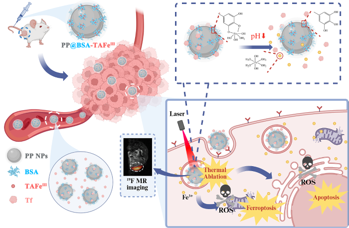
图1
PP@BSA-TAFeⅢ NPs的结构与作用原理示意图
Fig. 1
Illustration to demonstrate the structure and mechanism of PP@BSA-TAFeⅢ NPs
1 实验部分
1.1 实验试剂与材料
本研究使用的实验试剂与材料如表1所示.
表1 实验试剂与材料
Table 1
| 试剂/材料名称 | 生产厂商 | 规格 |
|---|---|---|
| 十二水合磷酸氢二钠 | 中国医药集团有限公司 | 500 g |
| 二水合磷酸二氢钠 | 中国医药集团有限公司 | 500 g |
| 二氯甲烷 | 国药集团化学试剂有限公司 | 500 mL |
| 甲醇 | 国药集团化学试剂有限公司 | 500 mL |
| 单宁酸(TA) | 西格玛奥德里奇贸易有限公司 | 100 g |
| 聚乙烯醇(PVA) | 西格玛奥德里奇贸易有限公司 | 250 g |
| 罗丹明B | 西格玛奥德里奇贸易有限公司 | 25 g |
| 牛血清白蛋白(BSA) | 阿达玛斯生命科学试剂有限公司 | 100 g |
| 全氟-15-冠-5-醚(PFCE) | 阿达玛斯试剂有限公司 | 1 g |
| 六水合三氯化铁 | 上海阿拉丁生化科技股份有限公司 | 500 g |
| 聚(D,L-乳酸-co-乙醇酸)(PLGA) | 上海阿拉丁生化科技股份有限公司 | 5 g |
| Calcein AM/PI细胞活性与细胞毒性检测试剂盒 | 上海碧云天生物技术股份有限公司 | 500 T |
| 苏木精&伊红(H&E)染色试剂盒 | 上海碧云天生物技术股份有限公司 | 200 T |
| TUNEL细胞凋亡检测试剂盒(绿色荧光) | 上海碧云天生物技术股份有限公司 | 20 T |
| 4',6-二脒基-2-苯基吲哚(DAPI)染色液 | 上海碧云天生物技术股份有限公司 | 10 mL |
| 细胞计数试剂盒-8(CCK-8) | 上海碧云天生物技术股份有限公司 | 2500 T |
| 异硫氰酸荧光素(FITC)标记鬼笔环肽 | 北京索莱宝科技有限公司 | 300 T |
| 2',7'-二氯荧光素二乙酸酯(DCFH-DA) | 北京索莱宝科技有限公司 | 25 mg |
| 无菌PBS | HyClone | 500 mL |
| RPMI 1640基础培养基 | Gibco Life Sciences | 500 mL |
| 青霉素-链霉素溶液 | Gibco Life Sciences | 100 mL |
| 胰蛋白酶 | Gibco Life Sciences | 100 mL |
| TRF ELISA检测试剂盒 | 上海江莱生物科技有限公司 | 48 T |
| 胎牛血清(FBS) | 长沙赛尔博克斯生物科技有限公司 | 50 mL |
| 4T1细胞 | 中国科学院上海细胞库 | TCM32 |
| Balb/c裸鼠 | 湖北贝恩特生物科技有限公司 | 6周龄 雌性 |
1.2 实验仪器
本研究使用的仪器如表2所示.
表2 实验仪器
Table 2
| 仪器名称 | 生产厂商 | 型号 |
|---|---|---|
| pH计 | METTLER TOLEDO | FE20 |
| 磁力加热搅拌器 | IKA | HS 7 |
| 高速离心机 | Beckman Coulter | Advanti J-25 |
| 动态光散射仪 | Malvern | ZetaSizer Nano-ZS90 |
| 透射电子显微镜 | JEOL | JEM-2100 |
| 扫描电子显微镜 | Carl Zeiss AG | Zeiss Sigma |
| 紫外分光光度计 | Thermo Fisher Scientific | Evolution 220 |
| 傅里叶变换红外吸收光谱仪 | Thermo Fisher Scientific | Nicolet iS10 |
| 808 nm激光器 | 北京镭志威 | LWIRL808 |
| 红外热成像仪 | 武汉红视热像科技 | HS160 |
| 细胞培养箱 | Thermo Fisher Scientific | HeraCell 150i |
| 细胞计数仪 | 瑞沃德 | C100-SE/C100 |
| 无菌操作台 | Thermo Fisher Scientific | MCS Advantage |
| 激光扫描共聚焦显微镜 | Nikon | AIR/A1 |
| 酶标仪 | Molecular Devices | Spectro Max 190 |
| 核磁共振波谱仪 | Bruker | Ascend WB 500 MHz |
| 微成像核磁共振谱仪 | Bruker | Avance 400 |
1.3 PP@BSA-TAFeIII NPs的合成与表征
在PLGA-PFCE纳米颗粒(简称PP NPs)表面形成BSA蛋白冠,进一步通过生物矿化作用络合TA-FeIII,最终制备PP@BSA-TAFeIII NPs.
PP NPs的合成:利用水/油/水溶剂蒸发技术制备PP NPs.称取10 mg PLGA溶于500 μL二氯甲烷,然后向体系中加入100 μL PFCE和5 mL质量浓度为4%的PVA溶液.充分混合后,将混合物置于冰上,超声处理40 min,超声程序设置为1 s开-2 s关的交替.超声处理完成后,将充分均质化的乳液在室温下敞口搅拌4 h以蒸发二氯甲烷溶剂.随后,将混合物在4 ℃(14 000 rpm)下离心15 min,离心产物用超纯水洗涤数次以除去PVA,制备得到PP NPs,分散在1.5 mL超纯水中.罗丹明B标记的PP NPs制备方法如下:将1 mg罗丹明B溶于100 μL甲醇后与PLGA的二氯甲烷溶液混合制备,其余步骤与上述一致.
PP@BSA-TAFeIII NPs的合成:将所制备的PP NPs取100 μL加入200 μL质量浓度为20 mg/mL的BSA水溶液中,4 ℃下放置于摇床摇晃,使BSA吸附在PP NPs表面形成蛋白冠,使用截留分子量(MWCO)为100 kDa的超滤管,通过超滤去除游离BSA,得到PP@BSA NPs.然后将TA溶液(质量浓度5 mg/mL)和FeCl3溶液(质量浓度3.9 mg/mL)以TA : Fe3+的摩尔比为1 : 5的比例先后加入PP@BSA NPs水溶液中,调节pH至7,使用3 kDa MWCO超滤管,用超纯水纯化3个循环以去除多余的Fe3+离子.
PP@BSA-TAFeIII NPs的表征:使用透射电子显微镜(TEM)观察PP NPs和PP@BSA-TAFeIII NPs的形貌、尺寸和元素组成.使用纳米激光粒度仪测定纳米颗粒的水合粒径和ζ电势.分别使用紫外-可见分光光度计(Ultraviolet-visible Absorption Spectra,UV-vis spectra)和傅里叶变换红外吸收光谱仪(Fourier Transform Infrared Absorption Spectra, FT-IR spectra)检测纳米颗粒的紫外-可见吸收光谱和傅里叶红外变换吸收光谱.
1.4 PP@BSA-TAFeIII NPs的体外性能评估
PP@BSA-TAFeⅢ NPs的pH响应性能:通过普鲁士蓝的紫外可见吸光光度法测定PP@BSA-TAFeIII NPs在不同pH条件下释放的Fe3+含量.将PP@BSA-TAFeIII NPs在不同pH条件下孵育,并在不同的时间点通过超滤离心收集释放的Fe3+离子,然后在pH 2.0下加入K4[Fe(CN)6]反应产生普鲁士蓝Fe4[Fe(CN)6]3,通过测定Fe4[Fe(CN)6]3在740 nm处的紫外吸光度来计算Fe3+的浓度.
PP@BSA-TAFeIII NPs的光热性能:分别将不同Fe3+浓度(0、10、20、40、70、100 μg/mL)的PP@BSA-TAFeIII NPs水溶液置于功率为1 W·cm-2的808 nm激光下,使用红外热成像仪实时监测溶液温度变化.然后分别对0.14 mg/mL的PP@BSA-TAFeIII NPs溶液进行多次激光照射-冷却循环,评估PP@BSA-TAFeIII NPs的光热稳定性.
PP@BSA-TAFeIII NPs吸附转铁蛋白:将PP@BSA-TAFeIII NPs与对照组PP@BSA-TA NPs、PP@BSA NPs在相同条件下与胎牛血清(FBS)孵育,随后进行凝胶电泳实验和酶联免疫吸附实验(ELISA),分析其蛋白冠的组成.
PP@BSA-TAFeIII NPs的MRI性能:将不同浓度的PP@BSA-TAFeIII NPs水溶液(PFCE:0、4、8、12、16、20 mmol/L)置于5 mm核磁共振样品管,进行19F MRI成像实验,以评估PP@BSA-TAFeIII NPs的19F MRI成像性能.
1.5 PP@BSA-TAFeIII NPs的细胞实验
小鼠乳腺癌细胞(4T1)培养于含10% FBS的RPMI 1640培养基中,细胞培养基中补充1%青霉素-链霉素溶液.将细胞置于37 ℃恒温、5%的CO2条件下培养.
PP@BSA-TAFeIII NPs的细胞靶向性:使用罗丹明B标记PP@BSA-TAFeIII NPs和PP@BSA NPs,将4T1细胞分别在有PP@BSA-TAFeIII NPs和胎牛血清(FBS)、有PP@BSA-TAFeIII NPs无FBS、有PP@BSA NPs和FBS的RPMI 1640培养基中孵育4 h.然后除去培养基,固定细胞,使用4',6-二脒基-2-苯基吲哚(4',6-Diamidino-2-phenylindole,DAPI)染色液对细胞核进行荧光染色,使用异硫氰酸荧光素(Fluorescein Isothiocyanate,FITC)标记鬼笔环肽对细胞骨架进行荧光染色,然后通过激光扫描共聚焦显微镜观察细胞对纳米颗粒的摄取情况.
PP@BSA-TAFeIII NPs的细胞毒性:使用细胞计数试剂盒-8(Cell Counting Kit-8, CCK-8)检测不同处理方式下纳米颗粒对4T1细胞的毒性.将细胞以5×10³/孔接种于96孔细胞培养板中,其中一列不接种,为空白组,将细胞置于37 ℃恒温、5%的CO2条件下培养过夜.分别向两个96孔板中加入含有不同Fe3+浓度的PP@BSA-TAFeIII NPs (0.5、1、5、10、15、20 μg/mL)的RPMI 1640完全培养基,同时,其中一列不加入任何药物,为空白对照组,每孔200 μL,并与细胞在37 ℃恒温、5%的CO2条件下再孵育4 h.将其中一个96孔板置于功率为1 W·cm-2的808 nm激光下照射10 min,随后移除培养基,并用PBS清洗细胞3次.向每孔加入100 μL的RPMI 1640培养基和10 μL的CCK-8试剂,再次放入培养箱孵育1 h.使用酶标仪测定450 nm处的吸光度,并按以下公式计算细胞存活率C:
其中,As为实验组吸光度,Ab为空白组吸光度,Ac为空白对照组的吸光度.
随后,分别向两个96孔板中加入含不同浓度PP@BSA NPs(0.5、1、5、10、15、20 μg/mL)的RPMI 1640完全培养基,重复以上操作,得到PP@BSA NPs的细胞毒性数据.
PP@BSA-TAFeIII NPs促肿瘤细胞活性氧(ROS)生成:将4T1细胞以约3×10⁵/皿接种到35 mm玻底培养皿中,在37 ℃恒温、5%的CO2条件下培养24 h,加入含有不同浓度PP@BSA-TAFeIII NPs(Fe3+浓度:0、10、20、40 μg/mL)的RPMI 1640完全培养基继续孵育4 h后,移除培养基,使用DCFH-DA荧光探针检测各组4T1细胞的ROS生成情况.
细胞TEM:将细胞以7×10⁵/瓶接种于T25细胞培养瓶中,在37 ℃恒温、5%的CO2条件下培养过夜,将4T1细胞分别在含PP@BSA-TAFeIII NPs和胎牛血清(FBS)的RPMI 1640培养基中孵育0、2、4 h,然后除去培养基,固定细胞,使用透射电子显微镜(TEM)观察细胞的铁死亡情况.
PP@BSA-TAFeIII NPs的细胞光热毒性:将4T1细胞以约3×10⁵/皿接种到35 mm玻底培养皿中,在37 ℃恒温、5%的CO2条件下培养24 h,向培养皿中分别加入含有不同浓度PP@BSA-TAFeIII NPs(Fe3+浓度:0、5、10、15、20 μg/mL)的RPMI 1640完全培养基继续孵育4 h,分别置于功率为1 W·cm-2的808 nm激光下照射10 min后,移除培养基,并用PBS清洗细胞3次.随后,为了在体外光热治疗处理后区分活细胞和死细胞,使用Calcein-AM(钙黄绿素)和PI(碘化丙啶)荧光染色试剂盒,分别对活细胞和死细胞进行染色,使用激光扫描共聚焦显微镜观察光热治疗处理后细胞的死亡情况.
1.6 PP@BSA-TAFeIII NPs的活体成像与治疗实验
所有动物实验均按照中国科学院精密测量科学与技术创新研究院动物伦理审查委员会针对该动物研究的伦理审查批准(APM21013T)进行.收集对数生长期的4T1细胞并用PBS重悬得到浓度为5×10⁶/mL细胞悬液.选取25只6周龄雌性Balb/c裸鼠为造模对象,向小鼠右后腿皮下注射200 μL细胞悬液,造模7天后,观察到小鼠肿瘤体积达150 mm3左右,此时开展活体相关实验.
PP@BSA-TAFeIII NPs的活体肿瘤MRI:分别向两组(每组1只)荷瘤裸鼠尾静脉注射200 μL PFCE浓度为100 mmol/L的PP@BSA-TAFeIII NPs和PP NPs溶液,注射后12 h,利用400 M磁共振成像仪采集荷瘤小鼠的19F MRI图像.19F MRI采集参数为:快速自旋回波(Rapid Acquisition with Refocused Echoes,RARE)序列,重复时间(Time of Repetition,TR)=800 ms,回波时间(Time of Echo,TE)=3 ms,平均次数(Number of Average)=1 800,RARE因子(RARE Factor)=4,矩阵大小(Matrix Size)=256×256,层厚(Slice Thickness)=15 mm.
PP@BSA-TAFeIII NPs的活体肿瘤光热成像:对三组(每组1只)4T1皮下瘤小鼠分别静脉注射PBS、PP@BSA NPs和PP@BSA-TAFeIII NPs,使用功率为1 W·cm-2的808 nm激光照射不同时间,通过红外热摄像机监测肿瘤区域的升温情况.
PP@BSA-TAFeIII NPs的活体肿瘤光热治疗:将肿瘤体积为150 mm3左右的荷瘤小鼠随机分为4组(1. PBS;2. PP@BSA NPs + 808 nm激光;3. PP@BSA-TAFeIII NPs;4. PP@BSA-TAFeIII NPs + 808 nm激光,每组各5只)进行以下操作:在第0天、第2天、第4天、第6天进行治疗.具体治疗方式为:第1组注射200 μL PBS,第2组注射200 μL PP@BSA NPs(PFCE浓度30 mmol/L)+808 nm激光(功率1 W·cm-2)照射10 min,第3组注射200 μL PP@BSA-TAFeIII NPs(PFCE浓度30 mmol/L),第4组注射200 μL PP@BSA-TAFeIII NPs(PFCE浓度30 mmol/L)+808 nm激光(功率1 W·cm-2)照射10 min.治疗期间,每两天测量一次肿瘤的体积并记录小鼠的体重.在治疗的第14天,对各组小鼠进行安乐死处理,收集每组小鼠的肿瘤组织,按照不同治疗组进行定位,并用标尺参考进行拍照记录.通过苏木精和伊红(H&E)染色以及TUNEL(TdT-mediated dUTP Nick-End Labeling)法对收集到的各组肿瘤组织进行组织病理学研究和组织化学分析.
2 结果与讨论
2.1 PP@BSA-TAFeIII NPs的表征
首先在PP NPs表面吸附BSA形成蛋白冠,随后TA和Fe3+在BSA蛋白冠表面进行生物矿化得到PP@BSA-TAFeIII NPs.在该体系中,Fe3+同时与TA和HO-/H2O配位,形成不饱和配位结构,使得Fe3+更容易在弱酸性环境中释放[38].TEM结果显示(图2(a)),PP NPs呈球形结构,分散均匀,平均粒径为103.9 nm;包裹BSA蛋白冠后,TEM图像虽不能直接观察到蛋白冠的结构,但纳米颗粒之间出现明显粘连;进一步,纳米颗粒表面负载TA-FeIII络合物后,纳米颗粒平均粒径增加到123.6 nm,外圈出现明显的包裹层,形貌基本不变.TEM Mapping结果显示(图2(b)),PP@BSA-TAFeIII NPs中除了来自PLGA和PFCE的碳、氧、氟之外,还同时存在氮、硫、铁元素,其中氮和硫来自蛋白冠,铁来自TA-FeIII络合物,表明TA-FeIII络合物以受控的方式在PP@BSA NPs表面形成.此外,动态光散射仪(DLS)的测定结果显示(图2(c)),PP NPs的水合粒径为164.2 nm,形成BSA蛋白冠后水合粒径增加到177.2 nm,加入TA和Fe3+后,纳米颗粒的水合粒径增加到190.1 nm.表面电位结果显示(图2(d)),吸附BSA后,纳米颗粒的ζ电势从-14.9 mV增加到-3.2 mV,进一步负载TA-FeIII后则增加到6.45 mV.紫外-可见吸收光谱显示(图2(e)),PP@BSA-TAFeIII NPs在560 nm处产生TA-FeIII的特征吸收峰.红外吸收光谱显示(图2(f)),加入Fe3+后,PP@BSA-TAFeIII NPs在3 425 cm-1处的O-H伸缩振动带变宽,强度减弱,这是由于羟基参与配位后O-H键断裂,单宁酸的氢键网络被破坏;1 750 cm-1处的C=O伸缩振动带减弱且略微发生红移,这是由于单宁酸中的羰基氧参与配位后,C=O键被削弱,键级降低;471 cm-1处出现新的吸收峰,这是由于产生了新的Fe-O配位键.以上结果进一步证明了PP@BSA-TAFeIII NPs的成功合成.
图2
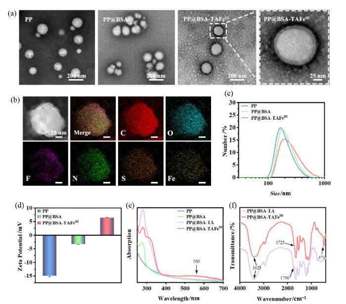
图2
PP@BSA-TAFeIII NPs的表征. (a) PP NPs、PP@BSA NPs、PP@BSA-TAFeIII NPs的TEM图;(b) PP@BSA-TAFeIII NPs的TEM mapping图;(c, d) PP NPs、PP@BSA NPs、PP@BSA-TAFeIII NPs的(c)水合粒径分布图和(d)ζ电势; (e)不同纳米颗粒(PFCE浓度为2 mmol/L)的紫外可见吸收光谱;(f) PP@BSA-TA NPs和PP@BSA-TAFeIII NPs(PFCE浓度为100 mmol/L)的红外吸收光谱
Fig. 2
Characterization of PP@BSA-TAFeIII NPs. (a) TEM images of PP NPs, PP@BSA NPs and PP@BSA-TAFeIII NPs; (b) TEM mapping image of PP@BSA-TAFeIII NPs; (c) Size distribution and (d) zeta potential of PP NPs, PP@BSA NPs and PP@BSA-TAFeIII NPs; (e) UV-vis absorption spectra of different nanoparticles (concentration of PFCE is 2 mmol/L); (f) FT-IR spectra of PP@BSA-TA NPs and PP@BSA-TAFeIII NPs (concentration of PFCE is 100 mmol/L)
2.2 PP@BSA-TAFeIII NPs的体外性能评估
图3
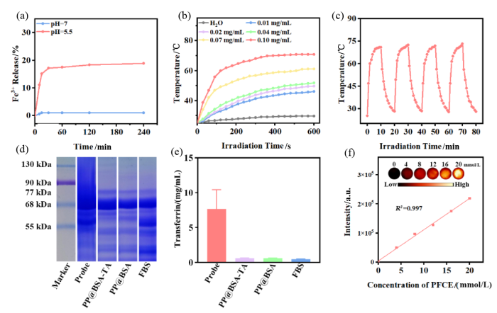
图3
PP@BSA-TAFeIII NPs的体外性能评估. (a) PP@BSA-TAFeIII NPs溶液在不同pH下Fe3+释放的速度;(b) 808 nm激光(1 W·cm-2)照射下,不同浓度的PP@BSA-TAFeIII NPs溶液和H2O的升温曲线图;(c) 808 nm激光(1 W·cm-2)循环照射下,PP@BSA-TAFeIII NPs溶液的温度变化曲线;(d) PP@BSA-TAFeIII NPs(Probe)、PP@BSA-TA NPs、PP@BSA NPs分别与FBS孵育后的凝胶电泳图;(e) ELISA定量检测吸附在不同纳米颗粒上的转铁蛋白含量;(f)不同浓度PP@BSA-TAFeIII NPs溶液的19F MRI图像,及其信号强度与浓度的线性拟合曲线
Fig. 3
In vitro characterization of PP@BSA-TAFeIII NPs. (a) pH-dependent release of Fe3+ by PP@BSA-TAFeIII NPs solution; (b) Temperature variation curves of PP@BSA-TAFeIII NPs solutions with different concentrations under the irradiation of 808 nm laser with 1.0 W·cm-2; (c) Temperature variation curves of PP@BSA-TAFeIII NPs solutions under the cyclic irradiation of 808 nm laser with 1.0 W·cm-2; (d) Gel electrophoresis of proteins adsorbed on PP@BSA-TAFeIII NPs (Probe), PP@BSA-TA NPs and PP@BSA NPs treated with FBS; (e) Quantitative detection of transferrin adsorbed on different nanoparticles by ELISA; (f) 19F MRI images of PP@BSA-TAFeIII NPs solutions at different concentrations, and the linear fitting curve of signal intensity versus concentration
PP@BSA-TAFeIII NPs具有良好的光热性能,如图3(b)所示,将Fe3+浓度为0~0.10 mg/mL的PP@BSA-TAFeIII NPs水溶液暴露于功率密度为1.0 W·cm-2的808 nm近红外激光下,随着PP@BSA-TAFeIII NPs浓度的增加,温度急剧升高,Fe3+浓度为0.10 mg/mL的PP@BSA-TAFeIII NPs在照射10 min后温度升高至70.7 ℃,而纯水的温度仅升高到29.1 ℃.对PP@BSA-TAFeIII NPs进行了多次激光照射-冷却循环,如图3(c)所示,根据该过程计算出PP@BSA-TAFeIII NPs的光热转换效率约为38.1%;在多次加热-冷却中,PP@BSA-TAFeIII NPs温度变化保持一致,证明其具有良好的光热稳定性.
为了证实PP@BSA-TAFeIII NPs能够通过配位不饱和FeIII吸附转铁蛋白,将PP@BSA NPs、PP@BSA-TA NPs、PP@BSA-TAFeIII NPs分别与胎牛血清(FBS)充分孵育后进行凝胶电泳测试.结果显示(图3(d)),PP@BSA-TAFeIII NPs携带的蛋白质主要为转铁蛋白(分子量为72.4~77.5 kDa)和牛血清白蛋白(65 kDa),同时这两种蛋白质也存在于在相同条件下用FBS处理的PP@BSA NPs和PP@BSA-TA NPs组中,但它们吸附转铁蛋白的数量远低于PP@BSA-TAFeIII NPs.ELISA定量结果(图3(e))进一步显示PP@BSA-TAFeIII NPs吸附的转铁蛋白含量远高于不含FeIII的对照组.这些结果表明,PP@BSA-TAFeIII NPs对转铁蛋白存在显著的特异性吸附,使其具有靶向肿瘤细胞的潜力.
为评估PP@BSA-TAFeIII NPs的19F MRI性能,使用Bruker Avance 400微成像核磁共振谱仪对不同浓度的PP@BSA-TAFeIII NPs水溶液(PFCE浓度依次为0、4、8、12、16、20 mmol/L)进行成像.实验结果如图3(f)所示,随着溶液浓度增加,探针的19F MRI信号逐渐增强,并且信号强度与其浓度呈良好的线性关系.
2.3 PP@BSA-TAFeIII NPs的体外抗肿瘤研究
为验证PP@BSA-TAFeIII NPs对肿瘤细胞的靶向效果,用罗丹明B对纳米颗粒进行标记,分别将PP@BSA-TAFeIII NPs和PP@BSA NPs加入含有FBS的RPMI 1640培养基,与4T1细胞共孵育4 h,并以未加入FBS的PP@BSA-TAFeIII NPs作为对照组.细胞荧光共聚焦实验结果(图4)表明,与FBS预先孵育的PP@BSA-TAFeIII NPs,细胞对其摄取远高于另外两组,这是由于PP@BSA-TAFeIII NPs充分吸附了FBS中的转铁蛋白,使得其能够与肿瘤表面过表达的转铁蛋白受体发生特异性吸附,提高肿瘤细胞摄取率.未经过FBS处理的PP@BSA-TAFeIII NPs表面不含转铁蛋白,而PP@BSA NPs由于并不含铁成分,经FBS处理后也无法吸附转铁蛋白,因此对肿瘤细胞均不具有靶向性.
图4
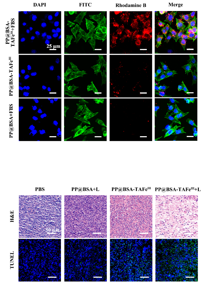
图4
4T1细胞分别与PP@BSA-TAFeIII NPs+FBS、PP@BSA-TAFeIII NPs、PP@BSA NPs+FBS孵育后的荧光共聚焦图像.细胞核使用DAPI进行染色,细胞骨架使用FITC标记鬼笔环肽进行染色
Fig. 4
CLSM images of 4Tl cells treated with PP@BSA-TAFeIII NPs+FBS, PP@BSA-TAFeIII NPs and PP@BSA NPs + FBS. Cell nuclei was stained by DAPI, and cytoskeleton was stained by FITC- phalloidin
CCK-8细胞毒性结果显示(图5(a)),PP@BSA NPs处理和PP@BSA NPs + 808 nm激光处理几乎不会对细胞的活性产生影响,相比之下,PP@BSA-TAFeIII NPs(IC50=94.8 μg/mL)处理和PP@BSA-TAFeIII NPs + 808 nm激光(IC50=11.2 μg/mL)处理均对4T1细胞产生了明显的毒性作用.当PP@BSA-TAFeIII NPs中Fe3+的浓度为20 μg/mL时,施加808 nm激光,4T1细胞的存活率仅为8.47%,说明联合治疗表现出了较高的抗肿瘤效率.使用ROS荧光探针(DCFH-DA)对不同浓度PP@BSA-TAFeIII NPs处理后的4T1细胞进行染色,以评估其诱导肿瘤细胞产生ROS的能力,结果如图5(b)所示,随着PP@BSA-TAFeIII NPs浓度升高,细胞内绿色荧光信号增强,这表明PP@BSA-TAFeIII NPs可以产生过量ROS,进一步诱导肿瘤细胞死亡.细胞TEM结果如图5(c)所示,PP@BSA-TAFeIII NPs处理2 h后,线粒体开始发生皱缩,体积变小,膜变暗,同时线粒体的嵴也开始减少,处理4 h后,线粒体的嵴完全消失,且外膜发生破裂,与细胞铁死亡的形态学特征一致,表明PP@BSA-TAFeIII NPs能够通过诱导铁死亡的方式造成肿瘤细胞损伤.除了铁死亡途径外,PP@BSA-TAFeIII NPs还可以通过光热效应(PTT)杀死肿瘤细胞,如图5(d)所示,在808 nm激光照射区域内(白色虚线右侧范围),PP@BSA-TAFeIII NPs的Fe3+浓度高于15 μg/mL时,细胞显示出强烈的红色荧光,而邻近未照射区域的细胞则显示出绿色荧光,表明肿瘤细胞因PP@BSA-TAFeIII NPs诱导的光热效应受到了严重损伤.综上所述,PP@BSA-TAFeIII NPs可以实现对肿瘤细胞的铁死亡与光热联合治疗.
图5
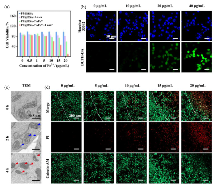
图5
PP@BSA-TAFeIII NPs的体外抗肿瘤性能. (a)不同纳米颗粒处理后4T1细胞的存活率;(b)不同Fe3+浓度的PP@BSA-TAFeIII NPs处理后细胞内ROS水平的共聚焦荧光图像(细胞核使用Hoechst 33342进行染色);(c) PP@BSA-TAFeIII NPs处理不同时间后4T1细胞的TEM图像;(d)不同Fe3+浓度的PP@BSA-TAFeIII NPs处理并用808 nm激光(1 W·cm-2)照射10 min后,对4T1细胞进行Calcein-AM/PI染色的共聚焦荧光图像(绿色荧光为活细胞,红色荧光为死细胞)
Fig. 5
In vitro antitumor efficacy of PP@BSA-TAFeIII NPs. (a) Viabilities of 4T1 cells treated with different nanoparticles; (b) CLSM images of ROS levels in 4Tl cells treated with different concentrations of Fe3+ in PP@BSA-TAFeIII NPs (cell nuclei was stained by Hoechst 33342); (c) TEM images of 4T1 cells treated with PP@BSA-TAFeIII NPs for different times; (d) CLSM images of 4T1 cells stained with Calcein-AM/PI (green fluorescence for live cells, red fluorescence for dead cells) after treatment with different concentrations of Fe3+ in PP@BSA-TAFeIII NPs and irradiation with 808 nm laser (1 W·cm-2) for 10 min
2.4 PP@BSA-TAFeIII NPs的活体成像与抗肿瘤研究
为进一步评估PP@BSA-TAFeIII NPs对活体肿瘤的靶向成像效果,分别将相同浓度的PP@BSA-TAFeIII NPs和PP NPs尾静脉注射至4T1荷瘤小鼠体内,12 h后采集小鼠的19F MRI图像.结果如图6(a)所示,注射PP@BSA-TAFeIII NPs 后肿瘤部位呈现明显的19F MRI信号,而注射PP NPs的小鼠肿瘤部位无信号,表明PP@BSA-TAFeIII NPs可以通过TA-FeIII介导吸附血液中的转铁蛋白,从而有效靶向肿瘤.
图6
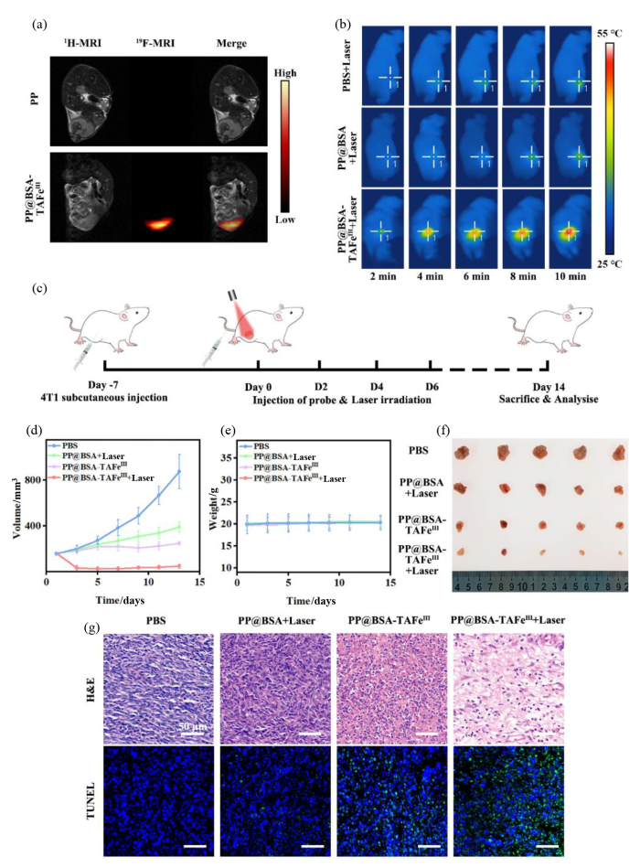
图6
PP@BSA-TAFeIII NPs的活体成像与抗肿瘤性能. (a)静脉注射PP@BSA-TAFeIII NPs和PP NPs 12 h后4T1荷瘤小鼠的19F MRI图像;(b)静脉注射PBS、PP@BSA NPs、PP@BSA-TAFeIII NPs至4T1荷瘤小鼠体内,经808 nm激光(1 W·cm-2)照射后肿瘤部位的光热成像;(c)活体抗肿瘤治疗示意图;不同治疗组小鼠的(d)肿瘤体积生长曲线、(e)体重变化曲线和(f)肿瘤照片;(g)不同治疗组小鼠肿瘤组织的H&E染色和TUNEL染色图像
Fig. 6
In Vivo tumor MR imaging and antitumor effect of PP@BSA-TAFeIII NPs. (a) 19F MRI images of 4T1 tumor-bearing mice after intravenous injection of PP@BSA-TAFeIII NPs or PP NPs for 12 h; (b) Infrared thermal photographs of the 4T1 tumor-bearing mice after intravenous injection of PBS, PP@BSA NPs or PP@BSA-TAFeIII NPs, followed by irradiation with 808 nm laser (1 W·cm-2); (c) Schematic illustration of the in vivo antitumor therapy; (d) Tumor volume growth curves, (e) body weight change curves and (f) tumor photographs of mice in different treatment groups; (g) H&E staining and TUNEL staining images of tumor tissues from mice in different treatment groups
基于PP@BSA-TAFeIII NPs良好的光热和促ROS生成性能,我们进一步在荷瘤小鼠模型上研究了其对肿瘤的治疗效果.向三组荷瘤小鼠分别静脉注射PBS、PP@BSA NPs和PP@BSA-TAFeIII NPs,并使用 808 nm激光(功率为1 W·cm-2)照射肿瘤10 min.红外热成像图显示(图6(b)),注射PP@BSA-TAFeIII NPs的小鼠肿瘤部位温度可升高至51.8 ℃,这个温度能够消融大部分肿瘤细胞,而注射PBS和PP@BSA NPs的小鼠仅分别升高至36.4 ℃和37.6 ℃,显示了PP@BSA-TAFeIII NPs在肿瘤部位的有效积累和良好的光热性能.活体治疗过程如图6(c)所示,治疗实验分为4个组别:PBS、PP@BSA NPs + Laser、PP@BSA-TAFeIII NPs、PP@BSA-TAFeIII NPs + Laser,每组5只,每2天测量肿瘤体积和小鼠体重(图6(d),6(f)).治疗结束后,PP@BSA-TAFeIII NPs + 808 nm激光照射组小鼠平均肿瘤体积为49.6 mm3,相比于PBS处理小鼠(872.9 mm3),其肿瘤抑制率为94.3%.而PP@BSA-TAFeIII NPs处理小鼠(249.6 mm3)和PP@BSA NPs + 808 nm激光照射处理小鼠(390.0 mm3)的肿瘤抑制率分别为71.4%和55.3%.肿瘤组织切片的H&E染色和TUNEL染色结果(图6(g))进一步证实,上述联合治疗组表现出大量的坏死肿瘤细胞和最强的细胞凋亡水平.另外,治疗期间小鼠体重不变(图6(e)),表明探针在小鼠体内没有明显的毒副作用.综上所述,PP@BSA-TAFeIII NPs在实现有效的抗肿瘤联合治疗的同时,具备良好的生物安全性.
3 结论
本文以全氟化碳纳米颗粒为核心,通过BSA蛋白冠涂层将单宁酸-铁(TA-FeIII)络合到纳米颗粒表面,构建了一种pH响应型智能诊疗一体纳米探针(PP@BSA-TAFeIII NPs).全氟化碳具有19F MRI造影效果,TA-FeIII络合物中的不饱和配位Fe3+会吸附血液中的转铁蛋白,在纳米探针表面形成“杂交”蛋白冠,能够识别肿瘤细胞表面过表达的转铁蛋白受体,使得探针能够高效富集于肿瘤组织,实现对肿瘤的高灵敏19F-MRI成像.同时,TA-FeIII络合物在肿瘤微酸环境下部分解离,释放Fe3+诱导肿瘤细胞铁死亡,剩余未解离部分则具备优异的光热效应,可实现对肿瘤的光热及铁死亡协同治疗.综上所述,基于蛋白冠原位调控策略为靶向抗肿瘤药物的响应性可控释放和肿瘤精准诊疗应用提供了新的思路.
利益冲突
无
参考文献
In vivo magnetic resonance detection of cancer by using multifunctional magnetic nanocrystals
[J].DOI:10.1021/ja052337c URL [本文引用: 1]
Impact of nanotechnology on drug delivery
[J].
DOI:10.1021/nn900002m
PMID:19206243
[本文引用: 1]

Nanotechnology is the engineering and manufacturing of materials at the atomic and molecular scale. In its strictest definition from the National Nanotechnology Initiative, nanotechnology refers to structures roughly in the 1-100 nm size regime in at least one dimension. Despite this size restriction, nanotechnology commonly refers to structures that are up to several hundred nanometers in size and that are developed by top-down or bottom-up engineering of individual components. Herein, we focus on the application of nanotechnology to drug delivery and highlight several areas of opportunity where current and emerging nanotechnologies could enable entirely novel classes of therapeutics.
Gram-scale synthesis of coordination polymer nanodots with renal clearance properties for cancer theranostic applications
[J].An ultrasmall hydrodynamic diameter is a critical factor for the renal clearance of nanoparticles from the body within a reasonable timescale. However, the integration of diagnostic and therapeutic components into a single ultrasmall nanoparticle remains challenging. In this study, pH-activated nanodots (termed Fe-CPNDs) composed of coordination polymers were synthesized via a simple and scalable method based on coordination reactions among Fe3+, gallic acid and poly(vinylpyrrolidone) at ambient conditions. The Fe-CPNDs exhibited ultrasmall (5.3 nm) hydrodynamic diameters and electrically neutral surfaces. The Fe-CPNDs also exhibited pH-activatable magnetic resonance imaging contrast and outstanding photothermal performance. The features of Fe-CPNDs greatly increased the tumour-imaging sensitivity and facilitated renal clearance after injection in animal models in vivo. Magnetic resonance imaging-guided photothermal therapy using Fe-CPNDs completely suppressed tumour growth. These findings demonstrate that Fe-CPNDs constitute a new class of renal clearable nanomedicine for photothermal therapy and molecular imaging.
Biodegradable polymeric nanoparticles based drug delivery systems
[J].DOI:10.1016/j.colsurfb.2009.09.001 URL [本文引用: 1]
Activatable 19F MRI nanoparticle probes for the detection of reducing environments
[J].DOI:10.1002/anie.v54.3 URL [本文引用: 1]
Ultrahigh 19F loaded Cu1.75S nanoprobes for simultaneous 19F magnetic resonance imaging and photothermal therapy
[J].DOI:10.1021/acsnano.5b06759 URL [本文引用: 1]
Overview and progress of X-nuclei magnetic resonance imaging in biomedical studies
[J].
DOI:10.1016/j.mrl.2023.05.002
PMID:40919505
[本文引用: 1]
Proton nuclear (H) is the observed nucleus on which most magnetic resonance imaging (MRI) applications depend. Most traditional H MRI can provide structural and functional information about organisms, while various non-proton nuclei (X-nuclei) MRI can provide more metabolic information. However, due to the relatively poor signal-to-noise ratio (SNR) of X-nuclei MRI, their applications are quite rare compared to H. Benefit from the rapid developments of MRI hardware and software technologies, X-nuclei MRI has recently attracted increasing interests in biomedical research. This review firstly introduces some current methods to improve the SNR of X-nuclei MRI. Secondly, this review describes biomedical applications of X-nuclei MRI, especially focusing on the current use of X-nuclei (C, O, F, Na and P) MRI to study related diseases in different organs, including the brain, liver, kidney, heart and bone. Finally, perspectives studies on X-nuclei imaging and its potential applications are described in biomedical research.© 2023 The Authors.
19F magnetic resonance imaging (MRI): From design of materials to clinical applications
[J].DOI:10.1021/cr500286d URL [本文引用: 2]
A unique 19F MRI agent for the tracking of non phagocytic cells in vivo
[J].
Liquid perfluorocarbons as contrast agents for ultrasonography and 19F-MRI
[J].
High F-content perfluoropolyether-based nanoparticles for targeted detection of breast cancer by19F magnetic resonance and optical imaging
[J].DOI:10.1021/acsnano.8b03726 URL [本文引用: 1]
Switchable 19F MRI polymer theranostics: towards in situ quantifiable drug release
[J].
Nanoparticle-based activatable MRI probes for disease imaging and monitoring
[J].
Multifunctional 19F MRI/CT imaging and combined photothermo-/chemo-therapy nanoplatform for tumor diagnosis and treatment
[J].
多功能19F磁共振/CT成像及光热/化学联合治疗纳米平台用于肿瘤诊疗
[J].
Frontiers in 19F-MR imaging: nanofluorides and 19F-CEST as novel extensions to the 19F-MRI toolbox
[J].
DOI:10.1039/D3CC00562C
URL
[本文引用: 1]
Fluorine-containing materials have enriched the field of molecular and cellular MRI with unambiguous and quantitative detection capabilities.
Fluoropolymer-MOF hybrids with switchable hydrophilicity for 19F MRI-monitored cancer therapy
[J].DOI:10.1021/acsnano.3c00694 URL [本文引用: 1]
Synthesis and applications of colloidal nanobioconjugates with modular multivalency: A review
[J].DOI:10.1021/acsanm.4c01371 URL [本文引用: 1]
Designed synthesis and surface engineering strategies of magnetic iron oxide nanoparticles for biomedical applications
[J].
PMID:27812592
Iron oxide nanoparticles (NPs) hold great promise for future biomedical applications because of their magnetic properties as well as other intrinsic properties such as low toxicity, colloidal stability, and surface engineering capability. Numerous related studies on iron oxide NPs have been conducted. Recent progress in nanochemistry has enabled fine control over the size, crystallinity, uniformity, and surface properties of iron oxide NPs. This review examines various synthetic approaches and surface engineering strategies for preparing naked and functional iron oxide NPs with different physicochemical properties. Growing interest in designed and surface-engineered iron oxide NPs with multifunctionalities was explored in in vitro/in vivo biomedical applications, focusing on their combined roles in bioseparation, as a biosensor, targeted-drug delivery, MR contrast agents, and magnetic fluid hyperthermia. This review outlines the limitations of extant surface engineering strategies and several developing strategies that may overcome these limitations. This study also details the promising future directions of this active research field.
Biogenic synthesis of novel nanomaterials and their applications
[J].
DOI:10.1039/D3NR03843B
URL
[本文引用: 1]
Synthesis of nanoparticles of different shapes and sizes using biological precursors and their applications.
The nanoparticle-protein complex as a biological entity
[J].DOI:10.1016/j.cis.2007.04.021 URL [本文引用: 1]
Precise nanomedicine for intelligent therapy of cancer
[J].DOI:10.1007/s11426-018-9397-5 [本文引用: 1]
The crown and the scepter: Roles of the protein corona in nanomedicine
[J].DOI:10.1002/adma.v31.45 URL [本文引用: 2]
The janus of protein corona on nanoparticles for tumor targeting, immunotherapy and diagnosis
[J].DOI:10.1016/j.jconrel.2022.03.056 URL [本文引用: 1]
Protein corona: Friend or foe? Co-opting serum proteins for nanoparticle delivery
[J].DOI:10.1016/j.addr.2022.114635 URL [本文引用: 1]
Biological effects of formation of protein corona onto nanoparticles
[J].
DOI:10.1016/j.ijbiomac.2021.01.152
PMID:33508360
Administration of nanomaterials based medicinal and drug carrier systems into systemic circulation brings about interaction of blood components e.g. albumin and globulin proteins with these nanosystems. These blood or serum proteins either get loosely attached over these nanocarriers and form soft protein corona or are tightly adsorbed over nanoparticles and hard protein corona formation occurs. Formation of protein corona has significant implications over a wide array of physicochemical and medicinal attributes. Almost all pharmacological, toxicological and carrier characteristics of nanoparticles get prominently touched by the protein corona formation. It is this interaction of nanoparticle protein corona that decides and influences fate of nanomaterials-based systems. In this article, authors reviewed several diverse aspects of protein corona formation and its implications on various possible outcomes in vivo and in vitro. A brief description regarding formation and types of protein corona has been included along with mechanisms and pharmacokinetic, pharmacological behavior and toxicological profiles of nanoparticles has been described. Finally, significance of protein corona in context of its in vivo and in vitro behavior, involvement of biomolecules at nanoparticle plasma interface and other interfaces and effects of protein corona on biocompatibility characteristics have also been touched upon.Copyright © 2021 Elsevier B.V. All rights reserved.
Surface modification of polymer nanoparticles with native albumin for enhancing drug delivery to solid tumors
[J].
DOI:S0142-9612(18)30508-8
PMID:30048910
[本文引用: 1]
Albumin is a promising surface modifier of nanoparticulate drug delivery systems. Serving as a dysopsonin, albumin can protect circulating nanoparticles (NPs) from the recognition and clearance by the mononuclear phagocytic system (MPS). Albumin may also help transport the NPs to solid tumors based on the increased consumption by cancer cells and interactions with the tumor microenvironment. Several studies have explored the benefits of surface-bound albumin to enhance NP delivery to tumors. However, it remains unknown how the surface modification process affects the conformation of albumin and the performance of the albumin-modified NPs. We use three different surface modification methods including two prevalent approaches (physisorption and interfacial embedding) and a new method based on dopamine polymerization to modify the surface of poly(lactic-co-glycolic acid) NPs with albumin and compare the extent of albumin binding, conformation of the surface-bound albumin, and biological performances of the albumin-coated NPs. We find that the dopamine polymerization method preserves the albumin structure, forming a surface layer that facilitates NP transport and drug delivery into tumors via the interaction with albumin-binding proteins. In contrast, the interfacial embedding method creates NPs with denatured albumin that offers no particular benefit to the interaction with cancer cells but rather promotes the MPS uptake via direct and indirect interactions with scavenger receptor A. This study demonstrates that the surface-bound albumin can bring distinct effects according to the way they interact with NP surface and thus needs to be controlled in order to achieve favorable therapeutic outcomes.Copyright © 2018 Elsevier Ltd. All rights reserved.
Cloaking nanoparticles with protein corona shield for targeted drug delivery
[J].
DOI:10.1038/s41467-018-06979-4
PMID:30382085
[本文引用: 1]
Targeted drug delivery using nanoparticles can minimize the side effects of conventional pharmaceutical agents and enhance their efficacy. However, translating nanoparticle-based agents into clinical applications still remains a challenge due to the difficulty in regulating interactions on the interfaces between nanoparticles and biological systems. Here, we present a targeting strategy for nanoparticles incorporated with a supramolecularly pre-coated recombinant fusion protein in which HER2-binding affibody combines with glutathione-Stransferase. Once thermodynamically stabilized in preferred orientations on the nanoparticles, the adsorbed fusion proteins as a corona minimize interactions with serum proteins to prevent the clearance of nanoparticles by macrophages, while ensuring systematic targeting functions in vitro and in vivo. This study provides insight into the use of the supra-molecularly built protein corona shield as a targeting agent through regulating the interfaces between nanoparticles and biological systems.
Rational design of a transferrin-binding peptide sequence tailored to targeted nanoparticle internalization
[J].
DOI:10.1021/acs.bioconjchem.6b00611
PMID:27977155
[本文引用: 1]
The transferrin receptor (TfR) is a promising target in cancer therapy owing to its overexpression in most solid tumors and on the blood-brain barrier. Nanostructures chemically derivatized with transferrin are employed in TfR targeting but often lose their functionality upon injection in the bloodstream. As an alternative strategy, we rationally designed a peptide coating able to bind transferrin on suitable pockets not involved in binding to TfR or iron by using an iterative multiscale-modeling approach coupled with quantitative structure-activity and relationship (QSAR) analysis and evolutionary algorithms. We tested that selected sequences have low aspecific protein adsorption and high binding energy toward transferrin, and one of them is efficiently internalized in cells with a transferrin-dependent pathway. Furthermore, it promotes transferrin-mediated endocytosis of gold nanoparticles by modifying their protein corona and promoting oriented adsorption of transferrin. This strategy leads to highly effective nanostructures, potentially useful in diagnostic and therapeutic applications, which exploit (and do not suffer) the protein solvation for achieving a better targeting.
Targeting the transferrin receptor to transport antisense oligonucleotides across the mammalian blood-brain barrier
[J].
Targeting the transferrin receptor for brain drug delivery
[J].DOI:10.1016/j.pneurobio.2019.101665 URL [本文引用: 1]
pH- and glutathione-responsive release of curcumin from mesoporous silica nanoparticles coated using tannic acid-Fe(Ⅲ) complex
[J].DOI:10.1039/C5RA16004A URL [本文引用: 1]
MRI-traceable nanoenzyme for cascade catalysis-enhanced immunotherapy
[J].
MRI示踪的纳米酶用于级联反应增强的免疫治疗
[J].
A cancer nanovaccine based on an FeAl-layered double hydroxide framework for reactive oxygen species-augmented metalloimmunotherapy
[J].
DOI:10.1021/acsnano.3c11960
PMID:38436248
[本文引用: 1]
The complexity and heterogeneity of individual tumors have hindered the efficacy of existing therapeutic cancer vaccines, sparking intensive interest in the development of more effective vaccines. Herein, we introduce a cancer nanovaccine for reactive oxygen species-augmented metalloimmunotherapy in which FeAl-layered double hydroxide (LDH) is used as a delivery vehicle with dihydroartemisinin (DHA) as cargo. The LDH framework is acid-labile and can be degraded in the tumor microenvironment, releasing iron ions, aluminum ions, and DHA. The iron ions contribute to aggravated intratumoral oxidative stress injury by the synergistic Fenton reaction and DHA activation, causing apoptosis, ferroptosis, and immunogenic cell death in cancer cells. The subsequently released tumor-associated antigens with the aluminum adjuvant form a cancer nanovaccine to generate robust and long-term immune responses against cancer recurrence and metastasis. Moreover, Fe ion-enabled -weighted magnetic resonance imaging can facilitate real-time tumor therapy monitoring. This cancer-nanovaccine-mediated metalloimmunotherapy strategy has the potential for revolutionizing the precision immunotherapy landscape.
Ferroptosis nanomedicine: Clinical challenges and opportunities for modulating tumor metabolic and immunological landscape
[J].
DOI:10.1021/acsnano.3c04632
PMID:37573530
Ferroptosis, a type of regulated cell death driven by iron-dependent phospholipid peroxidation, has captured much attention in the field of nanomedicine since it was coined in 2012. Compared with other regulated cell death modes such as apoptosis and pyroptosis, ferroptosis has many distinct features in the molecular mechanisms and cellular morphology, representing a promising strategy for treating cancers that are resistant to conventional therapeutic modalities. Moreover, recent insights collectively reveal that ferroptosis is tightly connected to the maintenance of the tumor immune microenvironment (TIME), suggesting the potential application of ferroptosis therapies for evoking robust antitumor immunity. From a biochemical perspective, ferroptosis is intricately regulated by multiple cellular metabolic pathways, including iron metabolism, lipid metabolism, redox metabolism,, highlighting the importance to elucidate the relationship between tumor metabolism and ferroptosis for developing antitumor therapies. In this review, we provide a comprehensive discussion on the current understanding of ferroptosis-inducing mechanisms and thoroughly discuss the relationship between ferroptosis and various metabolic traits of tumors, which offer promising opportunities for direct tumor inhibition through a nanointegrated approach. Extending from the complex impact of ferroptosis on TIME, we also discussed those important considerations in the development of ferroptosis-based immunotherapy, highlighting the challenges and strategies to enhance the ferroptosis-enabled immunostimulatory effects while avoiding potential side effects. We envision that the insights in this study may facilitate the development and translation of ferroptosis-based nanomedicines for tumor treatment.
A multiple-response cascade nanoreactor for starvation and deep catalysis chemodynamic assisted near-infrared-II mild photothermal therapy
[J].DOI:10.1021/cbmi.2c00003 URL [本文引用: 1]
pH-responsive metal-phenolic network nanoparticles for synergistic chemo-photodynamic antibacterial therapy
[J].DOI:10.1021/acsanm.4c05449 URL [本文引用: 1]
Tri-model imaging of perfluorohexane nanoparticles coated with iron and tannic acid
[J].
单宁酸铁包裹的载全氟己烷纳米粒三模态成像的实验研究
[J].
Coordinatively unsaturated Fe3+ based activatable probes for enhanced MRI and therapy of tumors
[J].DOI:10.1002/anie.v58.32 URL [本文引用: 1]
Study of complexes of tannic acid with Fe(III) and Fe(II)
[J].
