


引言
电子顺磁共振(electron paramagnetic resonance,EPR)是一种能够直接探测和研究含有未成对电子物质的波谱技术.连续波EPR(continuous-wave EPR,CW-EPR)通过监测微波的连续吸收以获取谱线线型、g因子及弛豫信息.CW-EPR虽能有效表征顺磁中心的电子结构与动力学性质,但其频谱分辨率本质上受限于自旋体系的弛豫加宽,且难以分离并精确测量多种相互作用共存下的微弱各向异性耦合项.脉冲EPR(pulsed EPR)的建立与发展从根本上突破了这一局限,该技术通过施加精确控制的微波脉冲,实现对未成对电子自旋的相干操控与探测,并通过分析其时域响应获取样品的结构与动力学信息.脉冲EPR这一方法的引入,使研究者能够借助精心设计的脉冲序列对自旋响应进行“编辑”,从而选择性地提取特定相互作用.
脉冲EPR的突出优势在于其能够有效分离并高精度测量在CW-EPR谱中通常被掩盖的微弱相互作用.其中,电子自旋回波包络调制(electron spin echo envelope modulation,ESEEM)等技术,通过探测电子与周围核自旋(如1H、14N、2H)的超精细耦合及核四极矩相互作用,实现了对顺磁中心配位结构原子级分辨率的解析.与此同时,脉冲偶极耦合EPR技术(pulsed dipolar EPR,PD-EPR)通过利用电子-电子偶极耦合,可测定纳米尺度的距离分布,为解析生物大分子的复合物结构、构象变化及动态过程提供关键的结构约束.脉冲EPR还能够直接且准确地表征电子自旋的弛豫时间.其中,自旋-晶格弛豫时间或纵向弛豫时间(spin-lattice relaxation time,T1)是表征电子自旋系统与晶格之间能量交换恢复到平衡状态的时间尺度,它描述的是磁化矢量在外磁场方向上恢复平衡的过程;而自旋-自旋弛豫时间或横向弛豫时间(spin-spin relaxation time,T2)是描述自旋体系中相干性丧失的时间尺度,它主要由自旋间的相互作用和局部场不均匀性导致,决定了信号的退相干速度.脉冲EPR为理解自旋相干性、能级布局弛豫及分子运动动力学提供了重要参考.鉴于其独特的技术优势,脉冲EPR已成为从原子尺度解析分子结构、电子结构与动力学的强大工具,其应用遍及催化中心机理研究、量子材料自旋特性表征、光合作用反应中心探索以及蛋白质功能构象解析等物理、化学、材料与生命科学的前沿领域.
蛋白质的动态结构特征与其生物学功能密切相关.经典的结构生物学方法有X射线晶体学[1,2]、冷冻电镜[3,4]和核磁共振波谱学[5]等.X射线晶体学和冷冻电镜虽然能够提供高分辨率的静态结构信息,但在解析动态构象变化方面存在固有局限[6],且对于样品的结晶要求较高,柔性结构不易确定,所得到的数据难以反映其在溶液状态下的真实生理结构.核磁共振技术虽然可以得到近生理状态下的分子结构,但对于样品的分子量有限制.对于分子量较大的蛋白质,核磁共振谱图的信号重叠严重[7],通常需要特殊的标记手段.PD-EPR技术通过测量自旋探针间的磁偶极相互作用,能够获取蛋白质纳米尺度的距离和取向信息[8
基于上述背景,本文系统介绍了PD-EPR研究中常见的实验方法、基本原理以及常用的自旋探针和标记策略,并以蛋白质体系为例,总结了PD-EPR在蛋白质动态结构研究中的典型应用.本综述简要论述其在生物大分子动态结构与相互作用研究中的应用,为PD-EPR技术在生物体系中的合理应用提供参考.
1 PD-EPR技术的基本原理
偶极-偶极相互作用的完整形式为[17]:
其中,
该式引入了关键的角度变量
其中,偶极耦合常数
偶极耦合频率
上述基于各向同性g因子的处理方式是理想情况.当研究涉及具有显著g因子各向异性的顺磁中心(如Cu2+、低自旋Fe3+)以及高场情况下NO自由基时,情况变得更为复杂.此时,偶极耦合频率不仅依赖于r和θ,还与自旋-自旋矢量在g因子主轴坐标系中的取向紧密相关.考虑到g因子的各向异性,以张量形式
其中,
图1
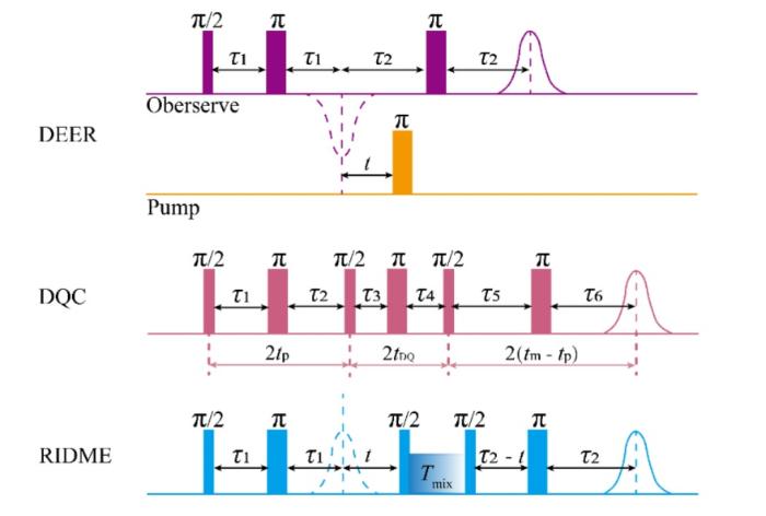
图1
DEER、DQC和RIDME实验的脉冲序列
Fig. 1
Pulse sequences used in DEER, DQC, and RIDME experiments
1.1 DEER
在典型的四脉冲DEER实验中,所记录的时域信号可用以下函数描述[8]:
该式中
DEER实验的精妙之处在于它巧妙地运用了两个不同频率的微波脉冲序列来分别独立地操控一对自旋(图2).其中一个频率,被称为观察频率,它施加一套通常包含探测脉冲和再聚焦脉冲的回波序列,专门用于激发和探测其中一个自旋(称为自旋A)的相干演化,并最终形成一个自旋回波信号作为检测的基准.在自旋A的演化过程中的某一个可变的时间点,DEER会在另一个不同的频率(称为泵浦频率)上施加一个额外的微波脉冲,这个脉冲有选择性地作用在另一个自旋(自旋B)上,使其发生180˚的翻转.这个受控的翻转动作瞬间改变了自旋B所产生的局部磁场的空间取向,从而直接调制了自旋A所感受到的总有效磁场,导致其拉莫进动频率发生一个微小的、依赖于两者相对取向和距离的偏移.这个频率偏移的累积效应最终会体现在自旋A回波信号的幅度上,使其随着泵浦脉冲施加时间点的变化而产生周期性的调制.通过系统地改变这个时间点并记录一系列回波幅度,DEER能得到一条衰减并振荡的时间轨迹.实验中获得的DEER时间轨迹如图2(d)所示,其信号由缓慢衰减的背景项和叠加其上的偶极调制项组成.背景信号主要来源于未被选择性激发的远程自旋或样品中随机分布的顺磁中心,而振荡调制部分则直接反映了自旋对之间的偶极耦合强度.通过对背景信号进行校正后,可以得到清晰的偶极调制轨迹,其调制深度λ反映了实际参与偶极耦合并被有效泵浦的自旋比例.对背景校正后的时间轨迹进行进一步的数据分析,可通过Tikhonov正则化等方法反演得到自旋之间的距离分布函数.DEER技术能够在约1.6~8 nm的距离范围内提供高精度的系综距离分布信息[24].
图2
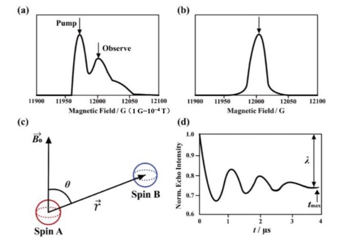
图2
偶极耦合脉冲EPR实验示意图. (a) DEER实验中泵浦脉冲与观测脉冲的位置关系示意图;(b) DQC和RIDME实验中的脉冲位置示意图;(c)实验中一对耦合电子自旋的空间构型示意,其中θ为自旋间距矢量
Fig. 2
Schematic illustration of pulsed dipolar EPR experiments. (a) Relative positions of pump and observer pulses in a DEER experiment; (b) Pulse position in DQC and RIDME experiments; (c) Geometry representation of a coupled electron spin pair, where θ denotes the angle between the inter-spin vector
DEER技术的主要优势在于方法成熟、结果可靠性高,并且拥有较为完善的商业化数据处理软件支持.然而,其实验实施对仪器硬件条件要求较高,通常需要配置两套独立的微波源及相应的放大器,增加了实验成本和操作复杂性.此外,为实现观测通道与泵浦通道的有效区分,两个自旋标记位点的顺磁共振谱线需具有足够的频率分离,这在某些标记位点选择或特定顺磁中心体系中可能较难满足.
1.2 DQC
DQC技术通过探测双量子相干信号来表征自旋对之间的偶极相互作用[25-
与DEER不同,标准的DQC实验可以在单一的微波频率下完成[21],这在一定程度上降低了对仪器频带宽度的要求.其核心在于一系列精心设计的微波脉冲序列,将自旋系统从热平衡混合态逐步驱动到高度关联的双量子相干态.在该状态下,两个自旋不再作为相互独立的个体演化,而是形成由自旋间相互作用主导的关联态,其联合量子态无法用单个自旋的状态来描述.双量子相干态在随后的演化时间内对自旋间偶极耦合强度高度敏感,同时能够有效抑制其他常见干扰因素,例如电子自旋与周围环境中原子核的超精细耦合,以及来自于未耦合的孤立顺磁中心的背景信号.演化结束后,另一组相位反转的脉冲序列将该不可直接观测的双量子相干态转换为可检测的单量子相干信号,即自旋回波.回波幅度包络中编码了关键的偶极相互作用信息.
通过改变序列中用于双量子相干演化的时间间隔,并记录相应的回波幅度变化,可以获得DQC时间轨迹,其振荡特征直接反映了自旋间距离分布.DQC技术最显著的优势在于其较高的理论灵敏度,在相同样品浓度下通常能获得优于DEER的信噪比,或实现对更低浓度样品的测量,这对于样品制备困难或表达量较低的蛋白质体系尤为重要.此外,由于其单频实验特性,DQC能够适用于两个自旋谱线严重重叠、DEER技术难以有效区分的情况.然而,这些优势以较高的实验复杂性为代价.DQC的脉冲序列通常包含六个或更多的微波脉冲,对各脉冲的时长、功率、相位以及脉冲间时间间隔均提出更为严格的要求,因此对谱仪的脉冲性能和稳定性构成了较大挑战.
1.3 RIDME
RIDME技术的核心在于利用自旋探针纵向弛豫过程中产生的随机自旋翻转效应[28]来探测电子自旋之间的偶极相互作用.其研究目标与DEER相同,均是通过偶极调制获取自旋间距离信息.但二者在实现自旋翻转的物理机制上存在本质差异.RIDME实验通常在单一微波频率下进行,其脉冲序列在形式上与四脉冲DEER实验相似,包含用于产生初始回波的探测脉冲序列.然而,与DEER不同的是,RIDME实验不包含第二个频率下主动施加的泵浦脉冲.
在RIDME实验中,关键过程发生在被称为弛豫混合时间(Tmix)的等待区间内.在该区间中不施加任何微波脉冲,自旋系统处于自由演化状态.此时,电子自旋固有的纵向弛豫过程发挥主导作用,该过程表现为自旋在能级之间的随机自发迁移,自旋从高能级弛豫回到低能级,或通过吸收晶格的热能进行反向跃迁.在Tmix区间内,一对耦合自旋中的自旋B有一定的概率因弛豫而发生状态翻转.尽管这种翻转是非受控的随机事件,但其效应与DEER实验中精确定时的泵浦脉冲类似,均会改变作用在自旋A上的局部磁场,从而在最终形成的自旋回波信号中引入依赖于自旋间距离的幅度调制.通过扫描Tmix的时长,即可获得RIDME的时间轨迹.
RIDME技术的显著优势在于实验实现相对简便.该方法无需双频微波通道,可在常规脉冲EPR谱仪上完成,显著降低了硬件要求和实验成本.更重要的是,RIDME完全规避了DEER技术对两个自旋频率分离度的要求,因此在两个标记位点谱线高度重叠的情况下,仍可实现有效的距离测量.此外,由于在较长的Tmix内,多个自旋都有可能发生弛豫翻转,有时会导致实验观测到的调制深度高于DEER.然而,RIDME技术也存在一定局限性.由于在Tmix区间内未施加微波激发,电子自旋不仅会相互耦合,还会与周围大量核自旋产生超精细相互作用,从而引入ESEEM信号.这些核调制信号会与电子-电子偶极调制信号严重叠加,使原始数据变得复杂,难以直接分离.为提高距离分布提取的可靠性,通常需要借助复杂的数据分析模型或在不同磁场条件下进行多组实验加以区分.因此,在定量测量的准确性和可靠性上,RIDME通常被认为略逊于DEER技术.
1.4 三种PD-EPR技术的比较
其中,
表1 自旋间距离r与最大偶极演化时间tmax的对应关系
Table 1
| r/nm | tmax/μs |
|---|---|
| 2 | 0.15 |
| 3 | 0.52 |
| 4 | 1.23 |
| 5 | 2.39 |
| 6 | 4.14 |
| 7 | 6.57 |
| 8 | 9.81 |
为便于对不同PD-EPR技术的技术特点和适用场景进行直观比较,表2总结了DEER、DQC和RIDME三种常用方法在仪器、样品要求、主要优缺点以及适用自旋探针类型等方面的主要差异,可根据目标体系的具体特征和实验条件选择合适的方法.
表2 三种PD-EPR技术的比较
Table 2
| 技术方法 | 微波频率要求 | 样品要求 | 主要优势 | 主要局限 | 适用自旋探针类型 |
|---|---|---|---|---|---|
| DEER | 双频微波 | 避免浓度过高导致背景增强 | 方法成熟,应用最广;软件支持完善 | 仪器要求高;要求自旋谱线具有足够频率分离 | 氮氧自由基、Mn2+、Gd3+等 |
| DQC | 单频微波 | 要求自旋具有较长相干时间 | 理论灵敏度高;适合低浓度样品;可用于谱线重叠体系 | 实验脉冲序列复杂;对样品相干时间敏感 | 氮氧自由基、三苯甲基自由基为主 |
| RIDME | 单频微波 | T1不宜过短 | 对宽谱、快速弛豫的体系具有显著优势 | 易受核调制干扰 | 顺磁性金属离子、三苯甲基自由基 |
2 数据处理与分析工具
PD-EPR实验获取的原始时间域信号需要经过系统的数据处理,才能提取可靠的自旋间距离分布信息.在实际分析过程中,背景信号校正、距离分布反演以及多构象态解析是数据处理中需要重点关注的几个问题.背景信号通常来源于未被选择性激发的远程顺磁中心或样品中随机分布的自旋,其形式和衰减特征会直接影响后续偶极调制信号的提取,因此合理选择背景模型和拟合区间是获得可靠距离分布的前提.
距离分布的反演通常采用正则化方法,其中以Tikhonov正则化应用最为广泛.该方法通过在拟合精度与分布平滑性之间取得平衡,从实验时间轨迹中反演得到物理意义合理的距离分布函数.然而,由于该反演过程本身具有一定的非唯一性,不同的正则化参数选择可能导致距离分布在宽度或峰形上的差异,尤其在存在多组分或构象高度重叠的体系中,这一问题更为突出.因此,在实际应用中通常需要结合误差分析、参数扫描或多模型比较等方式,对结果的稳健性进行评估.
目前,针对PD-EPR数据分析,已发展出多种成熟的软件工具.其中,DeerAnalysis[39]是应用最为广泛的分析平台之一,支持DEER及部分RIDME数据的背景校正、距离分布反演和误差评估,适用于大多数常规距离测量体系.近年来,基于机器学习的方法也被引入PD-EPR数据分析中,DEERNet[40]通过神经网络模型直接从时间域信号预测距离分布,在低信噪比或复杂体系中展现出较强的鲁棒性.DeerLab[41]则能够提供灵活的建模与拟合框架,支持多种背景模型、正则化策略以及贝叶斯分析方法,适用于多组分或构象异质性显著的体系分析.总体而言,数据处理方法的选择和参数设置在很大程度上影响PD-EPR距离分布结果的可信度.结合高质量实验数据、合理的物理模型以及利用多种分析手段进行交叉验证,是获得可靠结构信息的重要保障.
3 自旋探针
图3
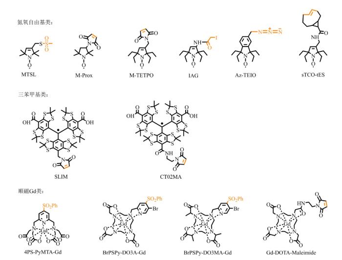
图3
PD-EPR实验中部分常用自旋探针的化学结构,其中与蛋白质反应基团以橙色标注
Fig. 3
Chemical structures of representative spin labels commonly used in PD-EPR experiments, in which the reactive moiety for protein modification was highlighted in yellow
3.1 氮氧自由基(NO)类探针
氮氧自由基作为PD-EPR研究中应用最为广泛的自旋探针,其核心结构中的NO自由基提供了稳定的顺磁中心.自由基两侧的季碳原子为其构象提供了良好的空间稳定性[42],未成对电子相对局域化,主要分布在N-O键上,提高了EPR测量的准确性.这类探针通常具有适中的电子自旋横向弛豫时间以及相对较窄的谱线宽度,因此在距离测量中表现出良好的综合性能.然而,NO自由基探针也存在一定局限性,其对还原性环境较为敏感,NO自由基容易被还原为羟胺[43].探针骨架中含氮杂环的结构特征会显著影响其还原稳定性.一般而言,六元哌啶环的稳定性低于五元环,不饱和吡咯环的稳定性低于饱和吡咯环[44,45].此外,通过用乙基取代NO自由基周围的甲基,可以显著提高NO自由基的抗还原能力,该策略已在细胞内实验中得到验证[45,46].
3.2 三苯甲基(trityl)类探针
三苯甲基类探针因其优异的化学稳定性,在特定研究场景中展现出独特优势.该类探针最显著的特征是具有较长的电子自旋横向弛豫时间[47],这一性质使其特别适用于RIDME等依赖较长弛豫混合时间的PD-EPR实验[38,48].同时,trityl自由基通常表现出极窄的EPR谱线宽度,有利于显著提升检测信噪比.此外,trityl类探针对还原性环境具有较强的耐受性,使其能够在活细胞等复杂生物体系中保持稳定[45]. 然而,该类探针分子体积相对较大,可能对生物分子的天然构象和动力学行为产生干扰,且其合成路线与标记策略相对复杂,这些因素在一定程度上限制了trityl类探针在蛋白质自旋标记中的应用.目前,这类探针主要用于长距离测量,或在活细胞原位EPR研究中发挥重要作用.
3.3 顺磁性金属离子探针
顺磁性金属离子探针为金属蛋白体系及特定实验条件下的PD-EPR研究提供了独特的研究手段.这类探针既包括天然存在的金属中心(如Cu²+、Fe³+等[49
4 蛋白质自旋标记方法
自旋标记技术是PD-EPR研究蛋白质动态结构的核心环节,其核心目标在于将顺磁探针特异且高效地引入目标分子的特定位点,从而利用电子自旋间的磁偶极相互作用获取纳米尺度的结构信息.理想的标记方法通常需要满足以下三个基本要求:(1)位点特异性,以避免非特异性标记干扰;(2)结构兼容性,尽可能减小对蛋白质天然构象和动力学行为的扰动;(3)较高的标记效率,以确保足够的信号强度.随着化学生物学与蛋白质工程技术的不断发展,自旋标记策略已由传统的半胱氨酸共价修饰方法,逐步拓展至基因编码的非天然氨基酸插入、金属结合肽标签等多种新型手段(图4).这些方法的出现不仅显著提升了自旋标记的灵活性和适用范围,也使研究者能够根据不同生物体系的特点设计PD-EPR实验方案.
图4
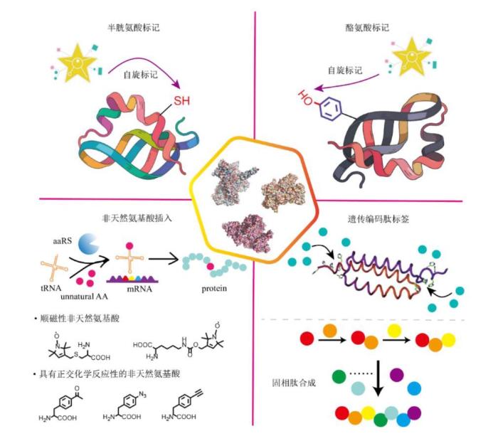
图4
蛋白质自旋标记的主要方法
Fig. 4
Schematic illustration of general strategies for site-specific modification of proteins with spin labels
4.1 半胱氨酸标记
半胱氨酸残基的巯基(-SH)因其高反应活性成为最常用的自旋标记位点.自旋探针通过携带的活性反应基团与蛋白质表面的游离巯基共价结合,这一方法具有标记效率高、反应条件温和的特点.为提高标记特异性,常采用分子生物学手段在目标蛋白中引入单一半胱氨酸突变,硫醇反应性探针如溴代乙酰胺和马来酰亚胺衍生物进一步拓展了反应选择性和稳定性,其中某些衍生物在生理pH条件下仍能保持良好反应活性,为活细胞内的原位标记提供了可能.值得注意的是,标记位点的选择需要综合考虑溶剂可及性、结构扰动等因素,通常需要借助分子建模或晶体结构数据进行理性设计,避免因空间位阻导致标记失败.
4.2 酪氨酸标记
4.3 非天然氨基酸插入
对于体系中存在多个半胱氨酸残基的蛋白质,可能需要进行数轮突变才能实现位点特异性的自旋标记.这既繁琐耗时,又增加了影响天然结构的风险.因此,开发高选择性的替代标记方法能够极大程度上扩展EPR的研究范围.基因密码子扩展技术的发展为蛋白质的定点自旋标记提供了全新途径.通过琥珀终止密码子和正交氨酰-tRNA合成酶/tRNA对,可以将精心设计的非天然氨基酸定点地插入蛋白质序列中[63,64]. 除了可以直接引入含有顺磁侧链的非天然氨基酸[64,65],还可以引入炔基、叠氮基等特殊官能团的非天然氨基酸[66-
4.4 其他标记方法
除上述方法外,一些具有代表性的特殊自旋标记策略同样值得关注,这些方法进一步拓展了EPR技术的应用范围.其中,遗传编码肽标签技术通过将特定金属结合序列基因编码至目标蛋白[68-
5 生物大分子动态结构研究中的应用
PD-EPR技术已经发展成为蛋白质动态结构研究领域的重要工具,其独特的时间与空间分辨率组合为理解蛋白质的构象动态与功能机制提供了全新的视角.为更直观地展示如何利用PD-EPR技术解析生物大分子的动态结构变化,图5给出了基于距离分布分析的动态结构研究示意图.该图以同一生物大分子在不同状态下的构象变化为例,展示了通过PD-EPR技术获取动态结构信息的基本思路.如图5所示,在不同状态下,生物大分子中自旋标记位点之间的距离分布特征会发生改变.PD-EPR实验能够测量这些状态下的自旋间距离分布函数.通过对比不同状态对应的距离分布,不仅可以识别构象转变所伴随的距离变化趋势,还能够揭示多种构象态的共存及其相对比例变化.图5中较窄的峰型对应着较为稳定和单一的构象状态,而较宽的峰型则反映了较高的动态性和多种构象共存的状态.与仅提供静态结构信息的方法相比,PD-EPR技术能够直接反映构象分布及其随条件变化的响应特征,为理解生物大分子功能相关的动态过程提供重要的结构约束.
图5
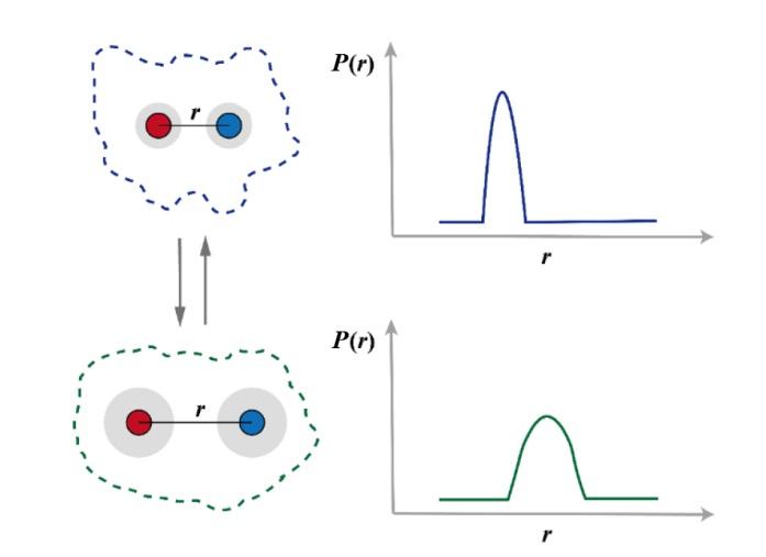
图5
基于PD-EPR的生物大分子动态结构研究示意图.展示标记到同一生物大分子情况下两个自旋标记位点之间的距离分布,其中灰色阴影表示两个自旋在不同条件下的分布变化.PD-EPR技术可以测量自旋间距离分布函数P(r),通过比较距离分布变化,研究生物大分子的结构变化
Fig. 5
Scheme of structural variations determined by PD-EPR measurements. This distance distribution measured by PD-EPR between two spin labels attached to a biomacromolecule, and the gray shading area represents the range of spin label relocations under different conditions. The PD-EPR technique measures the spin distance distribution function P(r). By comparing the changes in the distance distribution, it allows the study of conformational changes in biomolecules
近年来,随着高场谱仪技术的进步和新型脉冲序列的开发,该方法的应用广度和深度都得到了显著拓展[75,76].在技术方法学层面,现代PD-EPR不仅能够精确测定1.6~8 nm范围内的距离分布,更能通过多位点网络分析重构蛋白质的三维构象系综,这一能力在解析具有显著构象异质性的体系时展现出独特优势[75,77]. 特别值得关注的是,该技术在膜蛋白研究中的突破性应用,因为其能在近天然环境中解析传统方法难以捕捉的动态过程.通过开发针对完整大肠杆菌中外膜蛋白的原位标记与DEER测量策略,研究者实现了对天然膜中蛋白质构象动力学、寡聚化及配体-底物相互作用的高精度观测[78].更进一步,首次在完整细胞内追踪膜蛋白配体结合诱导的构象变化,避免了纯化过程可能引入的扰动[79].此外,通过脂质纳米盘技术和两亲性探针的巧妙设计,研究人员首次实现了对ABC转运蛋白在近天然膜环境中的构象循环进行实时监测,这些发现极大地深化了我们对跨膜转运分子机制的理解[80,81].
除了在蛋白质动态结构研究中的广泛应用外,PD-EPR技术在核酸、蛋白-核酸复合体等生物大分子中的研究也取得了重要进展.这些体系往往具有高度柔性、构象异质性强、难以结晶等特征,因此对结构技术提出了更高要求.PD-EPR凭借其纳米尺度的距离测量能力和对动态分布敏感的特点,为解析这些体系的构象和功能关系提供了关键手段.在RNA双链体系中,通过使用2,2,6,6-四甲基哌啶-1-氧自由基(2,2,6,6-Tetramethylpiperidin-1-oxyl,TEMPO)修饰的核苷构建一个RNA双螺旋并结合Q波段脉冲EPR,成功实现了间距达约28个碱基、长度约8 nm的距离分布测量,为双螺旋RNA的全局构象表征提供了关键参数.进一步结合分子动力学模拟,可将实验获得的距离分布直接对应到核酸骨架的空间间距,使得在无需严格刚性假设的条件下实现RNA长程构象的重建,为研究大型、柔性的RNA元件提供了有力工具[85].另一项研究利用能插入空位碱基对的鸟嘌呤衍生的硝基类标签,使其在RNA或DNA双链内稳定配对.这种标签对双螺旋结构干扰极小,并能提供与理论模拟高度一致的距离分布信息[86].借助非天然碱基对体系实现的长链RNA位点特异性标记策略,突破了传统RNA标记方法在分子长度与标记位置上的固有限制,并兼具高反应效率、简便纯化流程及无需变性条件等优势.这一技术的发展使得分子标尺技术能够应用于大尺寸RNA的结构与构象动态研究[87].在复杂RNA或RNA-蛋白复合体体系中,PD-EPR同样展现出强大的结构解析能力.通过在多个核苷位置引入自旋标签、测量多重距离约束,并结合计算建模,可以对大型RNA结构模块乃至RNA-蛋白复合体的整体折叠模型进行重建.这一EPR辅助结构解析方法在解决高度柔性的非编码RNA等难以被晶体学或传统NMR捕获的体系中尤为重要[88].对于DNA体系,PD-EPR也被用于测定不同序列、不同构型的DNA双链中的长程距离,从而区分弯曲、扭转及局部构象状态.相关研究表明,即使在含有多条结构异质的DNA双链混合样品中,也可以通过适当的反卷积方法从偶极信号中分离出多重距离分布.这使得EPR能够用于研究DNA药物结合过程等问题[89].
6 总结与展望
当前,PD-EPR技术已发展成为解析生物大分子动态结构的重要手段,但在实际应用中仍面临一些挑战.受电子自旋横向弛豫时间限制,目前多数PD-EPR实验仍需在低温条件下进行.样品冻结有助于提高信噪比和测量精度,但也可能在一定程度上影响分子在溶液态下的真实构象分布[90].此外,自旋标记的构象自由度及其对目标分子天然结构的潜在扰动,以及距离分布反演过程中存在的非唯一性问题,均对实验设计和数据解析提出了更高要求.
在此基础上,如何突破上述限制、拓展PD-EPR技术的适用范围,是该领域当前的重要研究方向之一.实现生理相关条件下,尤其是室温条件下的高灵敏度距离测量,仍是亟待解决的技术瓶颈.近年来,刚性自旋标记[91]、样品固定化策略以及高场高频谱仪的发展,为延长自旋相干时间和提升灵敏度提供了新的可能.随着研究对象向活细胞原位体系和高度复杂生物体系拓展,对实验灵敏度、重复性以及数据分析自动化程度的要求也在不断提高.此外,在数据解析过程中引入贝叶斯分析、机器学习等新方法,有望在多构象态共存和低信噪比条件下提升距离分布解析的可靠性[75].近期开发的DEERFold方法能够将实验获得的距离分布直接整合到神经网络架构中,仅通过稀疏的实验距离约束实现蛋白质构象集合的高效预测[80].
利益冲突
无
参考文献
Glimpse of structural biology through X-ray crystallography
[J].DOI:10.1016/j.cell.2014.10.051 URL [本文引用: 1]
How cryo-EM is revolutionizing structural biology
[J].DOI:10.1016/j.tibs.2014.10.005 URL [本文引用: 1]
Unravelling biological macromolecules with cryo-electron microscopy
[J].DOI:10.1038/nature19948 [本文引用: 1]
NMR characterization of the dynamics of biomacromolecules
[J].
Advancing time-resolved structural biology: latest strategies in cryo-EM and X-ray crystallography
[J].DOI:10.1038/s41592-025-02659-6 [本文引用: 1]
CRINEPT-TROSY NMR reveals p53 core domain bound in an unfolded form to the chaperone Hsp90
[J].The molecular chaperone Hsp90 sequesters oncogenic mutants of the tumor suppressor p53 that have unstable core domains. It is not known whether p53 is bound in an unfolded, partly folded, or distorted structure, as is unknown for the structure of any bound substrate of Hsp90. It is a particularly difficult problem to analyze in detail the structures of large complexes in which one component is (partly) unfolded. We have shown by transverse relaxation-optimized NMR spectroscopy combined with cross-correlated relaxation-enhanced polarization transfer (CRINEPT-TROSY) that p53 core domain bound in an approximately 200-kDa complex with Hsp90 was predominantly unfolded lacking helical or sheet secondary structure. This mode of binding might be a general feature of substrates of Hsp90.
Long-range distance determinations in biomacromolecules by EPR spectroscopy
[J].Electron paramagnetic resonance (EPR) spectroscopy provides a variety of tools to study structures and structural changes of large biomolecules or complexes thereof. In order to unravel secondary structure elements, domain arrangements or complex formation, continuous wave and pulsed EPR methods capable of measuring the magnetic dipole coupling between two unpaired electrons can be used to obtain long-range distance constraints on the nanometer scale. Such methods yield reliably and precisely distances of up to 80 A, can be applied to biomolecules in aqueous buffer solutions or membranes, and are not size limited. They can be applied either at cryogenic or physiological temperatures and down to amounts of a few nanomoles. Spin centers may be metal ions, metal clusters, cofactor radicals, amino acid radicals, or spin labels. In this review, we discuss the advantages and limitations of the different EPR spectroscopic methods, briefly describe their theoretical background, and summarize important biological applications. The main focus of this article will be on pulsed EPR methods like pulsed electron-electron double resonance (PELDOR) and their applications to spin-labeled biosystems.
Benchmark test and guidelines for DEER/PELDOR experiments on nitroxide-labeled biomolecules
[J].
DOI:10.1021/jacs.1c07371
PMID:34664948
[本文引用: 2]

Distance distribution information obtained by pulsed dipolar EPR spectroscopy provides an important contribution to many studies in structural biology. Increasingly, such information is used in integrative structural modeling, where it delivers unique restraints on the width of conformational ensembles. In order to ensure reliability of the structural models and of biological conclusions, we herein define quality standards for sample preparation and characterization, for measurements of distributed dipole-dipole couplings between paramagnetic labels, for conversion of the primary time-domain data into distance distributions, for interpreting these distributions, and for reporting results. These guidelines are substantiated by a multi-laboratory benchmark study and by analysis of data sets with known distance distribution ground truth. The study and the guidelines focus on proteins labeled with nitroxides and on double electron-electron resonance (DEER aka PELDOR) measurements and provide suggestions on how to proceed analogously in other cases.
DEER distance measurements on proteins
[J].
DOI:10.1146/annurev-physchem-032511-143716
PMID:22404592
Distance distributions between paramagnetic centers in the range of 1.8 to 6 nm in membrane proteins and up to 10 nm in deuterated soluble proteins can be measured by the DEER technique. The number of paramagnetic centers and their relative orientation can be characterized. DEER does not require crystallization and is not limited with respect to the size of the protein or protein complex. Diamagnetic proteins are accessible by site-directed spin labeling. To characterize structure or structural changes, experimental protocols were optimized and techniques for artifact suppression were introduced. Data analysis programs were developed, and it was realized that interpretation of the distance distributions must take into account the conformational distribution of spin labels. First methods have appeared for deriving structural models from a small number of distance constraints. The present scope and limitations of the technique are illustrated.
From in vitro towards in situ: structure-based investigation of ABC exporters by electron paramagnetic resonance spectroscopy
[J].DOI:10.1002/feb2.v594.23 URL [本文引用: 1]
Role of membrane mimetics on biophysical EPR studies of membrane proteins
[J].DOI:10.1016/j.bbamem.2023.184138 URL [本文引用: 1]
Film-electrochemical EPR spectroscopy to investigate electron transfer in membrane proteins in their native environment
[J].
Stationary phase EPR spectroscopy for monitoring membrane protein refolding by conformational response
[J].
DOI:10.1021/acs.analchem.8b04542
PMID:30474972
[本文引用: 1]
Protein production remains a major bottleneck in membrane protein structural biology. In many cases, large-scale recombinant protein expression is either unfeasible or impossible, driving structural biologists to explore new production avenues. Several membrane proteins have been successfully refolded from solubilized E. coli inclusion bodies. In recent years, a structure of the G-protein-coupled receptor CXCR1 was obtained using refolded material from E. coli inclusion bodies. However, aggregation during the refolding process is a common difficulty, which is often addressed by immobilization of the protein onto a solid support. Most spectroscopic methods are incompatible with these light-scattering matrices, which renders automated buffer exchange to screen refolding conditions impossible. This work explores a potential approach to overcome this problem by utilizing site-directed spin labeling and electron paramagnetic resonance (EPR) spectroscopy of protein bound to standard, commercially available Ni-NTA agarose resin. With this approach, the correct protein fold is determined by activity, which is inferred from a protein conformational response to a known stimulant. EPR spectra at each state of the refolding workflow of spin-labeled Haloarcula marismortui bacteriorhodopsin-I (HmbRI) are obtained, and refolded fractions of HmbRI with this platform are quantitated using both protein from inclusion bodies and denatured recombinant protein from E. coli membranes. The stimulant used for HmbRI is visible light. The solid support allows for multiple refolding trials through buffer exchanges, and the EPR spectra are collected on the order of seconds under ambient conditions.
Accurate extraction of nanometer distances in multimers by pulse EPR
[J].
DOI:10.1002/chem.201505143
PMID:26865468
[本文引用: 1]
Pulse electron paramagnetic resonance (EPR) is gaining increasing importance in structural biology. The PELDOR (pulsed electron-electron double resonance) method allows extracting distance information on the nanometer scale. Here, we demonstrate the efficient extraction of distances from multimeric systems such as membrane-embedded ion channels where data analysis is commonly hindered by multi-spin effects. © 2016 The Authors. Published by Wiley-VCH Verlag GmbH & Co. KGaA.
Pulsed dipolar EPR spectroscopy and metal ions: Methodology and biological applications
[J].
DOI:10.1002/cplu.201900705
PMID:31950648
[本文引用: 2]
Pulsed dipolar electron paramagnetic resonance spectroscopy (PDS) is a valuable method for determining biomolecular structures and their conformational changes during function. Often, this method is applied to paramagnetic metal ions that are either intrinsic co-factors of biomolecules or introduced into biomolecules as spin labels. Here, the key achievements and the remaining challenges of PDS on metal ions are summarized and critically discussed. The first part of the Review describes the methodology of PDS experiments and the related data analysis for each of the biologically relevant paramagnetic metal centers. The second part highlights applications with the aim to give an idea about what kind of questions from structural biology can be answered by PDS-based distance measurements involving metal centers.© 2020 The Authors. Published by Wiley-VCH Verlag GmbH & Co. KGaA.
Electron spin resonance of dipole-coupled anisotropic pairs in disordered systems
[J].DOI:10.1006/jmra.1995.1193 URL [本文引用: 1]
Site-directed spin labeling EPR spectroscopy in protein research
[J].
DOI:10.1515/hsz-2013-0155
PMID:23912220
[本文引用: 1]
Site-directed spin labeling (SDSL) in combination with electron paramagnetic resonance (EPR) spectroscopy has emerged as an efficient tool to elucidate the structure and the conformational dynamics of proteins under conditions close to the native state. This review article summarizes the basics as well as the recent progress in SDSL and EPR methods, especially for investigations on protein structure, protein function, and interaction of proteins with other proteins or nucleic acids. Labeling techniques as well as EPR methods are introduced and exemplified with applications to systems that have been studied in the author's laboratory in the past 15 years, headmost the sensory rhodopsin-transducer complex mediating the photophobic response of the halophilic archaeum Natronomonas pharaonis. Further examples underline the application of SDSL EPR spectroscopy to answer specific questions about the system under investigation, such as the nature and influence of interactions of proteins with other proteins or nucleic acids. Finally, it is discussed how SDSL EPR can be combined with other biophysical techniques to combine the strengths of the different methodologies.
EPR of site-directed spin-labeled proteins: A powerful tool to study structural flexibility
[J].DOI:10.1016/j.abb.2020.108323 URL [本文引用: 1]
Pulsed electron double resonance (PELDOR) and its applications in free-radicals research
[J].DOI:10.1007/BF03161886 URL [本文引用: 2]
A straightforward approach to the analysis of double electron-electron resonance data
[J].
DOI:10.1016/bs.mie.2015.07.031
PMID:26478498
[本文引用: 1]
Double electron-electron resonance (DEER) is now widely utilized to measure distance distributions in the 20-70Å range. DEER is frequently applied to biological systems that have multiple conformational states leading to complex distance distributions. These complex distributions raise issues regarding the best approach to analyze DEER data. A widely used method utilizes a priori background correction followed by Tikhonov regularization. Unfortunately, the underlying assumptions of this approach can impact the analysis. In this chapter, a method of analyzing DEER data is presented that is ideally suited to obtain these complex distance distributions. The approach allows the fitting of raw experimental data without a priori background correction as well as the rigorous determination of uncertainties for all fitting parameters. This same methodological approach can be used for the simultaneous or global analysis of multiple DEER data sets using variable ratios of a common set of components, thus allowing direct correlation of distance components with functionally relevant conformational and biochemical states. Examples are given throughout to highlight this robust fitting approach. © 2015 Elsevier Inc. All rights reserved.
Application of ELDOR in electron-spin echo for paramagnetic center space distribution in solids
[J].
Distance measurements on spin-labelled biomacromolecules by pulsed electron paramagnetic resonance
[J].
DOI:10.1039/b614920k
PMID:17431518
[本文引用: 3]
The biological function of protein, DNA, and RNA molecules often depends on relative movements of domains with dimensions of a few nanometers. This length scale can be accessed by distance measurements between spin labels if pulsed electron paramagnetic resonance (EPR) techniques such as electron-electron double resonance (ELDOR) and double-quantum EPR are used. The approach does not require crystalline samples and is well suited to biomacromolecules with an intrinsic flexibility as distributions of distances can be measured. Furthermore, oligomerization or complexation of biomacromolecules can also be studied, even if it is incomplete. The sensitivity of the technique and the reliability of the measured distance distribution depend on careful optimization of the experimental conditions and procedures for data analysis. Interpretation of spin-to-spin distance distributions in terms of the structure of the biomacromolecules furthermore requires a model for the conformational distribution of the spin labels.
Multiple-quantum ESR and distance measurements
[J].DOI:10.1016/S0009-2614(99)00972-0 URL [本文引用: 2]
Double quantum two-dimensional Fourier transform electron spin resonance: Distance measurements
[J].DOI:10.1016/0009-2614(96)00075-9 URL
Theory of double quantum two-dimensional electron spin resonance with application to distance measurements
[J].
DOI:10.1063/1.474490
URL
[本文引用: 1]
A formulation is presented for calculating double quantum two dimensional electron spin resonance (DQ-2D ESR) spectra in the rigid limit that correspond to recent experimental DQ-2D ESR spectra obtained from a nitroxide biradical. The theory includes the dipolar interaction between the nitroxide moieties as well as the fully asymmetric g and hyperfine tensors and the angular geometry of the biradical. The effects of arbitrary pulses (strong but not truly nonselective pulses) are included by adapting the recently introduced split Hamiltonian theory for numerical simulations. It is shown how arbitrary pulses in magnetic resonance create “forbidden” coherence pathways, and their role in DQ-2D ESR is delineated. The high sensitivity of these DQ-2D ESR signals to the strength of the dipolar interaction is demonstrated and rationalized in terms of the orientational selectivity of the “forbidden” pathways. It is further shown that this selectivity also provides constraints on the structural geometry (i.e., the orientations of the nitroxide moieties) of the biradicals. The theory is applied to the recent double quantum modulation (DQM) experiment on an end-labeled poly-proline peptide biradical. A distance of 18.5 Å between the ends is found for this biradical. A new two pulse double quantum experiment is proposed (by analogy to recent NMR experiments), and its feasibility for the ESR case is theoretically explored.
RIDME spectroscopy: New topics beyond the determination of electron spin-spin distances
[J].DOI:10.1021/acs.jpclett.4c02667 URL [本文引用: 1]
RIDME distance measurements using Gd(III) tags with a narrow central transition
[J].
DOI:10.1039/c6cp03299k
PMID:27355583
[本文引用: 2]
Methods based on pulse electron paramagnetic resonance allow measurement of the electron-electron dipolar coupling between two spin labels. Here we compare the most popular technique, Double Electron-Electron Resonance (DEER or PELDOR), with the dead-time free 5-pulse Relaxation-Induced Dipolar Modulation Enhancement (RIDME) method for Gd(iii)-Gd(iii) distance measurements at W-band (94.9 GHz, ≈3.5 T) using Gd(iii) tags with a small zero field splitting (ZFS). Such tags are important because of their high EPR sensitivity arising from their narrow central transition. Two systems were investigated: (i) a rigid model compound with an inter-spin distance of 2.35 nm, and (ii) two mutants of a homodimeric protein, both labeled with a DOTA-based Gd(iii) chelate and characterized by an inter-spin distance of around 6 nm, one having a narrow distance distribution and the other a broad distribution. Measurements on the model compound show that RIDME is less sensitive to the complications arising from the failure of the weak coupling approximation which affect DEER measurements on systems characterized by short inter-spin distances between Gd(iii) tags having a narrow central transition. Measurements on the protein samples, which are characterized by a long inter-spin distance, emphasize the complications due to the appearance of harmonics of the dipolar interaction frequency in the RIDME traces for S > 1/2 spin systems, as well as enhanced uncertainties in the background subtraction. In both cases the sensitivity of RIDME was found to be significantly better than DEER. The effects of the experimental parameters on the RIDME trace are discussed.
PELDOR and RIDME measurements on a high-spin manganese(II)bisnitroxide model complex
[J].DOI:10.1021/acs.jpca.6b00716 URL [本文引用: 1]
RIDME spectroscopy on high-spin Mn2+ centers
[J].DOI:10.1039/C6CP05239H URL [本文引用: 1]
Improving the accuracy of Cu(II)-nitroxide RIDME in the presence of orientation correlation in water-soluble Cu(II)-nitroxide rulers
[J].
DOI:10.1039/c8cp06573j
PMID:31025988
[本文引用: 1]
Orientation selection is a challenge in distance determination with double electron electron resonance (DEER) spectroscopy of rigid molecules. The problem is reduced when applying the Relaxation-Induced Dipolar Modulation Enhancement (RIDME) experiment. Here we present an in-depth study on nitroxide-detected RIDME in Cu(ii)-nitroxide spin pairs using two Cu(ii)-nitroxide rulers that are both water soluble and have comparable spin-spin distances. They differ in the type of the ligand (TAHA and PyMTA) for the Cu(ii) ion which results in different contributions of exchange coupling. Both rulers feature substantial orientation correlation between the molecular frames of the Cu(ii) complex and the nitroxide. We discuss how the spin-spin couplings can be accurately measured and how they can be correlated to the nitroxide resonance frequencies. In that, we pay particular attention to the suppression of nuclear modulation and of echo crossing artefacts, to background correction, and to orientation averaging. With a nitroxide observer sequence based on chirp pulses, we achieve wideband detection of all nitroxide orientations. Two-dimensional Fourier transformation of data obtained in this manner affords observer-EPR correlated RIDME spectra that enable visual understanding of the orientation correlation. The syntheses of the Cu(ii)-nitroxide rulers are presented. The synthetic route is considered to be of general use for the preparation of [metal ion complex]-nitroxide rulers, including water soluble ones.
Sub-micromolar pulse dipolar EPR spectroscopy reveals increasing CuII-labelling of double-histidine motifs with lower temperature
[J].DOI:10.1002/anie.v58.34 URL [本文引用: 1]
Comparison of PELDOR and RIDME for distance measurements between nitroxides and low-spin Fe(III) ions
[J].DOI:10.1021/acs.jpcb.5b02118 URL [本文引用: 1]
Mapping the structure of metalloproteins with RIDME
[J].
DOI:10.1016/bs.mie.2015.06.031
PMID:26478488
Distance measurements in biological macromolecules represent a very active field of application of pulsed electron paramagnetic resonance (EPR) spectroscopy. The relatively recently introduced pulsed EPR method of relaxation-induced dipolar modulation enhancement (RIDME) is conceptually similar to the popular double electron-electron resonance (DEER), but is much more suitable for studying the structures of metalloproteins while using their native paramagnetic metal centers as structural reference points. In particular, RIDME can largely alleviate the sensitivity and orientational selectivity problems that limit the application of DEER to such systems. In this contribution, the theoretical principles, implementation, optimization, and available experimental examples of RIDME are described with the purpose of enhancing the familiarity with this technique and promoting its application. © 2015 Elsevier Inc. All rights reserved.
Pulse dipolar electron paramagnetic resonance spectroscopy reveals buffer-modulated cooperativity of metal-templated protein dimerization
[J].
DOI:10.1021/acs.jpclett.2c01719
PMID:35976741
[本文引用: 1]
Self-assembly of protein monomers directed by metal ion coordination constitutes a promising strategy for designing supramolecular architectures complicated by the noncovalent interaction between monomers. Herein, two pulse dipolar electron paramagnetic resonance spectroscopy (PDS) techniques, pulse electron-electron double resonance and relaxation-induced dipolar modulation enhancement, were simultaneously employed to study the Cu-templated dimerization behavior of a model protein (sp. group G, protein G B1 domain) in both phosphate and Tris-HCl buffers. A cooperative binding model could simultaneously fit all data and demonstrate that the cooperativity of protein dimerization across α-helical double-histidine motifs in the presence of Cu is strongly modulated by the buffer, representing a platform for highly tunable buffer-switchable templated dimerization. Hence, PDS enriches the family of techniques for monitoring binding processes, supporting the development of novel strategies for bioengineering structures and stable architectures assembled by an initial metal-templated dimerization.
Room-temperature distance measurements using RIDME and the orthogonal spin labels trityl/nitroxide
[J].
DOI:10.1039/c8cp01093e
PMID:29594278
[本文引用: 1]
Electron paramagnetic resonance (EPR) based nanometer distance measurements at ambient temperatures are of particular interest for structural biology applications. The nitroxide spin labels commonly used in EPR reveal relatively short transverse relaxation under these conditions, which limits their use for detecting static dipolar interactions. At the same time, the longitudinal relaxation of nitroxide spin labels is still long enough to allow using them as 'pumped' species in the relaxation induced dipolar modulation enhancement (RIDME) experiment where the detection is carried out on the slower relaxing triarylmethyl (TAM) spin labels. In the present study, we report the first demonstration of room-temperature RIDME distance measurements in nucleic acids using TAM as the slow-relaxing detected species and traditional nitroxide as the fast-relaxing partner spin. Two types of immobilizers, glassy trehalose and the modified silica gel Nucleosil, were used for immobilization of the spin-labeled biomolecules. The room-temperature RIDME-based distance distributions are in good agreement with those measured at 80 K by other techniques. Room-temperature RIDME on the spin pairs trityl/nitroxide may become a useful method for the structural characterization of biomacromolecules and biomolecular complexes at near physiological temperatures.
Electron dipole-dipole interaction in ESEEM of nitroxide biradicals
[J].DOI:10.1016/S0009-2614(01)00721-7 URL [本文引用: 2]
DeerAnalysis2006—a comprehensive software package for analyzing pulsed ELDOR data
[J].DOI:10.1007/BF03166213 URL [本文引用: 1]
Neural network interpretation using descrambler groups
[J].
DOI:10.1073/pnas.2016917118
URL
[本文引用: 1]
Artificial neural networks are famously opaque—it is often unclear how they work. In this communication, we propose a group-theoretical way of finding out. It reveals considerable internal sophistication, even in simple neural networks: our nets apparently invented an elegant digital filter, a regularized integral transform, and even Chebyshev polynomials. This is a step toward saving reductionism. For centuries, the philosophical approach to science has been to find fundamental laws that govern reality, to test those laws, and to use their predictive power. Black-box neural networks amount to blasphemy within that school, but they are irresistible because they “just work.” Explaining how they work is a notoriously difficult problem, to which this paper offers a partial solution.
DeerLab: a comprehensive software package for analyzing dipolar electron paramagnetic resonance spectroscopy data
[J].
DOI:10.5194/mr-1-209-2020
URL
[本文引用: 1]
. Dipolar electron paramagnetic resonance (EPR) spectroscopy (DEER and other techniques) enables the structural characterization of macromolecular and biological systems by measurement of distance distributions between unpaired electrons on a nanometer scale. The inference of these distributions from the measured signals is challenging due to the ill-posed nature of the inverse problem. Existing analysis tools are scattered over several applications with specialized graphical user interfaces. This renders comparison, reproducibility, and method development difficult. To remedy this situation, we present DeerLab, an open-source software package for analyzing dipolar EPR data that is modular and implements a wide range of methods. We show that DeerLab can perform one-step analysis based on separable non-linear least squares, fit dipolar multi-pathway models to multi-pulse DEER data, run global analysis with non-parametric distributions, and use a bootstrapping approach to fully quantify the uncertainty in the analysis.
Advances in the synthesis of nitroxide radicals for use in biomolecule spin labelling
[J].
DOI:10.1039/c6cs00550k
PMID:29192696
[本文引用: 1]
EPR spectroscopy is an increasingly useful analytical tool to probe biomolecule structure, dynamic behaviour, and interactions. Nitroxide radicals are the most commonly used radical probe in EPR experiments, and many methods have been developed for their synthesis, as well as incorporation into biomolecules using site-directed spin labelling. In this Tutorial Review, we discuss the most practical methods for the synthesis of nitroxides, focusing on the tunability of their structures, the manipulation of their sidechains into spin labelling handles, and their installation into biomolecules.
Kinetics and mechanism of hydroxyl radical and OH-adduct radical reactions with nitroxides and with their hydroxylamines
[J].Stable nitroxide radicals are potent antioxidants and are among the most effective non-thiol radioprotectants, although they react with hydroxyl radicals more slowly than typical phenolic antioxidants or thiols. Surprisingly, the reduced forms of cyclic nitroxides, cyclic hydroxylamines, are better reductants yet have no radioprotective activity. To clarify the reason for this difference, we studied the kinetics and mechanisms of the reactions of nitroxides and their hydroxylamines with (*)OH radicals and with OH-adducts by using pulse radiolysis, fluorimetric determination of phenolic radiation products, and electron paramagnetic resonance spectrometric determination of nitroxide concentrations following radiolysis. Competition kinetics with phenylalanine as a reference compound in pulse radiolysis experiments yielded rate constants of (4.5 +/- 0.4) x 10(9) M(-1) s(-1) for the reaction of (*)OH radical with 2,2,6,6-tetramethylpiperidine-N-oxyl (TPO), 4-hydroxy-TPO (4-OH-TPO), and 4-oxo-TPO (4-O-TPO), (3.0 +/- 0.3) x 10(9) M(-1) s(-1) for deuterated 4-O-TPO, and (1.0 +/- 0.1) x 10(9) M(-1) s(-1) for the hydroxylamine 4-OH-TPO-H. The kinetic isotope effect suggests the occurrence of both (*)OH addition to the aminoxyl moiety of 4-O-TPO and H-atom abstraction from the 2- or 6-methyl groups or from the 3- and 5-methylene positions. This conclusion was further supported by final product analysis, which demonstrated that (*)OH partially oxidizes 4-O-TPO to the corresponding oxoammonium cation. The rate constants for the reactions of the nitroxides with the OH-adducts of phenylalanine and terephthalate have been determined to be near 4 x 10(6) M(-1) s(-1), whereas the hydroxylamine reacted at least 50 times slower, if at all. These findings indicate that the reactivity toward (*)OH does not explain the differences between the radioprotective activities of nitroxides and hydroxylamines. Instead, the radioprotective activity of nitroxides, but not of hydroxylamines, can be partially attributed to their ability to detoxify OH-derived secondary radicals.
Synthesis of 2,5-Bis(spirocyclohexane)-substituted nitroxides of pyrroline and pyrrolidine series, including thiol-specific spin label: An analogue of MTSSL with long relaxation time
[J].The nitroxides of 7-azadispiro[5.1.5.2]pentadecane and 7-azadispiro[5.1.5.2]pentadeca-14-ene series have been prepared, including thiol-specific methane thiosulfonate spin label for site-directed spin labeling. The effect of spirocyclohexane moieties on chemical and spectral properties has been studied. The obtained temperature dependencies of electron spin relaxation parameters demonstrate that new nitroxides may be suitable for PELDOR distance measurements at 80-120 K. Moreover, the new nitroxides demonstrated much higher stability toward reduction by ascorbate than spirocyclohexane-substituted nitroxides of piperidine series and showed 1.3-3.14 times lower reduction rates compared to corresponding 2,2,5,5-tetramethyl nitroxides.
Synthesis and reduction kinetics of sterically shielded pyrrolidine nitroxides
[J].
DOI:10.1021/ol302506f
PMID:23050653
[本文引用: 3]
A series of sterically shielded pyrrolidine nitroxides were synthesized, and their reduction by ascorbate (vitamin C) indicate that nitroxide 3, a tetraethyl derivative of 3-carboxy-PROXYL, is reduced at the slowest rate among known nitroxides, i.e., at a 60-fold slower rate than that for 3-carboxy-PROXYL.
Sterically shielded spin labels for in-cell EPR spectroscopy: Analysis of stability in reducing environment
[J].DOI:10.3109/10715762.2014.979409 URL [本文引用: 1]
Room-temperature electron spin relaxation of triarylmethyl radicals at the X- and Q-bands
[J].DOI:10.1021/acs.jpcb.5b03027 URL [本文引用: 1]
A pulsed EPR method to determine distances between paramagnetic centers with strong spectral anisotropy and radicals: The dead-time free RIDME sequence
[J].
DOI:10.1016/j.jmr.2009.08.008
PMID:19758831
[本文引用: 1]
Methods to determine distances between paramagnetic metal centers and radicals are scarce. This is unfortunate because paramagnetic metal centers are frequent in biological systems and so far have not been employed much as distance markers. Successful pulse sequences that directly target the dipolar interactions cannot be applied to paramagnetic metal centers with fast relaxation rates and large g-anisotropy, if no echos can be detected and the excitation bandwidth is not sufficient to cover a sufficiently large part of the spectrum. The RIDME method Kulik et al. (2002) [20] circumvents this problem by making use of the T(1)-induced spin-flip of the transition-metal ion. Designed to measure distance between such a fast relaxing metal center and a radical, it suffers from a dead time problem. We show that this is severe because the anisotropy of the metal center broadens the dipolar curves, which therefore, only can be analyzed if the full curve is known. Here, we introduce five-pulse RIDME (5p-RIDME) that is intrinsically dead-time free. Proper functioning of the sequence is demonstrated on a nitroxide biradical. The distance between a low-spin Fe(III) center and a spin label in spin-labeled cytochrome f shows the complete dipolar trace of a transition-metal ion center and a spin label, yielding the distance expected from the structure.
Endogenous Cu(II) labeling for distance measurements on proteins by EPR
[J].DOI:10.1002/chem.v30.72 URL [本文引用: 1]
Differentiating between label and protein conformers in pulsed dipolar EPR spectroscopy with the dHis-Cu2+(NTA) motif
[J].
An optimal acquisition scheme for Q-band EPR distance measurements using Cu2+-based protein labels
[J].\n Herein we establish a cost-efficient acquisition scheme for orientational-independent distance measurements using Cu\n 2+\n –protein labels, using insights from Monte-Carlo modeling, MD simulations, and DEER EPR measurements.\n
Pulsed multifrequency electron paramagnetic resonance spectroscopy reveals key branch points for one- vs two-electron reactivity in Mn/Fe proteins
[J].
DOI:10.1021/jacs.1c13738
PMID:35786920
Traditionally, the ferritin-like superfamily of proteins was thought to exclusively use a diiron active site in catalyzing a diverse array of oxygen-dependent reactions. In recent years, novel redox-active cofactors featuring heterobimetallic Mn/Fe active sites have been discovered in both the radical-generating R2 subunit of class Ic (R2c) ribonucleotide reductases (RNRs) and the related R2-like ligand-binding oxidases (R2lox). However, the protein-specific factors that differentiate the radical reactivity of R2c from the C-H activation reactions of R2lox remain unknown. In this work, multifrequency pulsed electron paramagnetic resonance (EPR) spectroscopy and ligand hyperfine techniques in conjunction with broken-symmetry density functional theory calculations are used to characterize the molecular and electronic structures of two EPR-active intermediates trapped during aerobic assembly of the R2lox Mn/Fe cofactor. A Mn(μ-O)(μ-OH)Fe species is identified as the first EPR-active species and represents a common state between the two classes of redox-active Mn/Fe proteins. The species downstream from the Mn(μ-O)(μ-OH)Fe state exhibits unique EPR properties, including unprecedented spectral breadth and isotope-dependent g-tensors, which are attributed to a weakly coupled, hydrogen-bonded Mn(μ-OH)Fe species. This final intermediate precedes formation of the Mn/Fe resting state and is suggested to be relevant to understanding the endogenous reactivity of R2lox.
Cu(II)-based DNA labeling identifies the structural link between transcriptional activation and termination in a metalloregulator
[J].
DOI:10.1039/d1sc06563g
PMID:35282619
[本文引用: 1]
Understanding the structural and mechanistic details of protein-DNA interactions that lead to cellular defence against toxic metal ions in pathogenic bacteria can lead to new ways of combating their virulence. Herein, we examine the Copper Efflux Regulator (CueR) protein, a transcription factor which interacts with DNA to generate proteins that ameliorate excess free Cu(i). We exploit site directed Cu(ii) labeling to measure the conformational changes in DNA as a function of protein and Cu(i) concentration. Unexpectedly, the EPR data indicate that the protein can bend the DNA at high protein concentrations in the Cu(i)-free state. On the other hand, the bent state of the DNA is accessed at a low protein concentration in the presence of Cu(i). Such bending enables the coordination of the DNA with RNA polymerase. Taken together, the results lead to a structural understanding of how transcription is activated in response to Cu(i) stress and how Cu(i)-free CueR can replace Cu(i)-bound CueR in the protein-DNA complex to terminate transcription. This work also highlights the utility of EPR to measure structural data under conditions that are difficult to access in order to shed light on protein function.This journal is © The Royal Society of Chemistry.
High sensitivity in-cell EPR distance measurements on proteins using an optimized Gd(III) spin label
[J].
DOI:10.1021/acs.jpclett.8b02663
PMID:30277780
[本文引用: 1]
Distance measurements by electron-electron double resonance (DEER) carried out on spin-labeled proteins delivered into cells provide new insights into the conformational states of proteins in their native environment. Such measurements depend on spin labels that exhibit high redox stability and high DEER sensitivity. Here we present a new Gd(III)-based spin label, BrPSPy-DO3A-Gd(III), which was derived from an earlier label, BrPSPy-DO3MA-Gd(III), by removing the methyl group from the methyl acetate pending arms. The small chemical modification led to a reduction in the zero-field splitting and to a significant increase in the phase memory time, which together culminated in a remarkable improvement of in-cell DEER sensitivity, while maintaining the high distance resolution. The excellent performance of BrPSPy-DO3A-Gd(III) in in-cell DEER measurements was demonstrated on doubly labeled ubiquitin and GB1 delivered into HeLa cells by electroporation.
Small Gd(III) tags for Gd(III)-Gd(III) distance measurements in proteins by EPR spectroscopy
[J].
DOI:10.1021/acs.inorgchem.8b00133
PMID:29629761
The C7-Gd and C8-Gd tags are compact hydrophilic cyclen-based lanthanide tags for conjugation to cysteine residues in proteins. The tags are enantiomers, which differ in the configuration of the 2-hydroxylpropyl pendant arms coordinating the lanthanide ion. Here, we report the electron paramagnetic resonance (EPR) performance of the C7-Gd (S configuration) and C8-Gd (R configuration) tags loaded with Gd(III) on two mutants of the homodimeric ERp29 protein. The W-band EPR spectra were found to differ between the tags in the free state and after conjugation to the protein. In addition, the spectra were sensitive to the labeling position, which may originate from an environment-dependent charge density on the Gd(III)-coordinating oxygens. This is in agreement with previous NMR experiments with different lanthanide ions, which suggested sensitivity to H-bonding. W-band H-ENDOR (electron-electron double resonance) experiments detected effects from orientation selection in the central transition, due to a relatively narrow distribution in the ZFS parameters as indicated by simulations. In contrast, the distance distributions derived from DEER (double electron-electron resonance) measurements were insensitive to the R or S configuration of the tags and did not exhibit any orientation selection effects. The DEER measurements faithfully reflected the different widths of the distance distributions at the different protein sites in agreement with previous DEER measurements using other Gd(III) tags. Due to their small size, short tether to the protein, and a broad central EPR transition, the C7-Gd and C8-Gd tags are attractive Gd(III) tags for measurements of relatively short (<4 nm) distances by EPR spectroscopy.
In cell Gd3+-based site-directed spin labeling and EPR spectroscopy of eGFP
[J].
Exploring the dynamics and structure of PpiB in living Escherichia coli cells using electron paramagnetic resonance spectroscopy
[J].
New developments in spin labels for pulsed dipolar EPR
[J].
DOI:10.3390/molecules191016998
PMID:25342554
[本文引用: 1]
Spin labelling is a chemical technique that enables the integration of a molecule containing an unpaired electron into another framework for study. Given the need to understand the structure, dynamics, and conformational changes of biomacromolecules, spin labelling provides a relatively non-intrusive technique and has certain advantages over X-ray crystallography; which requires high quality crystals. The technique relies on the design of binding probes that target a functional group, for example, the thiol group of a cysteine residue within a protein. The unpaired electron is typically supplied through a nitroxide radical and sterically shielded to preserve stability. Pulsed electron paramagnetic resonance (EPR) techniques allow small magnetic couplings to be measured (e.g., <50 MHz) providing information on single label probes or the dipolar coupling between multiple labels. In particular, distances between spin labels pairs can be derived which has led to many protein/enzymes and nucleotides being studied. Here, we summarise recent examples of spin labels used for pulse EPR that serve to illustrate the contribution of chemistry to advancing discoveries in this field.
Gd(III)-Gd(III) relaxation-induced dipolar modulation enhancement for in-cell electron paramagnetic resonance distance determination
[J].
DOI:10.1021/acs.jpclett.9b00340
PMID:30864799
[本文引用: 1]
In-cell distance determination by electron paramagnetic resonance (EPR) spectroscopy reveals essential structural information about biomacromolecules under native conditions. We demonstrate that the pulsed EPR technique RIDME (relaxation induced dipolar modulation enhancement) can be utilized for such distance determination. The performance of in-cell RIDME has been assessed at Q-band using stiff molecular rulers labeled with Gd(III)-PyMTA and microinjected into Xenopus laevis oocytes. The overtone coefficients are determined to be the same for protonated aqueous solutions and inside cells. As compared to in-cell DEER (double electron-electron resonance, also abbreviated as PELDOR), in-cell RIDME features approximately 5 times larger modulation depth and does not show artificial broadening in the distance distributions due to the effect of pseudosecular terms.
Enlarging the panoply of site-directed spin labeling electron paramagnetic resonance (SDSL-EPR): Sensitive and selective spin-labeling of tyrosine using an isoindoline-based nitroxide
[J].
DOI:10.1021/bc4000542
PMID:23642211
[本文引用: 1]
Site-directed spin labeling (SDSL) combined with electron paramagnetic resonance (EPR) spectroscopy has emerged as a powerful approach to study structure and dynamics in proteins. One limitation of this approach is the fact that classical spin labels are functionalized to be grafted on natural or site-directed mutagenesis generated cysteine residues. Despite the widespread success of cysteine-based modification strategies, the technique becomes unsuitable when cysteine residues play a functional or structural role in the protein under study. To overcome this limitation, we propose an isoindoline-based nitroxide to selectively target tyrosine residues using a Mannich type reaction, the feasibility of which has been demonstrated in a previous study. This nitroxide has been synthesized and successfully grafted successively on p-cresol, a small tetrapeptide and a model protein: a small chloroplastic protein CP12 having functional cysteines and a single tyrosine. Studying the association of the labeled CP12 with its partner protein, we showed that the isoindoline-based nitroxide is a good reporter to reveal changes in its local environment contrary to the previous study where the label was poorly sensitive to probe structural changes. The successful targeting of tyrosine residues with the isoindoline-based nitroxide thus offers a highly promising approach, complementary to the classical cysteine-SDSL one, which significantly enlarges the field of applications of the technique for probing protein dynamics.
Orthogonal tyrosine and cysteine site-directed spin labeling for dipolar pulse EPR spectroscopy on proteins
[J].
DOI:10.1021/acs.jpclett.7b02220
PMID:28933855
[本文引用: 1]
Site-directed spin labeling of native tyrosine residues in isolated domains of the protein PTBP1, using a Mannich-type reaction, was combined with conventional spin labeling of cysteine residues. Double electron-electron resonance (DEER) EPR measurements were performed for both the nitroxide-nitroxide and Gd(III)-nitroxide label combinations within the same protein molecule. For the prediction of distance distributions from a structure model, rotamer libraries were generated for the two linker forms of the tyrosine-reactive isoindoline-based nitroxide radical Nox. Only moderate differences exist between the spatial spin distributions for the two linker forms of Nox. This strongly simplifies DEER data analysis, in particular, if only mean distances need to be predicted.
Tyrosine-targeted spin labeling and EPR spectroscopy: An alternative strategy for studying structural transitions in proteins
[J].DOI:10.1002/anie.201102539 PMID:21919142 [本文引用: 1]
A general method for site-specific incorporation of unnatural amino acids into proteins
[J].A new method has been developed that makes it possible to site-specifically incorporate unnatural amino acids into proteins. Synthetic amino acids were incorporated into the enzyme beta-lactamase by the use of a chemically acylated suppressor transfer RNA that inserted the amino acid in response to a stop codon substituted for the codon encoding residue of interest. Peptide mapping localized the inserted amino acid to a single peptide, and enough enzyme could be generated for purification to homogeneity. The catalytic properties of several mutants at the conserved Phe66 were characterized. The ability to selectively replace amino acids in a protein with a wide variety of structural and electronic variants should provide a more detailed understanding of protein structure and function.
Site-specific incorporation of biophysical probes into proteins
[J].Biophysical probes which can detect structural changes in proteins and the interaction of proteins with other macromolecules are important tools in studying protein function. Many difficulties remain, however, in introducing probes into proteins site-specifically. Here we report the successful site-specific incorporation of a spin-labeled, a fluorescent, and a photoactivatible amino acid into a variety of surface and internal sites in bacteriophage T4 lysozyme by using unnatural amino acid mutagenesis. In addition, we report the purification and spectral characterization of T4 lysozyme mutants containing the spin-labeled amino acid and the fluorescent amino acid. The ability to incorporate these probes site-specifically allows for novel studies of protein structure and dynamics. Moreover, this work demonstrates that the Escherichia coli protein biosynthetic machinery can tolerate unnatural amino acids with little resemblance to the natural amino acids.
A genetically encoded spin label for electron paramagnetic resonance distance measurements
[J].
DOI:10.1021/ja411535q
PMID:24428347
[本文引用: 1]
We report the genetic encoding of a noncanonical, spin-labeled amino acid in Escherichia coli. This enables the intracellular biosynthesis of spin-labeled proteins and obviates the need for any chemical labeling step usually required for protein electron paramagnetic resonance (EPR) studies. The amino acid can be introduced at multiple, user-defined sites of a protein and is stable in E. coli even for prolonged expression times. It can report intramolecular distance distributions in proteins by double-electron electron resonance measurements. Moreover, the signal of spin-labeled protein can be selectively detected in cells. This provides elegant new perspectives for in-cell EPR studies of endogenous proteins.
Addition of the keto functional group to the genetic code of Escherichia coli
[J].
DOI:10.1073/pnas.0234824100
URL
[本文引用: 1]
\n Although the keto group is the most versatile of the functional groups in organic chemistry, it is absent in the genetically encoded amino acids. To overcome this natural limitation on protein biosynthesis, we have evolved an orthogonal tRNA-synthetase pair that makes possible the efficient incorporation of a keto amino acid,\n p\n -acetyl-\n l\n -phenylalanine, into proteins in\n E. coli\n with high translational fidelity in response to the amber nonsense codon. To demonstrate the utility of this keto amino acid, we have used it to modify a protein selectively with a small molecule fluorophore and biotin derivative. This additional genetically encoded amino acid should greatly expand our ability to manipulate protein structure and function both\n in vitro\n and in living cells.\n
Synthesis of new spin labels for Cu-free click conjugation
[J].DOI:10.1016/j.tetlet.2011.03.077 URL
Site-directed spin labeling of a genetically encoded unnatural amino acid
[J].
DOI:10.1073/pnas.0912009106
PMID:19995976
[本文引用: 2]
The traditional site-directed spin labeling (SDSL) method, which utilizes cysteine residues and sulfhydryl-reactive nitroxide reagents, can be challenging for proteins that contain functionally important native cysteine residues or disulfide bonds. To make SDSL amenable to any protein, we introduce an orthogonal labeling strategy, i.e., one that does not rely on any of the functional groups found in the common 20 amino acids. In this method, the genetically encoded unnatural amino acid p-acetyl-L-phenylalanine (p-AcPhe) is reacted with a hydroxylamine reagent to generate a nitroxide side chain (K1). The utility of this scheme was demonstrated with seven mutants of T4 lysozyme, each containing a single p-AcPhe at a solvent-exposed helix site; the mutants were expressed in amounts qualitatively similar to the wild-type protein. In general, the EPR spectra of the resulting K1 mutants reflect higher nitroxide mobilities than the spectra of analogous mutants containing the more constrained disulfide-linked side chain (R1) commonly used in SDSL. Despite this increased flexibility, site dependence of the EPR spectra suggests that K1 will be a useful sensor of local structure and of conformational changes in solution. Distance measurements between pairs of K1 residues using double electron electron resonance (DEER) spectroscopy indicate that K1 will also be useful for distance mapping.
Pulse dipolar EPR reveals double-histidine motif CuII-NTA spin-labeling robustness against competitor ions
[J].DOI:10.1021/acs.jpclett.1c00211 URL
The use of Mn(II) bound to His-tags as genetically encodable spin-label for nanometric distance determination in proteins
[J].
DOI:10.1021/acs.jpclett.6b00362
PMID:26938795
[本文引用: 1]
A genetically encodable paramagnetic spin-label capable of self-assembly from naturally available components would offer a means for studying the in-cell structure and interactions of a protein by electron paramagnetic resonance (EPR). Here, we demonstrate pulse electron-electron double resonance (DEER) measurements on spin-labels consisting of Mn(II) ions coordinated to a sequence of histidines, so-called His-tags, that are ubiquitously added by genetic engineering to facilitate protein purification. Although the affinity of His-tags for Mn(II) was low (800 μM), Mn(II)-bound His-tags yielded readily detectable DEER time traces even at concentrations expected in cells. We were able to determine accurately the distance between two His-tag Mn(II) spin-labels at the ends of a rigid helical polyproline peptide of known structure, as well as at the ends of a completely cell-synthesized 3-helix bundle. This approach not only greatly simplifies the labeling procedure but also represents a first step towards using self-assembling metal spin-labels for in-cell distance measurements.
Going the dHis-tance: Site-directed Cu2+ labeling of proteins and nucleic acids
[J].DOI:10.1021/acs.accounts.0c00761 URL [本文引用: 1]
TOPP: A novel nitroxide-labeled amino acid for EPR distance measurements
[J].DOI:10.1002/anie.201103315 PMID:21898726 [本文引用: 1]
Phospholamban structural dynamics in lipid bilayers probed by a spin label rigidly coupled to the peptide backbone
[J].
PMID:15448204
We have used chemical synthesis and electron paramagnetic resonance to probe the structural dynamics of phospholamban (PLB) in lipid bilayers. Derivatives of monomeric PLB were synthesized, each of which contained a single spin-labeled 2,2,6,6,-Tetramethyl-piperidine-N-oxyl-4-amino-4-carboxylic acid amino acid, with the nitroxide-containing ring covalently and rigidly attached to the alpha-carbon, providing direct insight into the conformational dynamics of the peptide backbone. 2,2,6,6,-tetramethyl-piperidine-N-oxyl-4-amino-4-carboxylic acid was attached at positions 0, 11, and 24 in the cytoplasmic domain or at position 46 in the transmembrane domain. The electron paramagnetic resonance spectrum of the transmembrane domain site (position 46) indicates a single spectral component corresponding to strong immobilization of the probe, consistent with the presence of a stable and highly ordered transmembrane helix. In contrast, each of the three cytoplasmic domain probes has two clearly resolved spectral components (conformational states), one of which indicates nearly isotropic nanosecond dynamic disorder. For the probe at position 11, an N-terminal lipid anchor shifts the equilibrium toward the restricted component, whereas Mg(2+) shifts it in the opposite direction. Relaxation enhancement, due to Ni(2+) ions chelated to lipid head-groups, provides further information about the membrane topology of PLB, allowing us to confirm and refine a structural model based on previous NMR data. We conclude that the cytoplasmic domain of PLB is in a dynamic equilibrium between an ordered conformation, which is in direct contact with the membrane surface, and a dynamically disordered form, which is detached from the membrane and poised to interact with its regulatory target.
Incorporation of spin-labelled amino acids into proteins
[J].DOI:10.1002/(ISSN)1097-458X URL [本文引用: 1]
Protein modeling with DEER spectroscopy
[J].DOI:10.1146/biophys.2025.54.issue-1 URL [本文引用: 3]
Research progress of EPR spectrometer under high frequency and high field
[J].
高频高场下EPR谱仪的研究进展
[J].
DOI:10.11938/cjmr20233051
[本文引用: 1]
电子顺磁共振(EPR)是一种研究磁性材料微观信息的测量手段.由于早期的EPR研究受到磁场强度和微波频率的限制,一些材料相关的微观信息并不能得到清晰地显示.近些年来,随着强磁场技术和微波技术的发展,连续波电子顺磁共振(cw-EPR)谱仪和脉冲电子顺磁共振(pulsed EPR)谱仪在高频高场下得到了充分的应用,同时谱仪的灵敏度和分辨率等技术指标也得到了比较好的提升.本文主要介绍了在高频高场下EPR谱仪的原理和构造,国内外发展历程和研究现状,以及在相关领域的最新应用.
Cross-validation of distance measurements in proteins by PELDOR/DEER and single-molecule FRET
[J].
DOI:10.1038/s41467-022-31945-6
PMID:35906222
[本文引用: 2]
Pulsed electron-electron double resonance spectroscopy (PELDOR/DEER) and single-molecule Förster resonance energy transfer spectroscopy (smFRET) are frequently used to determine conformational changes, structural heterogeneity, and inter probe distances in biological macromolecules. They provide qualitative information that facilitates mechanistic understanding of biochemical processes and quantitative data for structural modelling. To provide a comprehensive comparison of the accuracy of PELDOR/DEER and smFRET, we use a library of double cysteine variants of four proteins that undergo large-scale conformational changes upon ligand binding. With either method, we use established standard experimental protocols and data analysis routines to determine inter-probe distances in the presence and absence of ligands. The results are compared to distance predictions from structural models. Despite an overall satisfying and similar distance accuracy, some inconsistencies are identified, which we attribute to the use of cryoprotectants for PELDOR/DEER and label-protein interactions for smFRET. This large-scale cross-validation of PELDOR/DEER and smFRET highlights the strengths, weaknesses, and synergies of these two important and complementary tools in integrative structural biology.© 2022. The Author(s).
In situ observation of conformational dynamics and protein ligand-substrate interactions in outer-membrane proteins with DEER/PELDOR spectroscopy
[J].
DOI:10.1038/s41596-019-0182-2
PMID:31278399
[本文引用: 1]
Observation of structure and conformational dynamics of membrane proteins at high resolution in their native environments is challenging because of the lack of suitable techniques. We have developed an approach for high-precision distance measurements in the nanometer range for outer-membrane proteins (OMPs) in intact Escherichia coli and native membranes. OMPs in Gram-negative bacteria rarely have reactive cysteines. This enables in situ labeling of engineered cysteines with a methanethiosulfonate spin label (MTSL) with minimal background signals. Following overexpression of the target protein, spin labeling is performed with E. coli or isolated outer membranes (OMs) under selective conditions. The interspin distances are measured in situ, using pulsed electron-electron double resonance (PELDOR or DEER) spectroscopy. The residual background signals, which are problematic for in situ structural biology, contribute specifically to the intermolecular part of the signal and can be selectively removed to extract the desired interspin distance distribution. The initial cloning stage can take 5-7 d, and the subsequent protein expression, OM isolation, spin labeling, PELDOR experiment, and data analysis typically take 4-5 d. The described protocol provides a general strategy for observing protein ligand-substrate interactions, oligomerization, and conformational dynamics of OMPs in their native OM and intact E. coli.
Ligand induced conformational changes of a membrane transporter in E. coli cells observed with DEER/PELDOR
[J].
DOI:10.1021/jacs.5b13382
PMID:26795032
[本文引用: 1]
An unrealized goal in structural biology is the determination of structure and conformational change at high resolution for membrane proteins within the cellular environment. Pulsed electron-electron double resonance (PELDOR) is a well-established technique to follow conformational changes in purified membrane protein complexes. Here we demonstrate the first proof of concept for the use of PELDOR to observe conformational changes in a membrane protein in intact cells. We exploit the fact that outer membrane proteins usually lack reactive cysteines and that paramagnetic spin labels entering the periplasm are selectively reduced to achieve specific labeling of the cobalamin transporter BtuB in Escherichia coli. We characterize conformational changes in the second extracellular loop of BtuB upon ligand binding and compare the PELDOR data with high-resolution crystal structures. Our approach avoids detergent extraction, purification, and reconstitution usually required for these systems. With this approach, structure, function, conformational changes, and molecular interactions of outer membrane proteins can be studied at high resolution in the cellular environment.
Modeling protein conformational ensembles by guiding AlphaFold2 with double electron electron resonance (DEER) distance distributions
[J].DOI:10.1038/s41467-025-62582-4 [本文引用: 3]
Asymmetric conformations and lipid interactions shape the ATP-coupled cycle of a heterodimeric ABC transporter
[J].
DOI:10.1038/s41467-023-42937-5
PMID:37938578
[本文引用: 1]
Here we used cryo-electron microscopy (cryo-EM), double electron-electron resonance spectroscopy (DEER), and molecular dynamics (MD) simulations, to capture and characterize ATP- and substrate-bound inward-facing (IF) and occluded (OC) conformational states of the heterodimeric ATP binding cassette (ABC) multidrug exporter BmrCD in lipid nanodiscs. Supported by DEER analysis, the structures reveal that ATP-powered isomerization entails changes in the relative symmetry of the BmrC and BmrD subunits that propagates from the transmembrane domain to the nucleotide binding domain. The structures uncover asymmetric substrate and Mg binding which we hypothesize are required for triggering ATP hydrolysis preferentially in one of the nucleotide-binding sites. MD simulations demonstrate that multiple lipid molecules differentially bind the IF versus the OC conformation thus establishing that lipid interactions modulate BmrCD energy landscape. Our findings are framed in a model that highlights the role of asymmetric conformations in the ATP-coupled transport with general implications to the mechanism of ABC transporters.© 2023. The Author(s).
A bioresistant nitroxide spin label for in-cell EPR spectroscopy: In vitro and in oocytes protein structural dynamics studies
[J].
DOI:10.1002/anie.201710184
PMID:29227566
[本文引用: 1]
Approaching protein structural dynamics and protein-protein interactions in the cellular environment is a fundamental challenge. Owing to its absolute sensitivity and to its selectivity to paramagnetic species, site-directed spin labeling (SDSL) combined with electron paramagnetic resonance (EPR) has the potential to evolve into an efficient method to follow conformational changes in proteins directly inside cells. Until now, the use of nitroxide-based spin labels for in-cell studies has represented a major hurdle because of their short persistence in the cellular context. The design and synthesis of the first maleimido-proxyl-based spin label (M-TETPO) resistant towards reduction and being efficient to probe protein dynamics by continuous wave and pulsed EPR is presented. In particular, the extended lifetime of M-TETPO enabled the study of structural features of a chaperone in the absence and presence of its binding partner at endogenous concentration directly inside cells.© 2018 Wiley-VCH Verlag GmbH & Co. KGaA, Weinheim.
Darobactin B stabilises a lateral-closed conformation of the bam complex in E. coli cells
[J].DOI:10.1002/anie.v62.34 URL [本文引用: 1]
Selective high-resolution detection of membrane protein-ligand interaction in native membranes using trityl-nitroxide PELDOR
[J].
DOI:10.1002/anie.201606335
PMID:27511025
[本文引用: 1]
The orchestrated interaction of transmembrane proteins with other molecules mediates several crucial biological processes. Detergent solubilization may significantly alter or even abolish such hetero-oligomeric interactions, which makes observing them at high resolution in their native environment technically challenging. Dipolar electron paramagnetic resonance (EPR) techniques such as pulsed electro-electron double resonance (PELDOR) can provide very precise distances within biomolecules. To concurrently determine the inter-subunit interaction and the intra-subunit conformational changes in hetero-oligomeric complexes, a combination of different spin labels is required. Orthogonal spin labeling using a triarylmethyl (TAM) label in combination with a nitroxide label is used to detect protein-ligand interactions in native lipid bilayers. This approach provides a higher sensitivity and total selectivity and will greatly facilitate the investigation of multimeric transmembrane complexes employing different spin labels in the native lipid environment.© 2016 WILEY-VCH Verlag GmbH & Co. KGaA, Weinheim.
High-resolution measurement of long-range distances in RNA: pulse EPR spectroscopy with TEMPO-labeled nucleotides
[J].
DOI:10.1039/c5sc04631a
PMID:29997809
[本文引用: 1]
Structural information at atomic resolution of biomolecular assemblies, such as RNA and RNA protein complexes, is fundamental to comprehend biological function. Modern spectroscopic methods offer exceptional opportunities in this direction. Here we present the capability of pulse EPR to report high-resolution long-range distances in RNAs by means of a recently developed spin labeled nucleotide, which carries the TEMPO group directly attached to the nucleobase and preserves Watson-Crick base-pairing. In a representative RNA duplex with spin-label separations up to 28 base pairs (≈8 nm) we demonstrate that the label allows for a model-free conversion of inter-spin distances into base-pair separation (Δbp) if broad-band pulse excitation at Q band frequencies (34 GHz) is applied. The observed distance distribution increases from ±0.2 nm for Δbp = 10 to only ±0.5 nm for Δbp = 28, consistent with only small deviations from the "ideal" A-form RNA structure. Molecular dynamics (MD) simulations conducted at 20 °C show restricted conformational freedom of the label. MD-generated structural deviations from an "ideal" A-RNA geometry help disentangle the contributions of local flexibility of the label and its neighboring nucleobases and global deformations of the RNA double helix to the experimental distance distributions. The study demonstrates that our simple but strategic spin labeling procedure can access detailed structural information on RNAs at atomic resolution over distances that match the size of macromolecular RNA complexes.
High-resolution EPR distance measurements on RNA and DNA with the non-covalent Ǵ spin label
[J].
DOI:10.1093/nar/gkz1096
PMID:31777925
[本文引用: 1]
Pulsed electron paramagnetic resonance (EPR) experiments, among them most prominently pulsed electron-electron double resonance experiments (PELDOR/DEER), resolve the conformational dynamics of nucleic acids with high resolution. The wide application of these powerful experiments is limited by the synthetic complexity of some of the best-performing spin labels. The recently developed $\bf\acute{G}$ (G-spin) label, an isoindoline-nitroxide derivative of guanine, can be incorporated non-covalently into DNA and RNA duplexes via Watson-Crick base pairing in an abasic site. We used PELDOR and molecular dynamics (MD) simulations to characterize $\bf\acute{G}$, obtaining excellent agreement between experiments and time traces calculated from MD simulations of RNA and DNA double helices with explicitly modeled $\bf\acute{G}$ bound in two abasic sites. The MD simulations reveal stable hydrogen bonds between the spin labels and the paired cytosines. The abasic sites do not significantly perturb the helical structure. $\bf\acute{G}$ remains rigidly bound to helical RNA and DNA. The distance distributions between the two bound $\bf\acute{G}$ labels are not substantially broadened by spin-label motions in the abasic site and agree well between experiment and MD. $\bf\acute{G}$ and similar non-covalently attached spin labels promise high-quality distance and orientation information, also of complexes of nucleic acids and proteins.© The Author(s) 2019. Published by Oxford University Press on behalf of Nucleic Acids Research.
Empowering the molecular ruler techniques with unnatural base pair system to explore conformational dynamics of flaviviral RNAs
[J].DOI:10.1016/j.sbi.2024.102944 URL [本文引用: 1]
EPR-aided approach for solution structure determination of large RNAs or protein-RNA complexes
[J].
DOI:10.1038/ncomms4669
PMID:24828280
[本文引用: 1]
High-resolution structural information on RNA and its functionally important complexes with proteins is dramatically underrepresented compared with proteins but is urgently needed for understanding cellular processes at the molecular and atomic level. Here we present an EPR-based protocol to help solving large RNA and protein-RNA complex structures in solution by providing long-range distance constraints between rigid fragments. Using enzymatic ligation of smaller RNA fragments, large doubly spin-labelled RNAs can be obtained permitting the acquisition of long distance distributions (>80 A) within a large protein-RNA complex. Using a simple and fast calculation in torsion angle space of the spin-label distributions with the program CYANA, we can derive simple distance constraints between the spin labels and use them together with short-range distance restraints derived from NMR to determine the structure of a 70 kDa protein-RNA complex composed of three subcomplexes.
Distance determination in heterogeneous DNA model systems by pulsed EPR
[J].Many biological systems, especially those based on nucleic acids, are structurally heterogeneous in solution. We demonstrate here the ability to measure multiple distances, of between 2 and 7 nm, from a heterogeneous mixture of double-spin-labeled DNA duplexes. We have constructed a DNA distance ruler based on the attachment of nitroxide spin labels to 2'-amino-modified nucleosides. The distribution of distances between the spin labels was obtained by Tikhonov regularization analysis of the dipolar coupling evolution data measured by using the electron paramagnetic resonance method, pulsed-electron double resonance (PELDOR). Optimization of the conditions and techniques used in the preparation of the samples has allowed us to increase the sensitivity and reduce aggregation artifacts. As a result, we have been able to demonstrate deconvolution of distances from structurally heterogeneous samples and show the limits of the technique by examining data derived from up to five DNA duplexes, in a single mixture, in which the concentration of each species was as low as 5 microM.
Submillisecond freezing permits cryoprotectant-free EPR double electron-electron resonance spectroscopy
[J].
DOI:10.1002/cphc.202000312
PMID:32383308
[本文引用: 1]
Double electron-electron resonance (DEER) EPR spectroscopy is a powerful method for obtaining distance distributions between pairs of engineered nitroxide spin-labels in proteins and other biological macromolecules. These measurements require the use of cryogenic temperatures (77 K or less) to prolong the phase memory relaxation time (T) sufficiently to enable detection of a DEER echo curve. Generally, a cryoprotectant such as glycerol is added to protein samples to facilitate glass formation and avoid protein clustering (which can result in a large decrease in T) during relatively slow flash freezing in liquid N. However, cryoprotectants are osmolytes and can influence protein folding/unfolding equilibria, as well as species populations in weak multimeric systems. Here we show that submillisecond rapid freezing, achieved by high velocity spraying of the sample onto a rapidly spinning, liquid nitrogen cooled copper disc obviates the requirement for cryoprotectants and permits high quality DEER data to be obtained in absence of glycerol. We demonstrate this approach on five different protein systems: protein A, the metastable drkN SH3 domain, urea-unfolded drkN SH3, HIV-1 reverse transcriptase, and the transmembrane domain of HIV-1 gp41 in lipid bicelles.© 2020 Wiley-VCH Verlag GmbH & Co. KGaA, Weinheim.
Rigid and stable nitroxide spin label for high-resolution distance measurements on proteins by DEER experiments
[J].
Dynamics of nucleic acids at room temperature revealed by pulsed EPR spectroscopy
[J].
DOI:10.1002/anie.201803682
PMID:29858557
[本文引用: 1]
The investigation of the structure and conformational dynamics of biomolecules under physiological conditions is challenging for structural biology. Although pulsed electron paramagnetic resonance (like PELDOR) techniques provide long-range distance and orientation information with high accuracy, such studies are usually performed at cryogenic temperatures. At room temperature (RT) PELDOR studies are seemingly impossible due to short electronic relaxation times and loss of dipolar interactions through rotational averaging. We incorporated the rigid nitroxide spin label Ç into a DNA duplex and immobilized the sample on a solid support to overcome this limitation. This enabled orientation-selective PELDOR measurements at RT. A comparison with data recorded at 50 K revealed averaging of internal dynamics, which occur on the ns time range at RT. Thus, our approach adds a new method to study structural and dynamical processes at physiological temperature in the <10 μs time range with atomistic resolution.© 2018 Wiley-VCH Verlag GmbH & Co. KGaA, Weinheim.
In situ electron paramagnetic resonance spectroscopy using single nanodiamond sensors
[J].
DOI:10.1038/s41467-023-41903-5
PMID:37805509
[本文引用: 1]
An ultimate goal of electron paramagnetic resonance (EPR) spectroscopy is to analyze molecular dynamics in place where it occurs, such as in a living cell. The nanodiamond (ND) hosting nitrogen-vacancy (NV) centers will be a promising EPR sensor to achieve this goal. However, ND-based EPR spectroscopy remains elusive, due to the challenge of controlling NV centers without well-defined orientations inside a flexible ND. Here, we show a generalized zero-field EPR technique with spectra robust to the sensor's orientation. The key is applying an amplitude modulation on the control field, which generates a series of equidistant Floquet states with energy splitting being the orientation-independent modulation frequency. We acquire the zero-field EPR spectrum of vanadyl ions in aqueous glycerol solution with embedded single NDs, paving the way towards in vivo EPR.© 2023. Springer Nature Limited.
Single-DNA electron spin resonance spectroscopy in aqueous solutions
[J].DOI:10.1038/s41592-018-0084-1 [本文引用: 1]


