


引言
dMRI通常通过扩散加权成像(Diffusion-Weighted Imaging,DWI)[7]实现数据采集,即在不同扩散梯度方向和不同b值条件下施加扩散敏感梯度场,从而获得一组扩散加权图像(DWIs),这些DWIs及其对应扩散信号构成了扩散张量估计和脑微结构建模的基础. 然而,扩散敏感梯度会引入指数型信号衰减,在高b值或高分辨率下尤为显著,造成信噪比大幅降低[8]. 此外,DWI中的噪声并非理想高斯白噪声,而是受组织微结构、纤维取向及采集参数调制,此类噪声在后续张量拟合中被放大[9],严重影响定量指标的准确性. 该问题在脑干与小脑等磁敏感伪影显著的区域尤为突出,常伴随信号衰减与几何畸变,加剧微结构解析难度. 为了获取完整的扩散信息,dMRI通常需要在多个扩散方向上进行重复采集[10],这不仅延长扫描时间,也更易受头动等生理运动干扰,引入运动伪影. 因此,有效降噪对保障微结构建模的稳定性与生物学可解释性至关重要.
早期dMRI降噪多采用基于图像冗余性、局部相似性或特定噪声模型的传统方法[11,12],虽在特定条件下有效,但依赖强噪声假设,难以兼顾结构保持与噪声抑制. 近年来,深度学习的兴起为dMRI降噪提供了全新视角[13]. 以卷积神经网络(Convolutional Neural Network,CNN)及其变体为主的深度神经网络方法通过数据驱动方式学习特征表示,在细节恢复方面展现出较大潜力. 针对dMRI数据的多方向、多b值及莱斯噪声特性,相关研究对网络结构与输入形式进行了针对性设计,以更好地利用空间邻域信息或扩散方向相关性;同时,为应对高质量真值数据匮乏的问题,自监督与无监督降噪策略逐渐受到关注.
本团队前期研究[14]主要围绕扩散张量成像(Diffusion Tensor Imaging,DTI)降噪问题,系统分析了莱斯噪声对张量估计及扩散参数稳定性的影响. 在此基础上,本文进一步以深度学习方法为核心,拓展至多种非高斯噪声条件下的原始扩散加权信号降噪研究,结合扩散编码与信号衰减特性,重点关注降噪对微结构建模可靠性的影响. 鉴于现有深度学习方法多遵循相似的端到端处理思路,本文从关键技术环节出发,对代表性方法进行系统梳理与展望. 本文第1节介绍dMRI的成像原理与噪声类型,分析dMRI中噪声的形成机制,并阐明降噪研究所面临的主要挑战;第2节阐述现有dMRI降噪算法的研究进展以及所存在的局限性;第3节介绍降噪效果的评估指标体系;第4节结合实验结果对不同算法的性能进行对比与讨论;第5节在总结全文研究工作的基础上,对现有dMRI降噪方法的局限性进行归纳,并从方法建模与应用需求角度讨论未来的发展趋势.
1 dMRI的降噪问题
1.1 dMRI原理
dMRI是一种能够在无创条件下观察组织中水分子微观扩散行为的成像方法[15]. 与传统MRI主要反映质子T1/T2弛豫特性不同,dMRI通过施加扩散敏感梯度脉冲,使磁共振信号对水分子扩散运动产生调制,从而反映组织微结构的完整性与方向性. 其信号衰减过程通常可由Stejskal-Tanner方程建模:
其中,S0为无扩散加权信号,D为表观扩散系数(ADC),而b值表示扩散敏感因子,决定对扩散运动的敏感程度. 不同的b值代表不同的扩散梯度,b值的定义如下:
其中,γ表示质子的旋磁比,G表示梯度强度,
为刻画组织在不同方向上的扩散特性,dMRI需在多个扩散方向及不同b值下进行采集,每个方向对应一幅扩散加权图像. 基于这些图像,可进一步重建DTI或更复杂的微结构模型,如取向分布函数(Orientation Distribution Function,ODF)及神经突取向离散与密度成像(Neurite Orientation Dispersion and Density Imaging,NODDI)等(见图1),逐步形成从低阶模型到高阶模型的层次化建模框架. 其中,NODDI是一种具有明确生物物理解释的多室模型,将体素内信号分解为细胞内、细胞外及各向同性自由水三类隔室,该模型的信号表达式可形式化为:
其中,fic表示神经突内体积分数(Intra-cellular Volume Fraction,ICVF),反映局部神经突的密度fec表示神经突外体积分数(Extra-cellular Volume Fraction,ECVF),反映神经突周围间隙占总体积的比例;fiso表示各向同性体积分数(Isotropic Volume Fraction,IVF),反映体素内自由水或脑脊液成分的比例;Sic即细胞内扩散信号,反映位于神经突内部的水分子信号;Sec即神经突外扩散信号,反映在神经突之间空隙中流动的水分子信号;Siso即各向同性扩散信号,反映体素内各向同性成分的信号;g为单位梯度方向向量;b为扩散敏感因子. g为单位梯度方向向量;b为扩散敏感因子. 为有效约束模型参数并确保解的唯一性,NODDI要求采用多壳层采集协议,并通过非线性最小二乘优化等数值方法联合估计fic和方向弥散指数(Orientation Dispersion Index,ODI)等微结构参数.
图1
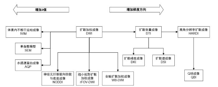
图1
扩散加权成像及其衍生模型的发展谱系
Fig. 1
The developmental history of DWI and its derivative models
需要指出的是,NODDI等高阶微结构模型对输入DWI的信噪比高度敏感. 已有研究表明,低信噪比条件下的噪声会导致ODI系统性高估、ICVF低估,从而削弱模型参数的生物学可解释性. 因此,高质量的降噪处理不仅是改善图像视觉质量的预处理步骤,更是保障微结构参数稳健估计的重要前提,这也凸显了降噪技术在dMRI分析流程中的基础性作用.
1.2 噪声来源
在dMRI中,观测信号的退化不仅源于硬件引入的热噪声和射频/梯度系统扰动,更受到采集策略、图像重建及扩散编码过程的多重调制,导致噪声呈现非高斯、空间相关与非平稳等复杂统计特性. 此外,涡流、运动等引起的结构性伪影虽非随机噪声,却常与噪声耦合,需明确区分. 表1给出了dMRI中噪声机制与伪影的系统性分类.
表1 dMRI中噪声机制和伪影的分类
Table 1
| 类型 | 分类 | 来源 | 典型特征 | 降噪是否关注 |
|---|---|---|---|---|
| 随机噪声 | 热噪声 | 由接收链路与硬件产生的随机电子噪声 | 在k空间中近似独立同分布的复高斯噪声 | √ |
| 采集放大的噪声 | 数据采集策略放大已有噪声的方差与相关性 | 噪声方差增加,空间相关性增强,非平稳性显著 | √ | |
| 重建后的统计噪声 | 由复值高斯噪声经过模值运算和多通道合成后形成的统计分布 | 非高斯,非对称,存在偏置 | √ | |
| 空间相关与非平稳噪声 | 由于线圈灵敏度、并行成像与重建过程导致的不再满足空间独立同分布的噪声 | 具有空间相关性,噪声强度随位置变化 | √ | |
| 扩散特有噪声效应 | 扩散编码导致的信号衰减与噪声统计之间的耦合效应 | 高b值下SNR急剧下降;噪声偏置主导信号 | √ | |
| 结构性伪影 | 采集相关伪影 | 由设备不完美、模型失配以及其他偶然性因素导致的系统性误差 | 具有系统性、非随机性;通常表现为空间几何畸变或相位错误 | × |
| 重建与后处理伪影 | 由算法假设或数值处理引入的确定性误差 | 与重建/后处理参数强相关,具有明显的结构模式 | × |
原始采集的MRI数据在k空间中是复数形式:
该非线性变换破坏了高斯噪声的对称性,使得重建后的幅度信号不再满足加性噪声假设. 在单线圈或等效单线圈条件下,幅度信号M遵循莱斯分布,其概率密度函数为:
其中,A为无噪声下的信号幅度,
与一般MRI不同,dMRI需针对多个不同b值的扩散方向采集信号,这使噪声特性进一步复杂化. 由于所有方向数据来源于同一个k空间采集与重建系统,这使得噪声会呈现相关性,主要包括:空间相关性,诸如敏感度编码(SENSitivity Encoding,SENSE)和广义自校准部分并行采集(Generalized Autocalibrating Partially Parallel Acquisitions,GRAPPA)等并行成像技术,以及重建插值过程,会在图像域引入像素间噪声耦合;方向相关性,梯度切换过程中的系统噪声模式、涡流效应及梯度非理想性会在不同扩散方向间引入相关结构.
此外,由于b值控制扩散敏感性,随着b值增大,信号按指数衰减:
这会导致SNR急剧下降,使莱斯偏倚进一步被放大. 在微结构建模中,这种低SNR与幅度图像中呈现的非高斯统计噪声分布共同作用,会导致主扩散方向估计不稳定、各向异性分数(Fractional Anisotropy,FA)被低估、平均扩散率(Mean Diffusivity,MD)被高估,尤其在纤维交叉或细小结构区域中更为显著.
1.3 dMRI降噪挑战
首先,噪声分布复杂且信号依赖性强. 与传统MRI不同,DWIs通常为复数模值图像,其噪声服从非高斯统计噪声分布,并随空间位置、b值及并行成像因子而变化. 尤其在高b值条件下,信号显著衰减,噪声偏倚效应加剧,导致信号被系统性高估. 这种非平稳、非高斯的噪声特性,使得假设高斯噪声的传统滤波方法难以直接适用,也对噪声统计建模与降噪策略设计提出了更高要求.
其次,多方向与多b值数据的高维结构带来降噪一致性的难题. dMRI需要在多个扩散方向及不同b值下采集图像,各方向信号受噪声影响程度不同. 若对各方向图像独立降噪,容易破坏方向间的内在一致性,并在张量拟合或高阶模型中放大误差,表现为主扩散方向不稳定,FA偏低或MD偏高等问题. 因此,如何在降噪过程中同时保持空间结构与方向相关性,是当前算法设计中的关键挑战.
最后,缺乏高质量金标准数据限制了监督式学习方法的推广. 医学影像采集中难以获得无噪声的真值图像,而合成数据往往难以真实反映复杂噪声特征及组织结构,导致深度学习模型的泛化能力不足. 即使是自监督与无监督方法,也容易受到噪声独立性假设的限制,在极低信噪比条件下表现不稳.
为应对上述挑战,研究者从传统图像滤波、统计建模到深度学习等多层次方法展开了探索.
2 dMRI降噪方法
基于莱斯噪声建模、空间滤波与低秩分解等方法虽提升了DWIs的质量,但在低SNR下仍面临细节丢失、过度平滑及鲁棒性不足等局限. 为此,具备强大特征表达能力的深度学习技术已成为降噪的新利器. 本节将综述传统算法的进展与瓶颈,并重点讨论深度学习算法的技术模块.
2.1 传统方法及其局限性
由于DWIs存在显著的莱斯噪声偏倚,并且不同方向间存在噪声相关性,这些特性在低SNR及高b值条件下尤为突出. 在深度学习兴起之前,研究者主要依赖两类互补的传统范式:基于局部/非局部相似性的数据驱动滤波,以及基于全局结构先验的模型驱动优化.
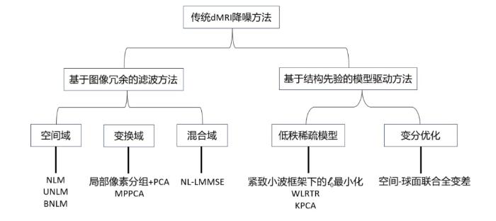
基于相似性的滤波方法假设图像存在大量重复或相似的结构,通过在空间域、变换域或混合域中聚合相似信息实现降噪. 空间域方法以非局部均值(Non-Local Means,NLM)[16]为代表,Manjón等人[17]首次将NLM算法应用到MRI领域,针对MRI特有的莱斯噪声,提出了无偏非局部均值(Unbiased NLM,UNLM),通过平方和重建图像消除低信号区域的偏倚. Coupé等人[18]进一步提出块状非局部均值(Blockwise NLM,BNLM),通过重叠分块显著降低计算成本. 在此基础上,研究者引入变换域策略[19]以增强稀疏性,结合局部像素分组与主成分分析(Principal Component Analysis,PCA),在平滑区域取得了良好的降噪效果;而基于Marchenko-Pastur的主成分分析(Marchenko-Pastur Principal Component Analysis,MPPCA)[20]则基于随机矩阵理论自动估计噪声水平并去除噪声主成分,无需预设参数. 此外,混合域方法如非局部线性最小均方误差(Non-Local Linear Minimum Mean Square Error,NL-LMMSE)结合非局部相似性与离散余弦变换,在复数域中同时处理幅度与相位信息,避免实数域降噪导致的相位丢失问题[21].
基于全局先验的方法则引入更强的全局结构先验,通过优化框架联合建模DWI数据的高维特性. 其中,低秩与稀疏模型利用多方向DWIs在梯度维度上的冗余性:例如,基于紧致小波框架的ℓ0最小化方法(即最稀疏约束优化)通过邻近方向协同降噪,有效提升信噪比[22];加权低秩张量恢复(Weighted Low-Rank Tensor Recovery,WLRTR)为不同奇异值分配自适应权重,更灵活地刻画张量结构;核主成分分析(Kernel PCA,KPCA)将线性PCA扩展至再生核希尔伯特空间,通过非线性映射提升对复杂解剖结构的表达能力[23]. 另一方面,变分优化方法结合物理噪声模型与几何正则化:McGraw等人针对高角度分辨率扩散成像(High Angular Resolution Diffusion Imaging,HARDI)提出空间-球面联合全变差模型,在保证三维空间平滑的同时保持方向分布连续性[24];后续工作进一步融合数据保真项与边缘保护约束,提升降噪真实性与结构保真度[25]. 本节对提到的传统算法按照不同的降噪处理策略进行了总结,如图2所示.
图2
尽管上述方法在各自框架内取得进展,但它们共同面临三大瓶颈:(1)手工设计的先验难以适应复杂解剖结构与多样噪声模式;(2)对噪声参数或高质量参考数据的依赖限制了临床泛化性;(3)计算复杂度高,难以扩展至全脑高分辨率或多壳层数据. 这些局限促使研究者转向端到端、数据驱动的深度学习框架,后者通过大规模学习隐式捕捉空间-方向联合特征,这已成为当前dMRI降噪的主流方向.
2.2 基于深度学习的降噪方法
针对DWIs数据的高维与非高斯噪声特性,深度学习降噪已形成相对统一的端到端范式,主要包含三个核心环节:(1)输入数据结构:为充分利用空间与q空间冗余,需优化输入维度(2D/3D/4D)、多方向编码(通道堆叠、图结构或球面表示)及辅助模态(如T1加权图像)的引入策略;(2)网络架构设计:旨在平衡局部细节与跨方向一致性. CNN擅长捕捉解剖边缘,图神经网络(Graph Neural Network,GNN)显式刻画q空间方向相关性,Transformer架构则利用自注意力机制建模长程依赖. 不同架构本质上反映了对“局部-全局”特征的权衡;(3)损失函数构建:为保障微结构参数的准确性,损失函数需超越像素级误差,融合噪声统计先验、无偏风险估计或显式的物理一致性约束.
2.2.1 网络输入层的数据结构
自深度学习引入dMRI降噪任务以来,如何在网络输入端合理组织多维扩散数据成为核心问题. 与常规MRI不同,DWIs数据在空间、梯度及b值维度上高度耦合,兼具跨方向b值的统计相关性与空间各向异性. 现有研究主要通过引入多通道、3D体块或方向联合建模策略,以增强网络对扩散特征的感知能力.
早期方法普遍从单b值或单扩散方向的二维切片中提取局部图像块. Jurek等人[26]提出基于复数卷积神经网络的监督降噪方法,在复数域联合建模幅度与相位信息,有效刻画了DWIs数据中的非高斯噪声特性,提升了重建保真度. 为缓解高b值条件下信噪比显著下降带来的结构退化问题,有研究[27,28]在降噪卷积神经网络(Denoising Convolutional Neural Network,DnCNN)框架下引入双通道2D策略,将高b值、低SNR的DWIs作为主输入,低b值、高SNR的DWIs作为引导通道(如图3所示);Pfaff等人[29]进一步融合多方向与多对比度图像,通过自监督噪声建模实现无真值降噪,解决了金标准数据稀缺问题.
图3
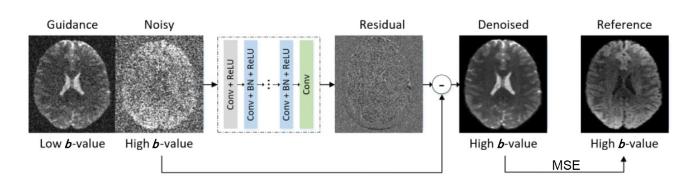
图3
DnCNN框架下的双通道输入模块. 图中缩写Conv为卷积(Convolution),BN为批量归一化(Batch Normalization),ReLU为修正线性单元(Rectified Linear Unit),MSE为均方误差(Mean Squared Error)
Fig. 3
Dual-channel input module in the DnCNN framework. Abbreviations: Conv, Convolution; BN, Batch Normalization; ReLU, Rectified Linear Unit; MSE, Mean Squared Error
然而,2D方法难以利用体素在三维空间中的连续结构与噪声相关性,易造成切片间信号不一致,进而影响扩散模型拟合与纤维追踪的稳定性. 为此,研究者转向3D体块输入. Ran等人[30]提出残差编解码Wasserstein生成对抗网络(Residual Encoder-Decoder Wasserstein Generative Adversarial Network,RED-WGAN),采用3D GAN架构,以32×32×6体素块为处理单元,通过3D卷积捕获层内纹理与层间连续性,并利用Wasserstein距离更稳定地优化生成过程,显著提升空间一致性. Wasserstein距离
其中,
4D输入理论上能捕捉跨方向冗余,尤其在低SNR条件下能进一步缓解方向独立降噪带来的不一致问题,降低张量拟合及高阶模型估计中的系统性误差. 但受限于计算开销与样本组织难度,直接端到端降噪的研究较少. 现有工作多采用间接融合策略,在模型内部通过特征提取、降维或结构变换的方式利用4D信息. Fadnavis等人[33]基于DWI的4D结构特性,提出了一种自监督降噪方法,利用其余扩散方向预测被留出的目标方向,从而在无需显式噪声模型和干净真值的条件下实现降噪;Du等人[34]提出的Node2Node模型则通过图结构建模与频域分解,将4D DTI数据的空间-方向关联性映射为图拓扑关系,在保持结构一致性的同时实现高效降噪. 相关实验表明,这类方法在多种数据条件下均能有效降低重建误差并提升纤维取向估计的稳定性.
不同成像模态及成像加权方式反映了组织在结构、功能及分子层面的不同特性,其所蕴含的信息既具有一定重叠性,又呈现出显著的互补性. 在dMRI降噪任务中,引入其他模态或不同加权方式的图像作为先验,有助于在低SNR条件下补充稳定的结构信息,从而缓解噪声抑制与结构保持之间的权衡问题. Ma等人[35]提出了一种双分支深度学习框架(如图4所示),通过联合稀疏采样的DWIs与T1加权图像的组织先验进行建模,有效提升了微结构参数的估计精度,并缓解了稀疏采样条件下的边界模糊问题. Niu等人[36]进一步将解剖模态引导策略扩展至超低剂量的正电子发射断层扫描(Positron Emission Tomography,PET)降噪任务,通过引入跨模态注意力机制实现多源特征的动态融合,验证了解剖先验在不同医学影像降噪场景中的普适价值.
图4
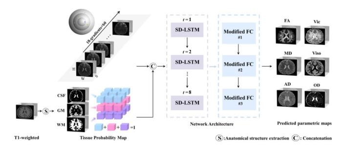
图4
双分支网络结构融合DWI与T1加权图像[35]. 图中缩写CSF为脑脊液(Cerebrospinal Fluid),GM为灰质(Gray Matter),WM为白质(White Matter),SD-LSTM为空间-方向长短期记忆网络(Spatial-Directional Long Short-Term Memory),FC为全连接(Fully Connected),AD为轴向扩散率(Axial Diffusivity),Vic表示ICVF,Viso为各向同性体积分数(Isotropic volume fraction),OD表示ODI
Fig. 4
Dual-branch network architecture integrating DWI and T1-weighted images[35]. Abbreviations: CSF, cerebrospinal fluid; GM, gray matter; WM, white matter; SD-LSTM, spatial-directional long short-term memory; FC, fully connected; AD, axial diffusivity; Vic, ICVF; Viso, isotropic volume fraction; OD, Orientation Dispersion Index
除跨模态融合外,在dMRI内部,不同b值或不同扩散方向的DWIs数据同样蕴含重要的互补信息. Naseem等人[37]提出的CMGDNet是跨模态引导降噪的代表性方法之一,其基于孪生网络(Siamese Network)结构的多分支特征提取与融合机制,在后续研究中被证明具有良好的可扩展性. 该模型通过引入特征剪枝策略,在保证跨模态特征一致性的同时减少冗余信息,为复杂模态条件下的稳健降噪提供了一种有效思路.
2.2.2 网络架构的设计与优化
在输入表示确定之后,网络架构设计成为影响dMRI降噪性能的关键因素. 该任务不仅要求有效抑制局部噪声,还需在低信噪比条件下保持扩散微结构的稳定. 早期方法多采用浅层CNN或残差结构以建模局部特征,随后逐步引入GAN、GNN及注意力机制等架构,以增强多尺度特征融合和长程依赖建模能力.
基于CNN的局部感受野与参数共享机制在低信噪比条件下具备稳定的特征提取能力. Zhang等人[38]提出了DnCNN(图3),通过前馈式结构结合残差学习与批量归一化,实现了不依赖噪声水平先验的盲降噪,在加性高斯噪声去除中表现出色,成为MRI降噪研究中的重要参考. 后续多个研究[13,39]将该模型引入DWI领域,在高b值条件下有效改善了边缘与纹理可见度. Cheng等人[13]将CNN引入DWIs数据的时间序列建模中,以同一空间位置在多次扩散加权采集中的信号序列作为网络输入,通过一维卷积沿时间维度提取相关特征,从而在保持DWIs数据时间连贯性的同时抑制随机噪声(图5). 该方法利用时间维度信号在高b值下的冗余性,实现了对噪声动态变化特性的建模,有效提升了高b值DWI图像的信噪比. 然而,标准CNN难以建模跨切片及跨方向的长程依赖关系. 为增强空间结构的一致性建模能力,研究者进一步发展了基于3D卷积和编码-解码结构的网络形式. DeepDTI[31]与SDnDTI[32]通过引入三维上下文信息并结合自监督学习策略,在无需高质量参考图像的条件下显著提升了张量估计的稳定性. 这类方法在一定程度上缓解了空间不连续问题,但在扩散方向间的统计依赖刻画上仍显不足.
图5
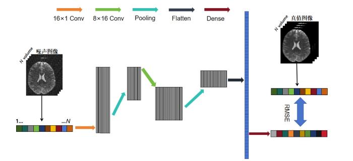
图5
1D-CNN降噪网络结构示意图. 网络以同一空间位置的N个高噪声体素构成的一维序列为输入,依次经过两层一维卷积(Conv)与池化操作(Pooling)进行特征提取. 第一层卷积通道数为16,核尺寸为1;第二层卷积通道数为32,核尺寸为8. 特征展平(Flatten)后通过全连接层(Dense)完成信号重构,并以均方根误差(Root Mean Squared Error, RMSE)损失作为损失函数进行监督训练
Fig. 5
Schematic diagram of the 1D-CNN denoising network architecture. The network takes a one-dimensional sequence composed of N highly noisy voxels at the same spatial location as input. It sequentially undergoes two layers of one-dimensional convolution (Conv) and Pooling operations for feature extraction. The first convolutional layer has 16 channels and a kernel size of 1; the second convolutional layer has 32 channels and a kernel size of 8. After feature Flatten, signal reconstruction is performed via a dense connected layer (Dense), with root mean squared error (RMSE) loss serving as the loss function for supervised training
图6

图6
生成器的网络架构图. 图中缩写Conv3D为3D卷积(3D Convolution),BatchNorm3D为3D批量归一化(3D Batch Normalization),LeakyReLU为带泄漏修正线性单元(Leaky Rectified Linear Unit)
Fig. 6
Architecture diagram of the generative network. Abbreviations: Conv3D, 3D Convolution; BatchNorm3D, 3D Batch Normalization; LeakyReLU, Leaky Rectified Linear Unit
图7
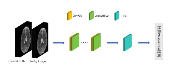
图7
判别器的网络架构图. 图中缩写Conv3D为3D卷积(3D Convolution),LeakyReLU为带泄漏修正线性单元(Leaky Rectified Linear Unit),FC为全连接层(Fully Connected)
Fig. 7
Architecture diagram of the discriminator network. Abbreviations: Conv3D, 3D Convolution; LeakyReLU, Leaky Rectified Linear Unit; FC, Fully Connected
近年来,扩散模型(Diffusion Models)通过显式建模从噪声到数据的逐步生成过程,在分布层面引入强先验约束,为解决上述问题提供了新的视角. 不同于GAN的对抗训练或CNN的判别式回归,扩散模型能够在多步反演过程中逐渐抑制噪声偏置,并对莱斯分布噪声及其信号依赖特性具有天然适应性. 基于这一优势,Xiang等人[41]提出的DDM2(Denoising Diffusion Model for dMRI)将扩散模型引入dMRI降噪任务,在无需高质量参考数据的条件下,实现了对复杂噪声分布与空间-方向一致性的联合建模,标志着dMRI降噪从判别式网络向生成式分布建模的重要转变.
2.2.3 损失函数的构建与设计
损失函数的设计是连接网络结构与物理约束的核心环节,直接影响dMRI降噪在噪声抑制、结构保真与扩散一致性之间的权衡. 受限于扩散加权幅度图像的莱斯分布噪声特性、高维扩散方向结构以及高质量参考数据稀缺等因素,单一像素级误差难以充分刻画降噪结果的物理合理性. 围绕上述挑战,相关研究逐步从基于像素重建误差的损失函数,发展到引入扩散模型、噪声统计及结构与方向一致性约束的多层级损失设计范式,以在保证降噪效果的同时提升微结构建模的稳定性与可靠性.
最为广泛的是基于像素级误差的损失函数,通过直接度量预测结果与参考图像在体素层面的差异来驱动网络优化,但这并不适用于dMRI的降噪过程. 为弥补纯像素级约束在物理一致性方面的不足,将扩散张量模型作为弱监督约束的损失设计逐渐受到关注. DeepDTI[31]在监督学习框架下采用L2损失逐步逼近高质量参考图像,有效降低了低方向数采集条件下张量估计的误差,并在提升信噪比方面展现出稳健的基线性能. SDnDTI[32]对每个子集进行张量拟合并生成高SNR的伪参考DWIs,随后以L2损失驱动网络预测结果在各子集间收敛到物理一致的解,并通过取均值使降噪结果更接近物理一致的张量重建结果,同时不依赖外部高质量真值数据. 在无参考或自监督降噪场景中,研究者通过引入噪声独立性假设[34],使传统像素级损失在无干净真值条件下仍保持无偏性,从而缓解训练数据稀缺问题.
然而,由于扩散加权的幅度图像通常服从莱斯分布,直接使用L2损失可能导致噪声偏倚被错误地解释为信号. 针对这一问题,部分研究[42]将莱斯或非中心卡方似然项显式引入损失函数或正则化项中,以更精确地刻画噪声统计特性,从而降低系统性误差的累积. 进一步地,Patch2Self[33]利用dMRI在q空间上的冗余性,提出了基于“留一原则”的自监督损失设计,在训练过程中随机遮蔽某一扩散方向的体素值,仅利用其余方向进行预测,并以L2或相似性度量作为优化目标. 另一类无真值损失来源于无偏风险估计,该方法可在幅度域直接构建损失函数,并结合空间噪声分布建模,在MRI降噪任务中取得了与基于均方误差(Mean Squared Error,MSE)损失相近的性能[43],为dMRI降噪的损失设计提供了参考.
其中,
综上,dMRI降噪损失函数的设计已从单一的像素级误差最小化,逐步发展为融合物理一致性、方向一致性与分布建模的多层次约束体系.
2.3 不同降噪策略的适用性与局限性
表2对本文涉及的基于dMRI的深度学习降噪方法进行了系统总结. 从整体发展脉络来看,dMRI降噪方法的演进反映了研究重点从“噪声统计建模”向“高维结构一致性建模”的逐步转移. 传统方法通过显式噪声模型和人工先验,在参数可解释性与稳定性方面具有优势,但其性能高度依赖噪声假设,在高b值和复杂纤维区域难以兼顾结构保持与噪声抑制. 深度学习方法则通过隐式学习空间与方向特征,在复杂噪声条件下展现出更强的灵活性,但也引入了新的不确定性来源.
表2 基于dMRI的深度学习降噪方法总结
Table 2
| 模型框架 | 对应方法 | 数据来源 | 优点 | 展望 |
|---|---|---|---|---|
| CNN | 复数域2DCNN[26] | BrainWeb模拟脑数据库 | 分离实部和虚部处理,较好保留DWI完整信息,优于仅处理幅值图像的方法 | 目前主要用于2D切片,未来可扩展至3D重构,并结合多b值/多方向DWIs进一步提升降噪性能 |
| Guided DnCNN[27] | 30名真实患者的前列腺MRI数据集 | 通过双输入引导和特征融合机制,利用低b值DWIs的解剖结构信息,提升高b值DWIs的降噪质量并减少过平滑 | 可进一步优化融合策略和注意力机制;结合k空间与图像域加速技术,有望实现更高效的加速重建 | |
| Residual DnCNN[28] | 真实采集的带噪声DWIs与合成数据集 | 用残差学习预测噪声映射,兼顾降噪效率与细节保留,验证了方法在高维任务中的可行性 | 可探索更少平均次数输入以提升速度,并与运动校正模块结合,以适应临床动态场景 | |
| Enhanced SURE-based Method[29] | fastMRI数据集和Siemens Healthineers数据集 | 仅需输入图像,无需额外噪声先验或扫描设定;利用相邻图像互补信息提升降噪效果 | 已在fastMRI和西门子7 T场强设备上验证,后续可推广到其他解剖部位,如腹部、膝部DWIs | |
| DeepDTI[31] | Human Connectome Project(HCP)数据集 | 仅需1个b0图像和6个DWIs即可生成与90个DWIs接近质量的DTI,显著加快采集过程 | 方向敏感性可能受个体差异和扫描设备影响,需在更多临床人群和不同扫描协议下验证其泛化能力与诊断价值 | |
| SDnDTI[32] | HCP数据集 | 通过子集划分进行损失约束,可在有限样本条件下有效利用7例DWIs进行自监督学习 | 对极高分辨率或超低信噪比DTI数据,可能需调整子集划分策略,并使用更多DWI方向或更先进的张量拟合方法 | |
| JD-CNN[39] | 真实采集的低SNR数据和病例数据以及模拟数据集 | 同时输入所有b值的DWIs,利用跨b值结构相关性联合降噪,增强高b值DWIs的降噪能力 | 虽未假设噪声分布,但主要在高斯噪声下测试;未来需验证其在更复杂噪声模型下的表现 | |
| 1D-CNN[13] | HCP数据集以及模拟数据 | 以重建图像作为监督目标进行训练和评估,可较好解决高b值DWI中SoS重建所引入的噪声 | 效果依赖高质量参考图像,限制了直接应用范围;后续需扩大数据规模并增强模型鲁棒性 | |
| SSECNN[44] | 心脏DTI数据 | 利用不同扩散方向DWI的结构相似性构建自监督信号,并设计Sobel加权损失,有效抑制过平滑 | 可结合深度学习模型提取更具语义的切片相似性度量,进一步提升匹配精度和降噪效果 | |
| MCNN[47] | 真实采集的健康受试者、多发性硬化症患者数据以及合成的训练数据 真实采集的健康受试者、多发性硬化症患者数据以及合成的训练数据 | 直接在幅度图像上完成训练和推理,输入与输出均为SoS图像;标准重建下对噪声峰值伪影更鲁棒 | 对重建顺序高度敏感;当重建流程变为“先切分后线圈组合”时性能明显下降,不推荐用于高加速场景 | |
| CCNN[47] | 在复数图像上直接训练与推理,处理实部和虚部信息,较适用于平滑相位并能适应重建顺序变化 | 基于编码算子的CNN具有较大潜力,可为更强深度学习重建模型提供方向 | ||
| GNN | Node2Node[34] | 离体猪心脏、在体人类心脏以及合成数据集 | 将不同方向DWIs视为图节点,并结合图卷积小波变换进行频谱分解与信息匹配,可同时滤除噪声和谐波干扰,提升结构感知能力 | 心脏成像易受运动影响,未来可结合运动校正;图信号处理计算开销较大,还需探索更高效实现以满足实时或准实时需求 |
| 扩散模型 | Di-Fusion[46] | Stanford HARDI、Sherbrooke 3-Shell和PPMI数据集 | 基于J-invariance优化,仅用成对噪声DWIs训练,并通过迭代式精炼实现高保真重建,在复杂噪声下仍可保留DWIs结构细节 | 可进一步探索将dMRI物理模型,融入扩散模型或损失函数中,以增强物理可解释性 |
| DDM2[41] | Sherbrooke 3-Shell、Stanford HARDI、PPMI以及真实采集数据 | 提出三阶段协同策略:先学习噪声分布,再桥接真实噪声与扩散过程,最后生成高质量降噪图像,实现由弱到强的降噪 | 推理耗时较长,难以满足实时需求;后续可通过推理加速、数据一致性约束及防止结构幻觉等策略进一步改进 |
具体而言,基于CNN的方法在计算效率与稳定性方面具备明显优势,适合在方向数较多、信噪比中等的条件下应用,但其对扩散方向间长程依赖的刻画能力有限;GAN类方法通过分布级约束改善了结构保真度,但训练过程不稳定、可重复性不足,限制了其临床推广;GNN通过构建梯度方向图建模非欧几里得空间的一致性,而Transformer则可视为具备全局感受野的广义图神经网络,能实现更深层的跨维度依赖捕捉. 但由于后者对计算资源要求极高,因此在dMRI降噪领域尚处于研究验证阶段.
尽管Transformer模型在dMRI降噪中尚处于起步阶段,但其核心的自注意力机制已被广泛集成于现代生成式扩散模型的架构中,为其提供了关键的全局特征提取能力. 生成式扩散模型在理论上为解决莱斯分布噪声与方向一致性问题提供了新的范式,其通过逐步反演过程显式抑制噪声偏置,在自监督条件下表现出较强潜力. 然而,该类方法的计算复杂度和推理时间仍是制约其实际应用的重要因素.
综合来看,不同方法在性能、稳定性与可推广性之间存在明显的权衡关系,尚不存在所有条件下均占优的统一解决方案. 这也表明,未来dMRI降噪的发展方向并非单一模型结构的演进,更有可能是融合物理先验与深度学习优势的混合建模框架.
3 降噪方法评价指标
在dMRI与DTI降噪研究中,评估指标不仅是衡量算法性能的工具,更是指导算法改进与优化的核心依据. 不同于自然图像降噪任务,DWIs数据具有更强的临床相关性和物理依赖性,因此降噪的成败不仅取决于图像的视觉质量,还要确保信号的定量准确性. 与此同时,图像降噪效果的评估也不能仅局限于图像本身的细节保留度和边缘平滑度,还需要兼顾降噪后的图像是否有利于扩散张量的拟合. 因此,本节将从视觉评估、量化指标以及扩散张量参数指标三方面来进行介绍.
3.1 视觉评估
视觉评估是在常规量化指标之外的一种主观评价方式,旨在通过直接观察降噪后的图像来判断方法的有效性与临床适用性. 其主要考察维度包括:
(1)噪声抑制与纹理保留:评估图像整体的平滑度与清晰度,重点权衡随机噪声的抑制程度与过度平滑风险,避免因降噪过强导致纹理细节丢失.
(2)解剖结构保真度:重点考察脑区结构、组织边界以及白质纤维走向等关键解剖特征的保留情况. 若降噪过强,可能引起边缘模糊或细节信息丢失,从而影响后续的定量分析和临床诊断.
(3)伪影与虚假信号:降噪过程中可能引入新的图像伪影,如条纹、块状伪影或不自然的纹理增强. 此类问题需要通过肉眼甄别,以确保降噪结果的真实性与可靠性.
3.2 量化评估
量化评估通过数值指标客观衡量降噪性能,重点考察噪声抑制、细节保真及结构一致性. 常用指标包括MSE、峰值信噪比(Peak Signal-to-Noise Ratio,PSNR)及结构相似性(Structural Similarity Index Measure,SSIM). 以下公式均基于二维图像数据.
(1)MSE:衡量的是降噪图像与参考图像之间的像素级差异,定义如下:
其中,
由于MSE的单位是原始数据单位的平方,而RMSE的单位与原始数据的单位完全相同,因此RMSE在实际应用中通常比MSE更直观、更容易理解.
(2) PSNR:基于MSE定义,用于评估降噪后图像的整体质量:
其中,L表示图像的最大像素值. PSNR数值越高,表示降噪效果越好.
(3) SSIM:从亮度、对比度和结构三个方面衡量图像的相似度,公式如下:
其中,
3.3 扩散特征参数评价
在DTI研究中,还需重点评估扩散张量参数的稳定性与准确性. 这些参数直接量化了组织微观环境与纤维结构特征,降噪算法必须确保其物理可靠性,以防止对后续临床分析引入系统性偏差.
常用的DTI指标包括MD、FA、轴向扩散率(Axial Diffusivity,AD)和径向扩散率(Radial Diffusivity,RD). 假设扩散张量的三个特征值分别为
(1) MD:表示张量三个特征值的平均值,反映了组织中总体的水分子扩散强度,常用于区分病灶和正常组织. 公式定义如下:
(2) FA:表征水分子扩散的各向异性程度,取值范围为[0,1]. FA越大,说明组织结构越有方向性;FA越小,则说明扩散接近各向同性,常用于刻画白质纤维完整性. 公式定义如下:
(3) AD:表示主扩散方向上的扩散强度,常对应沿白质纤维方向的扩散,主要与轴突完整性相关. 公式定义如下:
(4) RD:表示垂直于纤维方向的平均扩散强度,常与髓鞘完整性密切相关,公式定义如下:
值得注意的是,直接比较FA、MD等指标的全脑平均值往往存在误导性,因为图像中局部的高估和低估会在平均过程中相互抵消,导致即使局部失真严重,其全脑均值仍可能接近真值. 相比之下,采用逐体素平均绝对误差(Mean Absolute Percentage Error,MAPE)能够捕捉每个体素相对于真值的绝对偏离程度,避免了正负误差抵消的问题,从而能更严谨、更敏感地量化算法对微观结构细节的还原保真度. 因此对每个指标的处理如下:
其中,
4 实验结果对比与分析
本文先对当前主流的dMRI降噪算法进行了系统性梳理与归纳,涵盖了其基本原理、实现方式以及适用场景. 为了进一步验证这些方法在实际应用中的有效性与差异性,本节拟通过实验定量化地对比不同方法性能差异. 通过统一的评价指标与实验设置,本文对各类算法的降噪性能、微结构保持能力以及在不同噪声水平下的鲁棒性进行了定量评估,从而为后续方法的选择与改进提供更为客观和可靠的参考依据.
4.1 实验设计
为保证实验的可复现性与方法比较的公平性,本文采用经标准化预处理的HCP数据集进行公开评估. 原始数据已完成畸变、头动及涡流校正,该步骤主要消除几何伪影,未改变信号的统计噪声特性,因此可作为近似无噪参考. 随后进行脑组织掩膜提取并统一空间分辨率,以减少非扩散因素对降噪评价的影响. 每例DWI数据包含18个b=0方向以及各90个b=1 000和2 000 s/mm²的扩散加权方向,共198个方向.
本文的实验共使用2例受试者数据,其中1例用于监督式1D-CNN的训练与验证,另1例独立受试者数据用于测试. 为避免将同一受试者中具有相关性的扩散方向数据直接按比例随机拆分,训练集与验证集的划分并非基于198个扩散方向的简单8 : 2随机分配,而是以脑掩膜内体素对应的完整扩散信号向量作为样本单元,在保持各b值及扩散方向组合完整的前提下,于训练受试者内部按8 : 2随机划分为训练集与验证集,用于网络参数学习与模型选择. 对于无需训练的传统方法(PCA、MPPCA)及自监督方法(Patch2Self),其参数均沿用原文献或公开实现的默认配置,直接对测试集进行处理;对于监督式1D-CNN,模型采用Adam优化器(学习率为0.002,批大小为10 000)训练1 000个epoch,损失函数为MSE损失. 所有方法均在相同噪声水平下于同一测试受试者数据上进行评估,旨在受限训练数据条件下比较不同方法在统一实验设置下的相对定量表现与泛化能力. 需要说明的是,由于本文测试仅基于单例独立受试者,相关结果主要用于方法间的比较与趋势性分析,而不作为跨受试者群体统计推断的依据.
为模拟真实的莱斯噪声,首先在复数域信号的实部和虚部分别加入独立的零均值高斯噪声,即
其中,Sr和Si分别代表无噪声信号的实部和虚部,符号
通过该方式构建不同噪声水平下的含噪DWIs数据及其对应的参考真值数据.
本文选取三类代表性方法进行对比,涵盖了不同的降噪范式与建模假设. 其中,PCA与MPPCA代表传统低秩统计建模,利用局部冗余性直接处理含噪图像,作为无需训练的经典基线;Patch2Self代表自监督范式,仅依赖扩散方向间的冗余性在测试阶段降噪,无需高质量参考真值,具有较强实用性;1D-CNN则作为监督式深度学习代表,通过将同一空间位置的多次扩散加权信号构建为一维序列,利用CNN学习噪声与信号间的非线性映射,体现了数据驱动特征建模的优势. 上述方法覆盖了传统统计、监督与自监督三类主流策略,有助于系统评估不同方法在多噪声水平下的性能表现.
4.2 实验结果
表3 不同噪声水平下各降噪方法的SSIM指标对比
Table 3
| b值/(s/mm²) | 噪声水平 | 噪声图像 | PCA | MPPCA | Patch2Self | 1D-CNN |
|---|---|---|---|---|---|---|
| b=0 | 2% | 0.82 | 0.93 | 0.95 | 0.93 | 0.94 |
| 4% | 0.62 | 0.88 | 0.90 | 0.89 | 0.91 | |
| 6% | 0.47 | 0.81 | 0.85 | 0.84 | 0.86 | |
| 8% | 0.37 | 0.77 | 0.80 | 0.78 | 0.77 | |
| 10% | 0.33 | 0.73 | 0.75 | 0.73 | 0.78 | |
| b=1000 | 2% | 0.48 | 0.73 | 0.76 | 0.77 | 0.80 |
| 4% | 0.25 | 0.55 | 0.64 | 0.66 | 0.69 | |
| 6% | 0.16 | 0.43 | 0.54 | 0.56 | 0.57 | |
| 8% | 0.12 | 0.34 | 0.44 | 0.45 | 0.46 | |
| 10% | 0.09 | 0.29 | 0.35 | 0.36 | 0.41 | |
| b=2000 | 2% | 0.36 | 0.58 | 0.63 | 0.66 | 0.68 |
| 4% | 0.17 | 0.38 | 0.47 | 0.51 | 0.56 | |
| 6% | 0.10 | 0.26 | 0.33 | 0.38 | 0.40 | |
| 8% | 0.08 | 0.21 | 0.27 | 0.28 | 0.31 | |
| 10% | 0.07 | 0.19 | 0.20 | 0.23 | 0.25 |
表4 不同噪声水平下各降噪方法的PSNR指标对比
Table 4
| b值/(s/mm²) | 噪声水平 | 噪声图像 | PCA | MPPCA | Patch2Self | 1D-CNN |
|---|---|---|---|---|---|---|
| b=0 | 2% | 32.27 | 35.87 | 37.79 | 35.33 | 36.02 |
| 4% | 27.16 | 33.00 | 33.37 | 33.46 | 35.08 | |
| 6% | 23.72 | 29.49 | 29.67 | 30.19 | 31.77 | |
| 8% | 21.32 | 26.72 | 26.61 | 27.30 | 30.77 | |
| 10% | 19.40 | 24.29 | 24.06 | 24.73 | 27.58 | |
| b=1000 | 2% | 20.29 | 25.21 | 26.10 | 25.67 | 27.77 |
| 4% | 14.52 | 19.22 | 19.52 | 19.73 | 23.54 | |
| 6% | 10.90 | 14.58 | 14.72 | 15.08 | 20.46 | |
| 8% | 8.18 | 11.14 | 11.21 | 11.66 | 17.16 | |
| 10% | 5.97 | 8.48 | 8.42 | 8.91 | 15.96 | |
| b=2000 | 2% | 17.48 | 21.82 | 22.22 | 21.85 | 26.33 |
| 4% | 11.28 | 13.44 | 16.62 | 15.45 | 23.65 | |
| 6% | 7.32 | 9.70 | 12.80 | 10.63 | 20.51 | |
| 8% | 4.44 | 7.42 | 7.48 | 6.37 | 19.61 | |
| 10% | 2.19 | 5.95 | 3.58 | 4.21 | 17.92 |
表5 不同噪声水平下各降噪方法的RMSE指标对比
Table 5
| b值/(s/mm²) | 噪声水平 | 噪声图像 | PCA | MPPCA | Patch2Self | 1D-CNN |
|---|---|---|---|---|---|---|
| b=0 | 2% | 0.024 | 0.016 | 0.013 | 0.018 | 0.015 |
| 4% | 0.042 | 0.022 | 0.020 | 0.025 | 0.017 | |
| 6% | 0.060 | 0.028 | 0.027 | 0.030 | 0.025 | |
| 8% | 0.077 | 0.039 | 0.035 | 0.036 | 0.031 | |
| 10% | 0.082 | 0.046 | 0.041 | 0.042 | 0.037 | |
| b=1000 | 2% | 0.089 | 0.055 | 0.040 | 0.046 | 0.039 |
| 4% | 0.141 | 0.084 | 0.073 | 0.071 | 0.067 | |
| 6% | 0.167 | 0.101 | 0.082 | 0.075 | 0.071 | |
| 8% | 0.179 | 0.118 | 0.107 | 0.090 | 0.085 | |
| 10% | 0.186 | 0.122 | 0.128 | 0.107 | 0.095 | |
| b=2000 | 2% | 0.107 | 0.076 | 0.055 | 0.054 | 0.049 |
| 4% | 0.146 | 0.123 | 0.084 | 0.086 | 0.072 | |
| 6% | 0.184 | 0.146 | 0.106 | 0.113 | 0.095 | |
| 8% | 0.205 | 0.153 | 0.143 | 0.166 | 0.111 | |
| 10% | 0.232 | 0.159 | 0.182 | 0.178 | 0.123 |
在传统方法中,MPPCA通常优于PCA,这得益于Marchenko-Pastur定律对信号子空间维度的自适应估计,从而提高了信噪分离能力. 然而,在中高b值的较高噪声条件下,噪声主导特征分布,导致子空间估计偏差,MPPCA性能反而下降,低于更为稳健但简化的PCA. Patch2Self作为自监督方法,在无需额外训练数据的情况下能够取得相对稳定的降噪效果,但其基于邻域预测的机制在高噪声或较高b值条件下易引入过度平滑,导致细节纹理与边缘锐度下降. 低b值时,虽说1D-CNN容易造成过度平滑,导致SSIM大幅下降,但其在中高b值下展现出强大的结构保持能力. 图8给出了6%噪声水平下的视觉对比结果,进一步验证了上述结论. 表明深度学习方法能够在中高b值、高噪声下有效恢复关键解剖结构.
图8

图8
6%噪声水平下不同降噪方法的视觉对比结果. 从上至下依次给出b=0、b=1 000 s/mm²及b=2 000 s/mm² 条件下的图像,用以反映各方法在不同扩散敏感度下的表现差异
Fig. 8
Visual comparison of different denoising methods at a 6% noise level. From top to bottom, images under conditions of b=0, b=1 000 s/mm², and b=2 000 s/mm² are shown to reflect the performance differences of each method at varying diffusion sensitivities
考虑到DWI的核心价值在于下游定量建模,本研究进一步比较了降噪对DTI拟合结果的影响. 图9给出了不同降噪方法对FA参数的可视化结果,各方法均在不同程度上减弱了噪声引起的散点与条纹伪影. MPPCA方法能够显著降低背景噪声,但在组织边界处出现过平滑,导致局部细节损失;PCA降噪不充分,残留噪声明显. 相比之下,Patch2Self与1D-CNN在噪声抑制与结构保真之间取得了更合理的平衡,较好地保持了FA分布的空间一致性与各向异性特征.
图9
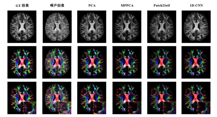
图9
4%噪声水平下不同降噪方法的FA图像(第一行)、FA的彩色编码图像(第二行)及局部细节对比图(第三行)(b=1 000 s/mm²)
Fig. 9
Comparison of FA maps (Row 1), color-coded FA maps (Row 2), and local details (Row 3) across different denoising methods at a 4% noise level (b=1 000 s/mm²)
为进一步评估降噪对DTI微观结构参数可靠性的影响,本研究进一步评估了四个DTI参数在降噪前后的误差变化,如表6所示. 在低噪声水平下,除各向异性FA(FAmape)外,其余扩散参数降噪后的MAPE略高于原始噪声图像. 这归因于降噪算法固有的信号平滑效应,有噪图像的解剖细节保留完好,而算法在去除微弱噪声的过程中,不可避免地对高频纹理细节造成了轻微损失或模糊. 随着噪声水平的增加,降噪算法在抑制噪声方面的收益迅速超过了信号损失的代价,从而表现出更低的误差. 尽管Patch2Self在视觉降噪方面表现出色,但定量分析显示其在微观结构参数的恢复上表现欠佳,该算法在利用q空间相关性进行信号预测时,倾向于抑制各向异性特征,导致扩散张量的方向选择性降低. 1D-CNN算法在中高噪声水平下对微观结构参数的恢复表现出卓越的鲁棒性. 这一结果表明深度学习算法在处理中高噪声条件下的扩散磁共振数据方面表现出一定优势,预示着其在未来高精度成像中的广阔应用前景.
表6 不同降噪方法在DTI微结构参数上的定量评估结果. 表中数值表示各微观结构参数(FA、MD、AD、RD)相对于金标准(Ground Truth)的MAPE. 数值越小表示降噪后的结果越接近真实值. 加粗数值表示各降噪方法的最优值
Table 6
| 指标 | 噪声水平 | Noisy | PCA | MPPCA | Patch2Self | 1D-CNN |
|---|---|---|---|---|---|---|
| FAMAPE | 2% | 0.40 | 0.25 | 0.36 | 0.40 | 0.28 |
| 4% | 0.81 | 0.45 | 0.53 | 0.71 | 0.43 | |
| 6% | 1.30 | 0.55 | 0.60 | 0.84 | 0.51 | |
| 8% | 1.86 | 0.61 | 0.60 | 0.88 | 0.56 | |
| 10% | 2.40 | 0.63 | 0.62 | 0.89 | 0.58 | |
| MDMAPE | 2% | 0.16 | 0.24 | 0.25 | 0.26 | 0.30 |
| 4% | 0.51 | 0.52 | 0.51 | 0.54 | 0.34 | |
| 6% | 0.70 | 0.69 | 0.64 | 0.72 | 0.36 | |
| 8% | 0.86 | 0.79 | 0.73 | 0.82 | 0.51 | |
| 10% | 0.92 | 0.85 | 0.84 | 0.89 | 0.62 | |
| ADMAPE | 2% | 0.16 | 0.26 | 0.29 | 0.31 | 0.30 |
| 4% | 0.44 | 0.56 | 0.57 | 0.61 | 0.28 | |
| 6% | 0.61 | 0.72 | 0.71 | 0.77 | 0.24 | |
| 8% | 0.87 | 0.78 | 0.77 | 0.85 | 0.55 | |
| 10% | 1.08 | 0.87 | 0.86 | 0.91 | 0.66 | |
| RDMAPE | 2% | 0.16 | 0.21 | 0.19 | 0.20 | 0.27 |
| 4% | 0.45 | 0.31 | 0.30 | 0.45 | 0.21 | |
| 6% | 0.67 | 0.45 | 0.43 | 0.69 | 0.38 | |
| 8% | 0.85 | 0.61 | 0.57 | 0.83 | 0.52 | |
| 10% | 1.08 | 0.87 | 0.82 | 0.91 | 0.64 |
5 结论与展望
dMRI降噪是保障扩散微结构建模可靠性的关键前提. 多数研究与本文实验结果均表明,在低信噪比、较高b值或扩散方向数受限的条件下,未经有效降噪的DWIs数据在张量拟合与微结构参数估计中容易产生系统性偏差,进而影响FA、MD等指标的稳定性与生物学可解释性. 合理的降噪处理不仅能够改善图像视觉质量,更直接决定了扩散模型参数估计与纤维追踪结果的可靠性.
通过对传统方法与深度学习方法的系统梳理与实验比较可以发现,基于明确噪声模型和统计假设的传统算法在中低噪声条件下仍具一定优势,尤其在参数可解释性和算法稳定性方面表现可靠;然而,在高噪声或复杂纤维结构区域,这类方法往往难以同时兼顾噪声抑制与结构保持,易出现过度平滑或方向一致性被破坏的问题. 相比之下,深度学习方法通过数据驱动方式学习隐式先验,在噪声抑制与结构保真之间展现出更优的灵活性,尤其是在引入多尺度特征提取、自监督学习或跨方向建模策略后,其在真实数据上的鲁棒性得到明显提升.
从建模范式上看,现有深度学习降噪主要以判别式模型(CNN/GAN)为代表,旨在学习含噪到干净信号的直接映射. 此类方法在中低噪声下效率较高,且已发展出基于噪声独立性或q空间冗余的自监督策略,以缓解真值稀缺问题. 然而,因缺乏对信号生成过程及噪声分布的显式刻画,其在高噪声或信号衰减显著时易受数据分布偏置影响,导致参数偏差. 针对现有判别式模型的上述局限,生成式扩散模型被视为未来该领域的重要演进方向. 尽管本研究尚未对其进行定量评估,但在理论上,该类方法通过学习噪声注入的逆过程实现对信号概率分布的建模,更契合莱斯噪声特性. 其多步反演机制在高噪声、高b值或复杂微结构区域展现出更强的灵活性,但同时也面临计算代价高、训练复杂及物理一致性约束设计难等挑战.
未来研究应着力于:(1)由判别式回归向分布级生成建模转变,基于扩散模型和得分匹配的方法通过显式刻画噪声到信号的逐步反演过程,在理论上更适配莱斯分布噪声等非高斯统计特性;(2)强化空间-方向联合建模,避免独立方向降噪破坏q空间一致性;(3)构建融合物理先验与深度表示能力的混合框架,在提升模型鲁棒性的同时增强结果的可解释性,从而提高方法在真实数据与临床应用场景中的可推广性.
利益冲突
无
参考文献
Cerebellar imaging with diffusion magnetic resonance imaging: approaches, challenges, and potential
[J].
An intrinsic anisotropic feature of DTI images derived by geometric properties on the Riemannian manifold
[J].DOI:10.1016/j.bspc.2023.105478 URL [本文引用: 1]
Mapping tissue microstructure of brain white matter in vivo in health and disease using diffusion MRI
[J].DOI:10.1162/imag_a_00344 URL [本文引用: 1]
The development of brain white matter microstructure
[J].DOI:10.1016/j.neuroimage.2017.12.097 URL [本文引用: 1]
Diffusion MRI of white matter microstructure development in childhood and adolescence: Methods, challenges and progress
[J].DOI:10.1016/j.dcn.2017.12.002 URL [本文引用: 1]
A time-dependent diffusion MRI signature of axon caliber variations and beading
[J].DOI:10.1038/s42003-020-1050-x [本文引用: 1]
A phantom for diffusion-weighted MRI (DW-MRI)
[J].
DOI:10.1002/jmri.23950
PMID:23576443
[本文引用: 1]

To develop tissue-equivalent diffusivity materials and build a spherical diffusion phantom which mimics the conditions typically found in biological tissues. Also, to assess the reproducibility of ADC measurements from a whole-body diffusion protocol.Nickel-doped agarose/sucrose gels were manufactured and used to build a spherical diffusion phantom with tissue-equivalent relaxation and diffusion compartments. The temporal stability of the gels was monitored for a period of 8 weeks and, using the same measurements, the reproducibility of ADC was assessed in a 1.5 Tesla (T) clinical system.The temporal stability of the nickel-doped agarose/sucrose gels diffusion properties was excellent (average coefficient of variation [CV] for ADC in all phantom compartments = 1.27%). The average CV for ADC measurements, excluding the phantom compartments affected by artifacts, was 0.76% showing that the reproducibility of ADC measurements using an EPI DW-MRI protocol is very good.Nickel-doped agarose/sucrose gels can be used as reference materials for MRI diffusion measurements and show excellent short-term stability with respect to ADC. A phantom made of these materials can be invaluable in optimizing DW-MRI protocols, developing novel pulse sequences for DW-MRI, or comparing ADC values among field strengths, vendors, and imaging centers.Copyright © 2013 Wiley Periodicals, Inc.
New method for diffusion-weighted images denoising based on patch-matching with higher-order singular value decomposition
[J].
DTI brain template construction based on gaussian averaging
[J].
基于高斯平均的DTI脑模板构建方法
[J].
DOI:10.11938/cjmr20212957
[本文引用: 1]
在获取被试的张量数据后通常对其进行多通道线性平均以得到张量模板.但线性平均不仅会忽略张量中的向量信息,还会使灰质和白质的交界处过于平滑,降低模板的分辨率.为了解决以上问题,本文引入了四元数及高斯加权平均来构建高斯扩散张量成像(Diffusion Tensor Imaging,DTI)脑模板.本文首先对55个健康被试的DTI数据进行预处理,使得数据伪影最小化;再通过扩散张量成像工具包(Diffusion Tensor Imaging ToolKit,DTI-TK)将预处理后的数据进行初步空间标准化;然后将张量通过特征分解得到特征向量和特征值;最后,将由特征向量转化的四元数标量和特征值分别进行高斯加权平均得到平均后的特征向量和特征值,并对其进行重建得到张量模板.实验结果表明相比于线性DTI模板,高斯DTI模板在DTED、COH、DVED、OVL、corr<sub>FA</sub>评估指标上表现更优,而IA指标较差,说明本文提出的高斯DTI模板在整体信息保留方面有所优化,但方向信息有所丢失.
Directional and inter-acquisition variability in diffusion-weighted imaging and editing for restricted diffusion
[J].
DOI:10.1002/mrm.29385
PMID:35861268
[本文引用: 1]
To evaluate and quantify inter-directional and inter-acquisition variation in diffusion-weighted imaging (DWI) and emphasize signals that report restricted diffusion to enhance cancer conspicuity, while reducing the effects of local microscopic motion and magnetic field fluctuations.Ten patients with biopsy-proven prostate cancer were studied under an Institutional Review Board-approved protocol. Individual acquisitions of DWI signal intensities were reconstructed to calculate inter-acquisition distributions and their statistics, which were compared for healthy versus cancer tissue. A method was proposed to detect and filter the acquisitions affected by motion-induced signal loss. First, signals that reflect restricted diffusion were separated from the acquisitions that suffer from signal loss, likely due to microscopic motion, by imposing a cutoff value. Furthermore, corrected apparent diffusion coefficient maps were calculated by employing a weighted sum of the multiple acquisitions, instead of conventional averaging. These weights were calculated by applying a soft-max function to the set of acquisitions per-voxel, making the analysis immune to acquisitions with significant signal loss, even if the number of such acquisitions is high.Inter-acquisition variation is much larger than the Rician noise variance, local spatial variations, and the estimates of diffusion anisotropy based on the current data, as well as the published values of anisotropy. The proposed method increases the contrast for cancers and yields a sensitivity of with a false positive rate of.Motion-induced signal loss makes conventional signal-averaging suboptimal and can obscure signals from areas with restricted diffusion. Filtering or weighting individual acquisitions prior to image analysis can overcome this problem.© 2022 The Authors. Magnetic Resonance in Medicine published by Wiley Periodicals LLC on behalf of International Society for Magnetic Resonance in Medicine.
Denoise diffusion-weighted images using higher-order singular value decomposition
[J].DOI:10.1016/j.neuroimage.2017.04.017 URL [本文引用: 1]
Diffusion weighted image denoising using overcomplete local PCA
[J].DOI:10.1371/journal.pone.0073021 URL [本文引用: 2]
Denoising diffusion weighted imaging data using convolutional neural networks
[J].
DOI:10.1371/journal.pone.0274396
URL
[本文引用: 5]
Diffusion weighted imaging (DWI) with multiple, high b-values is critical for extracting tissue microstructure measurements; however, high b-value DWI images contain high noise levels that can overwhelm the signal of interest and bias microstructural measurements. Here, we propose a simple denoising method that can be applied to any dataset, provided a low-noise, single-subject dataset is acquired using the same DWI sequence. The denoising method uses a one-dimensional convolutional neural network (1D-CNN) and deep learning to learn from a low-noise dataset, voxel-by-voxel. The trained model can then be applied to high-noise datasets from other subjects. We validated the 1D-CNN denoising method by first demonstrating that 1D-CNN denoising resulted in DWI images that were more similar to the noise-free ground truth than comparable denoising methods, e.g., MP-PCA, using simulated DWI data. Using the same DWI acquisition but reconstructed with two common reconstruction methods, i.e. SENSE1 and sum-of-square, to generate a pair of low-noise and high-noise datasets, we then demonstrated that 1D-CNN denoising of high-noise DWI data collected from human subjects showed promising results in three domains: DWI images, diffusion metrics, and tractography. In particular, the denoised images were very similar to a low-noise reference image of that subject, more than the similarity between repeated low-noise images (i.e. computational reproducibility). Finally, we demonstrated the use of the 1D-CNN method in two practical examples to reduce noise from parallel imaging and simultaneous multi-slice acquisition. We conclude that the 1D-CNN denoising method is a simple, effective denoising method for DWI images that overcomes some of the limitations of current state-of-the-art denoising methods, such as the need for a large number of training subjects and the need to account for the rectified noise floor.
Research progress of denoising algorithms for diffusion tensor images
[J].
扩散张量图像降噪算法研究进展
[J].
DOI:10.11938/cjmr20243087
[本文引用: 1]
扩散张量成像是研究组织大脑微结构与白质纤维束分布的重要手段,然而受扩散加权信号衰减与长回波时间的影响,扩散张量图像存在严重的低信噪比问题.因此,有效的去噪技术在提高图像质量方面发挥着重要的作用.本文首先阐述了扩散张量成像的原理及噪声类型;其次论述了经典的扩散张量图像去噪算法,包括基于传统图像处理方法与基于深度学习方法,并着重探讨了扩散张量图像去噪的研究现状及不足;接着介绍了去噪评估标准及常用的公开数据集;然后讨论分析了文中提及的扩散张量图像去噪方法;最后总结并对该领域未来的研究方向进行了展望.
Looking into the functional architecture of the brain with diffusion MRI
[J].DOI:10.1038/nrn1119 PMID:12778119 [本文引用: 1]
A non-local algorithm for image denoising
[C]//
MRI denoising using non-local means
[J].
DOI:10.1016/j.media.2008.02.004
PMID:18381247
[本文引用: 1]
Magnetic Resonance (MR) images are affected by random noise which limits the accuracy of any quantitative measurements from the data. In the present work, a recently proposed filter for random noise removal is analyzed and adapted to reduce this noise in MR magnitude images. This parametric filter, named Non-Local Means (NLM), is highly dependent on the setting of its parameters. The aim of this paper is to find the optimal parameter selection for MR magnitude image denoising. For this purpose, experiments have been conducted to find the optimum parameters for different noise levels. Besides, the filter has been adapted to fit with specific characteristics of the noise in MR image magnitude images (i.e. Rician noise). From the results over synthetic and real images we can conclude that this filter can be successfully used for automatic MR denoising.
An optimized blockwise nonlocal means denoising filter for 3-D magnetic resonance images
[J].
DOI:10.1109/TMI.2007.906087
PMID:18390341
[本文引用: 1]
A critical issue in image restoration is the problem of noise removal while keeping the integrity of relevant image information. Denoising is a crucial step to increase image quality and to improve the performance of all the tasks needed for quantitative imaging analysis. The method proposed in this paper is based on a 3-D optimized blockwise version of the nonlocal (NL)-means filter (Buades, et al., 2005). The NL-means filter uses the redundancy of information in the image under study to remove the noise. The performance of the NL-means filter has been already demonstrated for 2-D images, but reducing the computational burden is a critical aspect to extend the method to 3-D images. To overcome this problem, we propose improvements to reduce the computational complexity. These different improvements allow to drastically divide the computational time while preserving the performances of the NL-means filter. A fully automated and optimized version of the NL-means filter is then presented. Our contributions to the NL-means filter are: 1) an automatic tuning of the smoothing parameter; 2) a selection of the most relevant voxels; 3) a blockwise implementation; and 4) a parallelized computation. Quantitative validation was carried out on synthetic datasets generated with BrainWeb (Collins, et al., 1998). The results show that our optimized NL-means filter outperforms the classical implementation of the NL-means filter, as well as two other classical denoising methods [anisotropic diffusion (Perona and Malik, 1990)] and total variation minimization process (Rudin, et al., 1992) in terms of accuracy (measured by the peak signal-to-noise ratio) with low computation time. Finally, qualitative results on real data are presented.
Restoration of DWI data using a Rician LMMSE estimator
[J].DOI:10.1109/TMI.2008.920609 URL [本文引用: 1]
DWI filtering using joint information for DTI and HARDI
[J].DOI:10.1016/j.media.2009.11.001 URL [本文引用: 1]
Diffusion MRI noise mapping using random matrix theory
[J].
DOI:10.1002/mrm.26059
PMID:26599599
[本文引用: 1]
To estimate the spatially varying noise map using a redundant series of magnitude MR images.We exploit redundancy in non-Gaussian distributed multidirectional diffusion MRI data by identifying its noise-only principal components, based on the theory of noisy covariance matrices. The bulk of principal component analysis eigenvalues, arising due to noise, is described by the universal Marchenko-Pastur distribution, parameterized by the noise level. This allows us to estimate noise level in a local neighborhood based on the singular value decomposition of a matrix combining neighborhood voxels and diffusion directions.We present a model-independent local noise mapping method capable of estimating the noise level down to about 1% error. In contrast to current state-of-the-art techniques, the resultant noise maps do not show artifactual anatomical features that often reflect physiological noise, the presence of sharp edges, or a lack of adequate a priori knowledge of the expected form of MR signal.Simulations and experiments show that typical diffusion MRI data exhibit sufficient redundancy that enables accurate, precise, and robust estimation of the local noise level by interpreting the principal component analysis eigenspectrum in terms of the Marchenko-Pastur distribution. Magn Reson Med 76:1582-1593, 2016. © 2015 International Society for Magnetic Resonance in Medicine.© 2015 International Society for Magnetic Resonance in Medicine.
SNR-enhanced diffusion MRI with structure-preserving low-rank denoising in reproducing kernel Hilbert spaces
[J].DOI:10.1002/mrm.28752 URL [本文引用: 1]
Variational denoising of diffusion weighted MRI
[J].DOI:10.3934/ipi.2009.3.625 URL [本文引用: 1]
Denoising diffusion-weighted magnitude MR images using rank and edge constraints
[J].
DOI:10.1002/mrm.24728
PMID:23568755
[本文引用: 1]
To improve signal-to-noise ratio for diffusion-weighted magnetic resonance images.A new method is proposed for denoising diffusion-weighted magnitude images. The proposed method formulates the denoising problem as an maximum a posteriori} estimation problem based on Rician/noncentral χ likelihood models, incorporating an edge prior and a low-rank model. The resulting optimization problem is solved efficiently using a half-quadratic method with an alternating minimization scheme.The performance of the proposed method has been validated using simulated and experimental data. Diffusion-weighted images and noisy data were simulated based on the diffusion tensor imaging model and Rician/noncentral χ distributions. The simulation study (with known gold standard) shows substantial improvements in single-to-noise ratio and diffusion tensor estimation after denoising. In vivo diffusion imaging data at different b-values were acquired. Based on the experimental data, qualitative improvement in image quality and quantitative improvement in diffusion tensor estimation were demonstrated. Additionally, the proposed method is shown to outperform one of the state-of-the-art nonlocal means-based denoising algorithms, both qualitatively and quantitatively.The single-to-noise ratio of diffusion-weighted images can be effectively improved with rank and edge constraints, resulting in an improvement in diffusion parameter estimation accuracy.Copyright © 2013 Wiley Periodicals, Inc.
Supervised denoising of diffusion-weighted magnetic resonance images using a convolutional neural network and transfer learning
[J].DOI:10.1016/j.bbe.2022.12.006 URL [本文引用: 2]
Accelerating prostate diffusion-weighted MRI using a guided denoising convolutional neural network: retrospective feasibility study
[J].DOI:10.1148/ryai.2020200007 URL [本文引用: 2]
Accelerated in vivo cardiac diffusion-tensor MRI using residual deep learning-based denoising in participants with obesity
[J].
Enhancing diffusion-weighted prostate MRI through self-supervised denoising and evaluation
[J].
DOI:10.1038/s41598-024-75007-x
PMID:39414914
[本文引用: 2]
Diffusion-weighted imaging (DWI) is a magnetic resonance imaging (MRI) technique that provides information about the Brownian motion of water molecules within biological tissues. DWI plays a crucial role in stroke imaging and oncology, but its diagnostic value can be compromised by the inherently low signal-to-noise ratio (SNR). Conventional supervised deep learning-based denoising techniques encounter challenges in this domain as they necessitate noise-free target images for training. This work presents a novel approach for denoising and evaluating DWI scans in a self-supervised manner, eliminating the need for ground-truth data. By leveraging an adapted version of Stein's unbiased risk estimator (SURE) and exploiting a phase-corrected combination of repeated acquisitions, we outperform both state-of-the-art self-supervised denoising methods and conventional non-learning-based approaches. Additionally, we demonstrate the applicability of our proposed approach in accelerating DWI scans by acquiring fewer image repetitions. To evaluate denoising performance, we introduce a self-supervised methodology that relies on analyzing the characteristics of the residual signal removed by the denoising approaches.© 2024. The Author(s).
Denoising of 3D magnetic resonance images using a residual encoder-decoder Wasserstein generative adversarial network
[J].DOI:10.1016/j.media.2019.05.001 URL [本文引用: 3]
DeepDTI: High-fidelity six-direction diffusion tensor imaging using deep learning
[J].DOI:10.1016/j.neuroimage.2020.117017 URL [本文引用: 4]
SDnDTI: Self-supervised deep learning-based denoising for diffusion tensor MRI
[J].DOI:10.1016/j.neuroimage.2022.119033 URL [本文引用: 4]
Patch2Self: Denoising diffusion MRI with self-supervised learning
[C]//
Node2Node: self-supervised cardiac diffusion tensor image denoising method
[J].
DOI:10.3390/app131910829
URL
[本文引用: 5]
Although the existing cardiac diffusion tensor imaging (DTI) denoising methods have achieved promising results, most of them are dependent on the number of diffusion gradient directions, noise distributions, and noise levels. To address these issues, we propose a novel self-supervised cardiac DTI denoising network, Node2Node, which firstly expresses the diffusion-weighted (DW) image volumes along different directions as a graph, then the graph framelet transform (GFT) is implemented to map the DW signals into the GFT coefficients at different spectral bands, allowing us to accurately match the DW image pairs. After that, using the matched image pairs as input and target, a ResNet-like network is used to denoise in a self-supervised manner. In addition, a novel edge-aware loss based on pooling operation is proposed to retain the edge. Through comparison with several state-of-the-art methods on synthetic, ex vivo porcine, and in vivo human cardiac DTI datasets, we showed that the root mean square error (RMSE) of DW images and the average angular error (AAE) of fiber orientations obtained using Node2Node are the smallest, improved by 47.5% and 23.7%, respectively, on the synthetic dataset, demonstrating that Node2Node is not sensitive to the properties of the dataset.
3D anatomical structure-guided deep learning for accurate diffusion microstructure imaging
[C]//
Multicontrast MR-guided diffusion model for ultra-low-dose brain PET denoising in temporal lobe epilepsy
[J].
Cross-modal guidance assisted hierarchical learning based siamese network for MR image denoising
[J].
DOI:10.3390/electronics10222855
URL
[本文引用: 1]
Cross-modal medical imaging techniques are predominantly being used in the clinical suite. The ensemble learning methods using cross-modal medical imaging adds reliability to several medical image analysis tasks. Motivated by the performance of deep learning in several medical imaging tasks, a deep learning-based denoising method Cross-Modality Guided Denoising Network CMGDNet for removing Rician noise in T1-weighted (T1-w) Magnetic Resonance Images (MRI) is proposed in this paper. CMGDNet uses a guidance image, which is a cross-modal (T2-w) image of better perceptual quality to guide the model in denoising its noisy T1-w counterpart. This cross-modal combination allows the network to exploit complementary information existing in both images and therefore improve the learning capability of the model. The proposed framework consists of two components: Paired Hierarchical Learning (PHL) module and Cross-Modal Assisted Reconstruction (CMAR) module. PHL module uses Siamese network to extract hierarchical features from dual images, which are then combined in a densely connected manner in the CMAR module to finally reconstruct the image. The impact of using registered guidance data is investigated in removing noise as well as retaining structural similarity with the original image. Several experiments were conducted on two publicly available brain imaging datasets available on the IXI database. The quantitative assessment using Peak Signal to noise ratio (PSNR), Structural Similarity Index (SSIM), and Feature Similarity Index (FSIM) demonstrates that the proposed method exhibits 4.7% and 2.3% gain (average), respectively, in SSIM and FSIM values compared to other state-of-the-art denoising methods that do not integrate cross-modal image information in removing various levels of noise.
Beyond a gaussian denoiser: residual learning of deep CNN for image denoising
[J].
DOI:10.1109/TIP.2017.2662206
PMID:28166495
[本文引用: 1]
The discriminative model learning for image denoising has been recently attracting considerable attentions due to its favorable denoising performance. In this paper, we take one step forward by investigating the construction of feed-forward denoising convolutional neural networks (DnCNNs) to embrace the progress in very deep architecture, learning algorithm, and regularization method into image denoising. Specifically, residual learning and batch normalization are utilized to speed up the training process as well as boost the denoising performance. Different from the existing discriminative denoising models which usually train a specific model for additive white Gaussian noise at a certain noise level, our DnCNN model is able to handle Gaussian denoising with unknown noise level (i.e., blind Gaussian denoising). With the residual learning strategy, DnCNN implicitly removes the latent clean image in the hidden layers. This property motivates us to train a single DnCNN model to tackle with several general image denoising tasks, such as Gaussian denoising, single image super-resolution, and JPEG image deblocking. Our extensive experiments demonstrate that our DnCNN model can not only exhibit high effectiveness in several general image denoising tasks, but also be efficiently implemented by benefiting from GPU computing.
High-field MR diffusion-weighted image denoising using a joint denoising convolutional neural network
[J].
DOI:10.1002/jmri.26761
PMID:31012226
[本文引用: 2]
Low signal-to-noise ratio (SNR) has been a major limiting factor for the application of higher-resolution diffusion-weighted imaging (DWI). Most of the conventional denoising models suffer from the drawbacks of shallow feature extraction and hand-crafted parameter tuning. Although multiple studies have shown the promising applications of image denoising using convolutional neural networks (CNNs), none of them have considered denoising multiple b-value DWIs using a multichannel CNN model.To present a joint denoising CNN (JD-CNN) model to improve the SNR of multiple b-value DWI.Retrospective technical development.Twenty healthy rats and two rats with clinically confirmed focal cortical dysplasia were included to evaluate the performance of the proposed method.11.7T MRI, a multiple b-values DWI sequence.The total variation (TV) and BM3D denoising methods were also performed on the same dataset for comparison. Peak SNR (PSNR) and normalized mean square error (NMSE) were calculated for the assessment of image qualities.A paired Student's t-test was conducted to compare the diffusion parameter measurements between different approaches. P < 0.01 was considered statistically significant.Simulation results showed substantial improvement of image quality after JD-CNN denoising (PSNR of original image: 23.15 ± 1.77; PSNR of denoised image: 42.94 ± 2.12). The proposed method outperforms the state-of-the-art methods on high b-value DWIs in terms of PSNR (TV: 33.51 ± 0.83, BM3D: 35.12 ± 0.94, JD-CNN: 46.52 ± 0.98). In addition, the NMSE of the estimated apparent diffusion coefficient (ADC) reduces from 0.72 ± 0.13 to 0.45 ± 0.06 (P < 0.01) with the application of the JD-CNN model.The proposed method is able to remove noise with a wide range of noise levels in multiple b-value DWI and improve the diffusion parameter estimation. This shows potential clinical promise.2 Technical Efficacy Stage: 2 J. Magn. Reson. Imaging 2019;50:1937-1947.© 2019 International Society for Magnetic Resonance in Medicine.
Spatial adaptive and transformer fusion network (STFNet) for low-count PET blind denoising with MRI
[J].DOI:10.1002/mp.v49.1 URL [本文引用: 1]
DDM2: self-supervised diffusion MRI denoising with generative diffusion models
[C]//
Diffusion MRI data analysis assisted by deep learning synthesized anatomical images (DeepAnat)
[J].DOI:10.1016/j.media.2023.102744 URL [本文引用: 1]
Self-supervised MRI denoising: leveraging Stein's unbiased risk estimator and spatially resolved noise maps
[J].
DOI:10.1038/s41598-023-49023-2
[本文引用: 1]
Thermal noise caused by the imaged object is an intrinsic limitation in magnetic resonance imaging (MRI), resulting in an impaired clinical value of the acquisitions. Recently, deep learning (DL)-based denoising methods achieved promising results by extracting complex feature representations from large data sets. Most approaches are trained in a supervised manner by directly mapping noisy to noise-free ground-truth data and, therefore, require extensive paired data sets, which can be expensive or infeasible to obtain for medical imaging applications. In this work, a DL-based denoising approach is investigated which operates on complex-valued reconstructed magnetic resonance (MR) images without noise-free target data. An extension of Stein’s unbiased risk estimator (SURE) and spatially resolved noise maps quantifying the noise level with pixel accuracy were employed during the training process. Competitive denoising performance was achieved compared to supervised training with mean squared error (MSE) despite optimizing the model without noise-free target images. The proposed DL-based method can be applied for MR image enhancement without requiring noise-free target data for training. Integrating the noise maps as an additional input channel further enables the regulation of the desired level of denoising to adjust to the preference of the radiologist.
Self-supervised structural similarity-based convolutional neural network for cardiac diffusion tensor image denoising
[J].
DOI:10.1002/mp.16301
PMID:36775901
[本文引用: 2]
Diffusion tensor imaging (DTI) is a promising technique for non-invasively investigating the myocardial fiber structures of human heart. However, low signal-to-noise ratio has been a major limit of cardiac DTI to prevent us from detecting myocardium structure accurately. Therefore, it is important to remove the effect of noise on DW images.Although the conventional and deep learning-based denoising methods have shown the potential to deal with effectively the noise in diffusion weighted (DW) images, most of them are redundant information dependent or require the noise-free images as golden standard. In addition, the existed DW image denoising methods often suffer from problems of over-smoothing. To address these issues, we propose a self-supervised learning model, structural similarity based convolutional neural network with edge-weighted loss (SSECNN), to remove the noise effectively in cardiac DTI.Considering that the DW images acquired along different diffusion directions have structural similarity, and the noise in these DW images is independent and identically distributed, the structural similarity-based matching algorithm is proposed to search for the most similar DW images. Such similar noisy DW image pairs are then used as the input and target of the denoising network SSECNN, which consists of several convolutional and residual blocks. Through the self-supervised training with these image pairs, the network can restore the clean DW images and retain the correlations between the denoised DW images along different directions. To avoid the over-smoothing problem, we design a novel edge-weighted loss which enables the network to adaptively adjust the loss weights with iterations and therefore to improve the detail preserve ability of the model. To verify the superiority of the proposed method, comparisons with state-of-the-art (SOTA) denoising methods are performed on both synthetic and real acquired DTI datasets.Experimental results show that SSECNN can effectively reduce the noise in the DW images while preserving detailed texture and edge information and therefore achieve better performance in DTI reconstruction. For synthetic dataset, compared to the SOTA method, the root mean square error (RMSE), peak signal to noise ratio (PSNR) and structure similarity (SSIM) between the denoised DW images obtained with SSECNN and noise-free DW images are improved by 6.94%, 1.98% and 0.76% respectively when the noise level is 10%. As for the acquired cardiac DTI dataset, the SSECNN method could significantly improve signal to noise ratio (SNR) and contrast to noise ratio (CNR) of cardiac DW images and achieve more regular helix angle (HA) and transverse angle (TA) maps. The ablation experimental results validate that using the structure similarity-based method to search the similar DW image pairs yield the smallest loss, and with the help of the edge-weighted loss, the denoised DW images and diffusion metric maps can preserve more details.The proposed SSECNN method can fully explore the similarity between the DW images along different diffusion directions. Using such similarity and an edge-weighted loss enable us to denoise cardiac DTI effectively in a self-supervised manner. Our method can overcome the redundancy information dependence and over-smoothing problem of the SOTA methods. This article is protected by copyright. All rights reserved.This article is protected by copyright. All rights reserved.
Score-based self-supervised MRI denoising
[C]//
Self-supervised diffusion MRI denoising via iterative and stable refinement
[C]//
Training a neural network for Gibbs and noise removal in diffusion MRI
[J].DOI:10.1002/mrm.v85.1 URL [本文引用: 2]


