
Chinese Journal of Magnetic Resonance ›› 2023, Vol. 40 ›› Issue (1): 52-67.doi: 10.11938/cjmr20222971
• Articles • Previous Articles Next Articles
HAN Bing1,XU Jing2,WANG Yuanjun1,*( ),WANG Zhongling2,#()
Received:2022-01-18
Published:2023-03-05
Online:2022-04-04
Contact:
WANG Yuanjun,WANG Zhongling
E-mail:yjusst@126.com;zlwang138136@126.com
CLC Number:
HAN Bing,XU Jing,WANG Yuanjun,WANG Zhongling. Classification of BI-RADS 3-5 Breast Lesions Based on MRI Radiomics[J]. Chinese Journal of Magnetic Resonance, 2023, 40(1): 52-67.
Add to citation manager EndNote|Reference Manager|ProCite|BibTeX|RefWorks
Fig. 1
Experimental workflow of this research
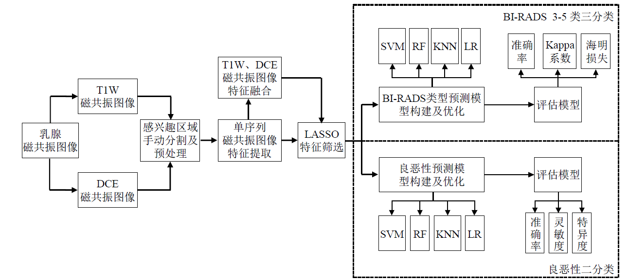
Fig. 2
Region of interest (ROI) segmentation of DCE magnetic resonance images. (a) Schematic diagram of ROI delineation at the largest layer of the lesion; (b) Schematic diagram of the lesion delineation
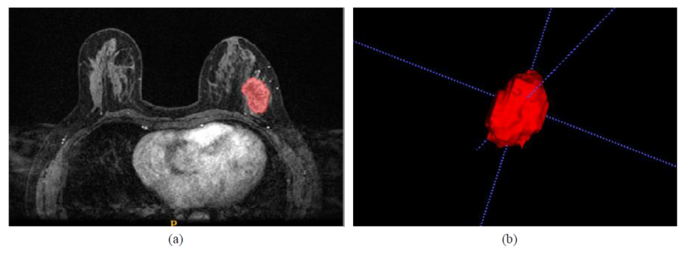

Table 1
Radiomics features of breast magnetic resonance images
| 特征类别 | 特征描述 | 特征数 | 特征输入 |
|---|---|---|---|
| shape | 表面积、伸长率、平面度、球形度、最大二维直径等 | 14 | 原始图像 |
| firstorder | 能量、熵、峰值、最大值、平均值、方差、第10百分位数、第90百分位数等 | 342 | 原始图像、派生图像 |
| GLCM | 自相关、联合平均、聚类突出度、聚类阴影、对比、相关、差熵等 | 418 | 原始图像、派生图像 |
| GLRLM | 长游程强调、短游程强调、灰度不均匀性、游程百分比、游程方差、游程熵等 | 304 | 原始图像、派生图像 |
| GLDM | 小依赖度、大依赖度、依赖不均匀性、依赖方差、依赖熵等 | 266 | 原始图像、派生图像 |
| GLSZM | 小区域强调、大区域强调、灰度不均匀性、区域百分比、区域方差、区域熵等 | 304 | 原始图像、派生图像 |
| NGTDM | 粗糙度、对比度、繁忙度、复杂度、强度 | 95 | 原始图像、派生图像 |
Table 2
The optimal radiomics features and corresponding coefficients of T1W images screened by LASSO algorithm
| 影像组学特征 | 系数 | 特征详情 |
|---|---|---|
| 联合熵(original_glcm_JointEntropy) | 0.010878 | 度量邻域强度值的可变性 |
| 平均值(log-sigma-5-0-mm-3D_firstorder_Mean) | 0.120571 | 描述肿瘤区域的平均灰度值 |
| 小区域低灰度级强调 (wavelet-HLH_glszm_SmallAreaLowGrayLevelEmphasis) | -0.110678 | 描述低灰度小尺寸区域体素的分布 |
| 归一化相关不均匀性 (wavelet-HHL_gldm_DependenceNonUniformityNormalized) | -0.089366 | 描述GLDM中体素相关关系的相似程度 |
| 依赖方差(wavelet-HHL_gldm_DependenceVariance) | 0.007990 | 描述GLDM中依赖大小的方差 |
| 相关性信息测度2(wavelet-HHH_glcm_Imc2) | -0.045300 | 量化纹理的复杂性 |
| 游程方差(wavelet-HHH_glrlm_RunVariance) | -0.059722 | 度量游程长度的方差 |
| 依赖熵(gradient_gldm_DependenceEntropy) | 0.019160 | 度量GLDM中依赖大小与灰度级分布的随机性程度 |
| 小依赖性低灰度级强调(gradient_gldm_SmallDependenceLowGrayLevelEmphasis) | -0.021736 | 描述体素小相关性与低阶灰度值的联合分布情况 |
| 相关性信息测度1(squareroot_glcm_Imc1) | 0.030811 | 量化纹理的复杂性 |
| 强度(exponential_ngtdm_Strength) | -0.055385 | 度量肿瘤图像的灰度变化程度 |
Table 3
The optimal radiomics features and corresponding coefficients of DCE images screened by LASSO algorithm
| 影像组学特征 | 系数 | 特征详情 |
|---|---|---|
| 原始图像最小值(original_firstorder_Minimum) | -0.013973 | 描述肿瘤区域的最小灰度值 |
| 大依赖高灰度级强调(original_gldm_LargeDependenceHighGrayLevelEmphasis) | 0.039997 | 测量具有较高灰度值和体素强相关关系的联合分布情况 |
| 偏度(wavelet-LHH_firstorder_Skewness) | 0.001540 | 度量强度平均值的分布不对称性 |
| 相关性信息测度1(wavelet-LHH_glcm_Imc1) | 0.057154 | 量化纹理的复杂性 |
| 小波变换LLL方向联合熵 (wavelet-LLL_glcm_JointEntropy) | 0.197864 | 度量邻域强度值的可变性 |
| 归一化逆差矩(gradient_glcm_Idmn) | 0.034171 | 度量肿瘤图像的局部平均程度 |
| 平方变换最小值(square_firstorder_Minimum) | 0.037978 | 描述肿瘤区域的最小灰度值 |
| 平方变换联合熵(square_glcm_JointEntropy) | 0.064132 | 度量邻域强度值的可变性 |
| 大区域低灰度级强调 (squareroot_glszm_LargeAreaLowGrayLevelEmphasis) | -0.014832 | 测量图像中具有较低灰度值的较大尺寸区域体素的分布 |
| 低灰度区域强调 (logarithm_glszm_LowGrayLevelZoneEmphasis) | -0.055083 | 描述低灰度级区域体素的分布 |
| 低灰度强调(logarithm_gldm_LowGrayLevelEmphasis) | -0.084834 | 描述低阶灰度体素的分布情况 |
Table 4
The optimal fusion features and corresponding coefficients screened by LASSO algorithm
| 影像组学特征 | 系数 | 特征详情 |
|---|---|---|
| 最大2D直径(列) (original_shape_Maximum2DDiameterColumn) | 0.081848 | 冠状平面中肿瘤表面网格顶点之间最大的欧几里得距离 |
| 相关性信息测度1(wavelet-LHL_glcm_Imc1) | 0.018552 | 量化纹理的复杂性 |
| 集群阴影(wavelet-LHH_glcm_ClusterShade) | -0.022909 | 描述图像的褶皱程度 |
| 小波变换HLL方向粗糙度 (wavelet-HLL_ngtdm_Coarseness) | -0.051320 | 度量中心体素与其邻域体素之间平均差异 |
| 小区域低灰度级强调 (wavelet-HLH_glszm_SmallAreaLowGrayLevelEmphasis) | -0.031217 | 描述图像中具有较低灰度值的较小尺寸区域体素分布 |
| 相关性信息测度2(wavelet-HHL_glcm_Imc2) | -0.066184 | 量化纹理的复杂性 |
| 逆方差(wavelet-HHH_glcm_InverseVariance) | 0.010646 | 度量肿瘤图像局部均匀程度 |
| 归一化灰度不均匀度 (wavelet-LLL_glszm_GrayLevelNonUniformityNormalized) | -0.138090 | 测量图像中灰度强度值的可变性 |
| 小波变换LLL方向粗糙度 (wavelet-LLL_ngtdm_Coarseness) | -0.086469 | 度量中心体素与其邻域体素之间平均差异 |
| 归一化区域大小不均匀度 (square_glszm_SizeZoneNonUniformityNormalized) | -0.022937 | 测量整个图像中大小区域体积的可变性 |
| 最小值(exponential_firstorder_Minimum) | -0.038634 | 描述肿瘤区域的最小灰度值 |
| 对比度(logarithm_ngtdm_Contrast) | -0.019143 | 度量体素灰度的空间变化率 |
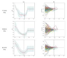
Fig. 3
The LASSO algorithm is used to screen the optimal T1W features, DCE features and fusion features, respectively. (a) The optimal λ is determined by adjusting λ to minimize the mean square error (MSE) of the model; (b) Convergence diagram of optimal characteristic coefficients
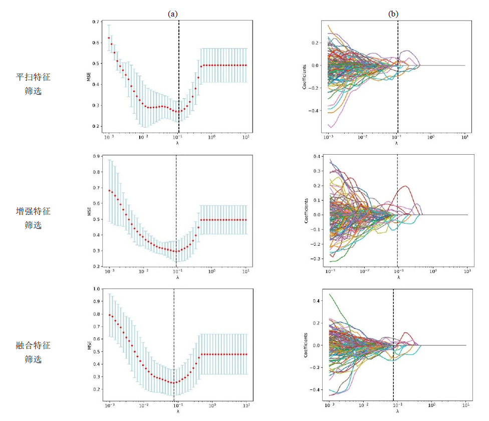
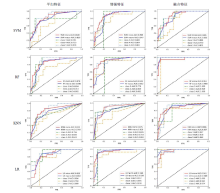
Fig. 4
ROC curves of four models based on different features (FPR: False positive rate, TPR: True positive rate)
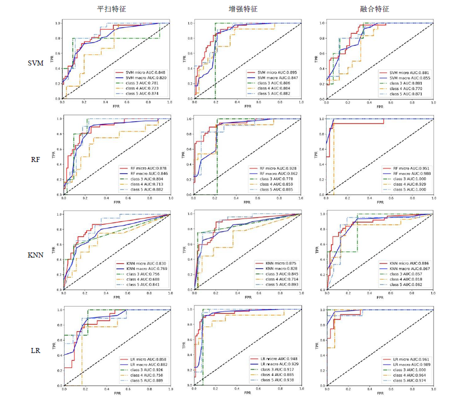
Table 5
Diagnostic efficacy of different models based on various features for classification of BI-RADS 3-5 breast lesions
| 特征 | 模型 | 准确率 | Kappa系数 | 海明损失 | Micro AUC | Macro AUC |
|---|---|---|---|---|---|---|
| 平扫特征 | SVM | 64.86% | 0.419 | 0.297 | 0.848 | 0.820 |
| RF | 70.27% | 0.421 | 0.333 | 0.878 | 0.846 | |
| KNN | 70.27% | 0.421 | 0.333 | 0.830 | 0.769 | |
| LR | 71.43% | 0.421 | 0.333 | 0.858 | 0.882 | |
| 增强特征 | SVM | 70.27% | 0.428 | 0.297 | 0.895 | 0.847 |
| RF | 86.49% | 0.428 | 0.297 | 0.928 | 0.862 | |
| KNN | 76.19% | 0.445 | 0.297 | 0.875 | 0.828 | |
| LR | 78.57% | 0.428 | 0.297 | 0.948 | 0.929 | |
| 融合特征 | SVM | 81.25% | 0.606 | 0.224 | 0.881 | 0.855 |
| RF | 87.50% | 0.676 | 0.188 | 0.951 | 0.988 | |
| KNN | 78.38% | 0.606 | 0.224 | 0.886 | 0.867 | |
| LR | 81.25% | 0.606 | 0.224 | 0.961 | 0.989 |
Table 6
The optimal radiomics features and corresponding coefficients of T1W images screened by LASSO algorithm
| 影像组学特征 | 系数 | 特征详情 |
|---|---|---|
| 联合能量(original_glcm_JointEnergy) | -0.074162 | 度量图像纹理中相邻灰度变换稳定程度 |
| 最大概率(original_glcm_MaximumProbability) | -0.049634 | 描述图像中出现次数最多的纹理特征 |
| 大依赖低灰度级强调(log-sigma-2-0-mm-3D_gldm_LargeDependenceLowGrayLevelEmphasis) | -0.029816 | 描述体素强相关关系与低阶灰度的联合分布情况 |
| 大区域低灰度级强调 (wavelet-LLH_glszm_LargeAreaLowGrayLevelEmphasis) | 0.001242 | 描述低灰度大尺寸区域体素的分布 |
| 小依赖低灰度级强调 (wavelet-HHL_gldm_SmallDependenceLowGrayLevelEmphasis) | -0.008748 | 描述体素小相关关系与低阶灰度的联合分布情况 |
| 小波变换HHL方向归一化区域大小不均匀度 (wavelet-HHL_glszm_SizeZoneNonUniformityNormalized) | -0.020799 | 测量整个图像中大小区域体积的可变性 |
| 强度(wavelet-HHL_ngtdm_Strength) | -0.083993 | 度量肿瘤图像的灰度变化程度 |
| 短游程低灰度级强调(square_glrlm_ShortRunLowGrayLevelEmphasis) | -0.088759 | 度量低阶灰度值与短游程长度的联合分布情况 |
| 小区域低灰度级强调(exponential_glszm_SmallAreaLowGrayLevelEmphasis) | 0.037705 | 描述低灰度小尺寸区域体素的分布 |
| 对数变换归一化区域大小不均匀度(logarithm_glszm_SizeZoneNonUniformityNormalized) | -0.004205 | 测量整个图像中大小区域体积的可变性 |
| 小依赖强调(logarithm_gldm_SmallDependenceEmphasis) | -0.011085 | 描述与领域内体素相关性较小的体素分布情况 |
Table 7
The optimum radiomics features and corresponding coefficients of DCE images screened by LASSO algorithm
| 影像组学特征 | 系数 | 特征详情 |
|---|---|---|
| 长轴长度(original_shape_MajorAxisLength) | 0.053568 | 度量肿瘤最长轴的长度 |
| 小区域强调(wavelet-LLH_glszm_SmallAreaEmphasis) | 0.007506 | 度量小尺寸区域的分布情况 |
| 大依赖低灰度级强调 (wavelet-LLH_gldm_LargeDependenceLowGrayLevelEmphasis) | -0.022168 | 描述体素强相关关系与低阶灰度的联合分布情况 |
| 强度(wavelet-LHL_ngtdm_Strength) | -0.002489 | 度量肿瘤图像的灰度变化程度 |
| 峰度(wavelet-LHH_firstorder_Kurtosis) | -0.038882 | 度量图像ROI中值分布的峰值 |
| 小依赖低灰度级强调 (wavelet-HLL_gldm_SmallDependenceLowGrayLevelEmphasis) | -0.003671 | 描述体素小相关关系与低阶灰度的联合分布情况 |
| 归一化灰度不均匀度 (wavelet-HLH_glszm_GrayLevelNonUniformityNormalized) | -0.117303 | 测量图像中灰度强度值的可变性 |
| 梯度变换联合熵(gradient_glcm_JointEntropy) | 0.001880 | 度量邻域强度值的可变性 |
| 平方变换联合熵(square_glcm_JointEntropy) | 0.058258 | 度量邻域强度值的可变性 |
| 强度(square_ngtdm_Strength) | -0.046019 | 度量肿瘤图像的灰度变化程度 |
| 归一化依赖不均匀性(exponential_gldm_DependenceNonUniformityNormalized) | -0.071656 | 测量整个图像中体素相关关系的相似程度 |
Table 8
The optimal fusion features and corresponding coefficients screened by LASSO algorithm
| 影像组学特征 | 系数 | 特征详情 |
|---|---|---|
| 长轴长度(original_shape_MajorAxisLength) | 0.050888 | 度量肿瘤最长轴的长度 |
| 联合能量(original_glcm_JointEnergy) | -0.016239 | 度量图像纹理中相邻灰度变换稳定程度 |
| 最大概率(original_glcm_MaximumProbability) | -0.071362 | 描述图像中出现次数最多的纹理特征 |
| 小波变换LLH方向-小区域强调 (wavelet-LLH_glszm_SmallAreaEmphasis) | 0.009840 | 度量小尺寸区域的分布情况 |
| 大依赖低灰度级强调 (wavelet-LLH_gldm_LargeDependenceLowGrayLevelEmphasis) | -0.014709 | 描述体素强相关关系与低阶灰度的联合分布情况 |
| 低灰度区域强调 (wavelet-LHH_glszm_LowGrayLevelZoneEmphasis) | -0.002699 | 描述低灰度级区域体素的分布 |
| 区域熵(wavelet-LHH_glszm_ZoneEntropy) | 0.090181 | 度量灰度区域大小与灰度级分布的不稳定性 |
| 相关性信息测度2(wavelet-HHL_glcm_Imc2) | -0.086920 | 量化纹理的复杂性 |
| 小波变换HHL方向-小区域强调 (wavelet-HHL_glszm_SmallAreaEmphasis) | -0.051711 | 度量小区域尺寸的分布情况 |
| 小依赖低灰度级强调 (wavelet-HHL_gldm_SmallDependenceLowGrayLevelEmphasis) | -0.012360 | 描述体素小相关关系与低阶灰度的联合分布情况 |
| 中位数(wavelet-HHH_firstorder_Median) | -0.023237 | 描述肿瘤区域的灰度中位数 |
| 逆方差(wavelet-HHH_glcm_InverseVariance) | 0.013141 | 度量肿瘤图像局部均匀程度 |
| 粗糙度(wavelet-LLL_ngtdm_Coarseness) | -0.014267 | 度量中心体素与其邻域体素灰度值的平均差异 |
| 短游程低灰度级强调(square_glrlm_ShortRunLowGrayLevelEmphasis) | -0.048257 | 度量低阶灰度值与短游程长度的联合分布情况 |
| 强度(square_ngtdm_Strength) | -0.002079 | 度量肿瘤图像的灰度变化程度 |
| 第10%分位值(squareroot_firstorder_10Percentile) | -0.003926 | 指肿瘤区域10%分位数的灰度值 |
| 小区域低灰度级强调(exponential_glszm_SmallAreaLowGrayLevelEmphasis) | 0.003075 | 描述低灰度小尺寸区域体素的分布 |
| 小依赖强调(logarithm_gldm_SmallDependenceEmphasis) | -0.002973 | 描述与领域内体素相关性较小的体素分布情况 |
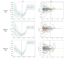
Fig. 5
The LASSO algorithm is used to screen the optimal T1W features, DCE features and fusion features, respectively. (a) The optimal λ is determined by adjusting λ to minimize the mean square error (MSE) of the model; (b) Convergence diagram of optimal characteristic coefficients
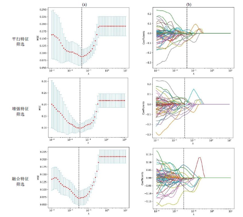
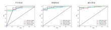
Fig. 6
ROC curves of four models based on different features
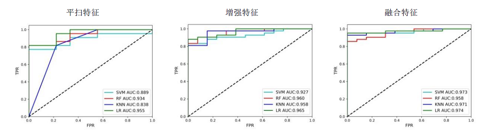
Table 9
Diagnostic efficacy of different models based on various features for benign and malignant breast lesions
| 特征 | 模型 | AUC | 准确率 | 灵敏度 | 特异度 |
|---|---|---|---|---|---|
| 平扫特征 | SVM | 0.889 | 80.65% | 66.67% | 86.67% |
| RF | 0.934 | 83.87% | 66.67% | 90.91% | |
| KNN | 0.838 | 80.65% | 77.78% | 81.82% | |
| LR | 0.955 | 83.87% | 77.78% | 86.36% | |
| 增强特征 | SVM | 0.927 | 85.45% | 92.31% | 83.33% |
| RF | 0.960 | 89.09% | 84.62% | 90.48% | |
| KNN | 0.958 | 85.45% | 84.62% | 85.71% | |
| LR | 0.965 | 89.09% | 84.62% | 90.48% | |
| 融合特征 | SVM | 0.973 | 90.91% | 78.57% | 95.19% |
| RF | 0.958 | 93.55% | 90.00% | 95.24% | |
| KNN | 0.971 | 92.73% | 92.86% | 92.68% | |
| LR | 0.974 | 94.55% | 85.71% | 97.56% |
Table 10
p values of Delong test results based on diagnostic efficiency of different models
| AUC | RF与SVM | RF与KNN | RF与LR | LR与SVM | LR与KNN | SVM与KNN | |
|---|---|---|---|---|---|---|---|
| BI-RADS分类 | Micro AUC | 0.041 | 0.058 | 0.134 | 0.069 | 0.071 | 0.317 |
| Macro AUC | 0.037 | 0.039 | 0.481 | 0.037 | 0.039 | 0.093 | |
| 良恶性分类 | AUC | 0.052 | 0.067 | 0.034 | 0.067 | 0.078 | 0.078 |
| [1] |
SIEGEL R L, MILLER K D, FUCHS H E, et al. Cancer statistics, 2021[J]. CA: A Cancer J Clin, 2021, 71(1): 7-33.
doi: 10.3322/caac.21654 |
| [2] |
FENG R M, ZONG Y N, CAO S M, et al. Current cancer situation in China: good or bad news from the 2018 global cancer statistics?[J]. Cancer Commun, 2019, 39(1): 22-33.
doi: 10.1186/s40880-019-0368-6 |
| [3] |
VALDORA F, HOUSSAMI N, ROSSI F, et al. Rapid review: radiomics and breast cancer[J]. Breast Cancer Res Treat, 2018, 169(2): 217-229.
doi: 10.1007/s10549-018-4675-4 |
| [4] |
MANN R M, CHO N, MOY L. Breast MRI: State of the art[J]. Radiology, 2019, 292(3): 520-536.
doi: 10.1148/radiol.2019182947 pmid: 31361209 |
| [5] |
WAKANA M, HYUNG W C, JOINES M M, et al. Quantitative predictors of response to neoadjuvant chemotherapy on dynamic contrast-enhanced 3T breast MRI[J]. J Breast Imaging, 2022, 4(2): 168-176.
doi: 10.1093/jbi/wbab095 |
| [6] |
SHAHAN C L, LAYNE G P. Advances in breast imaging with current screening recommendations and controversies[J]. Obstet Gynecol Clin North Am, 2022, 49(1): 1-33.
doi: 10.1016/j.ogc.2021.11.001 |
| [7] | LIU Y, CHEN J C, HU X Y, et al. Classification and localization of meningioma and acoustic neuroma in cerebellopontine angle based on mask RCNN[J]. Chinese J Magn Reson, 2021, 38(1): 58-68. |
| 刘颖, 陈静聪, 胡小洋, 等. 基于Mask RCNN的桥小脑角区脑膜瘤与听神经瘤分类定位研究[J]. 波谱学杂志, 2021, 38(1): 58-68. | |
| [8] |
WEAVER O, LEUNG J W T. Biomarkers and imaging of breast cancer[J]. AJR Am J Roentgenol, 2018, 210(2): 271-278.
doi: 10.2214/AJR.17.18708 |
| [9] |
RAWASHDEH M, LEWIS S, ZAITOUN M, et al. Breast lesion shape and margin evaluation: BI-RADS based metrics understate radiologists' actual levels of agreement[J]. Comput Biol Med, 2018, 96(1): 294-298.
doi: 10.1016/j.compbiomed.2018.04.005 |
| [10] |
SATAKE H, ISHIGAKI S, ITO R, NAGANAWA S. Radiomics in breast MRI: current progress toward clinical application in the era of artificial intelligence[J]. Radiol Med. 2022, 127(1): 39-56.
doi: 10.1007/s11547-021-01423-y |
| [11] |
LEITHNER D, HORVAT J V, OCHOA-ALBIZTEGUI R E, et al. Imaging and the completion of the omics paradigm in breast cancer[J]. Der Radiologe, 2018, 58(1): 7-13.
doi: 10.1007/s00117-018-0409-1 |
| [12] | WANG N, WANG Y J, LIAN P. Prediction of preoperative T staging of rectal cancer based on radiomics[J]. Chinese J Magn Reson, 2022, 39(1): 43-55. |
| 王楠, 王远军, 廉朋. 基于影像组学的直肠癌术前T分期预测[J]. 波谱学杂志, 2022, 39(1): 43-55. | |
| [13] |
IMBRIACO M, CUOCOLO R. Does texture analysis of MR images of breast tumors help predict response to treatment?[J]. Radiology, 2018, 286(2): 421-423.
doi: 10.1148/radiol.2017172454 pmid: 29356631 |
| [14] |
YIN X X, HADJILOUCAS S, ZHANG Y C, et al. MRI radiogenomics for intelligent diagnosis of breast tumors and accurate prediction of neoadjuvant chemotherapy responses — A review[J]. Comput Meth Prog Bio, 2021, 214: 106510.
doi: 10.1016/j.cmpb.2021.106510 |
| [15] |
NARANJO I D, GIBBS P, REINER J S, et al. Radiomics and machine learning with multiparametric breast MRI for improved diagnostic accuracy in breast cancer diagnosis[J]. Diagnostics, 2021, 11(6): 919-932.
doi: 10.3390/diagnostics11060919 |
| [16] | PRATIKSHA Y, SURBHI C. Effectivity of combined diffusion-weighted imaging and contrast-enhanced MRI in malignant and benign breast lesions[J]. Pol J Radiol, 2018, 83: e82-e93. |
| [17] | HU Q, WHITNEY H M, GIGER M L. Radiomics methodology for breast cancer diagnosis using multiparametric magnetic resonance imaging[J]. J Med Imaging, 2020, 7(4): 044502. |
| [18] | YE D M, WANG H T, YU T. The application of radiomics in breast MRI: A review[J]. Technol Cancer Res Tr, 2020, 19: 1-16. |
| [19] |
PAREKH V S, JACOBS M A. Multiparametric radiomics methods for breast cancer tissue characterization using radiological imaging[J]. Breast Cancer Res Tr, 2020, 180(2): 407-421.
doi: 10.1007/s10549-020-05533-5 pmid: 32020435 |
| [20] | DEMIRCIOGLU A, GRUENEISEN J, INGENWERTH M, et al. A rapid volume of interest-based approach of radiomics analysis of breast MRI for tumor decoding and phenotyping of breast cancer[J]. PLoS ONE, 2020, 15(6): e0234871. |
| [21] |
TSAROUCHI M I, VLACHOPOULOS G F, KARAHALIOU A N, et al. Multi-parametric MRI lesion heterogeneity biomarkers for breast cancer diagnosis[J]. Phys Medica, 2020, 80(2): 101-110.
doi: 10.1016/j.ejmp.2020.10.007 |
| [22] |
ZHANG Q, PENG Y S, LIU W, et al. Radiomics based on multimodal MRI for the differential diagnosis of benign and malignant breast lesions[J]. J Magn Reson Imaging, 2020, 52(2): 596-607.
doi: 10.1002/jmri.27098 pmid: 32061014 |
| [23] |
HAO W, GONG J, WANG S P, et al. Application of MRI radiomics-based machine learning model to improve contralateral BI-RADS 4 lesion assessment[J]. Front Oncol, 2020. doi: 10.3389/fonc.2020.531476.
doi: 10.3389/fonc.2020.531476 |
| [24] | QIAO M Y, LI C K, SUO S T, et al. Breast DCE-MRI radiomics: a robust computer-aided system based on reproducible BI-RADS features across the influence of datasets bias and segmentation methods[J]. Int J Comput Ass Rad, 2020, 15(5): 921-930. |
| [25] |
SCAPICCHIO C, GABELLONI M, BARUCCI A, et al. A deep look into radiomics[J]. La Radiologia Medica, 2021, 126: 1296-1311.
doi: 10.1007/s11547-021-01389-x |
| [26] | DOMINGUES I, ABREU P H, SANTOS J. Bi-Rads classification of breast cancer: A new pre-processing pipeline for deep models training[C]// IEEE International Conference of Image Processing (ICIP). IEEE, 2018.1378-1382. |
| [27] | SIDDEEQ S, LI J, BHATTI H M A, et al. Deep learning RN-BCNN model for breast cancer BI-RADS classification[C]. ICIGP 2021: 2021 The 4th International Conference on Image and Graphics Processing. 2021: 219-225. |
| [28] | WU P Q. Research progress of radiomics in lymph node metastasis of breast cancer[J]. Journal of Molecular Imaging, 2020, 43 (1): 31-35. |
| 吴佩琪. 影像组学在乳腺癌淋巴结转移中的研究进展[J]. 分子影像学杂志, 2020, 43(1): 31-35. |
| [1] | LI Na, XIAO Long, LI Sha, LIU Yudun, JIA Yushu, ZHANG Lei, CHEN Shizhen. 19F NMR Chemical Shift-encoded cRGD-targeted Dual-modal Probe for Multicolor Imaging [J]. Chinese Journal of Magnetic Resonance, 2026, 43(3): 279-290. |
| [2] | SU Yilin, LIU Yuanyuan, CUI Zhuoxu, LIANG Dong. Physics Guided Multi-contrast Magnetic Resonance Reconstruction Diffusion Model [J]. Chinese Journal of Magnetic Resonance, 2026, 43(3): 321-338. |
| [3] | CAI Yue, WANG Xuxia, LIU Sijie, GUO Haodong, CHEN Xi, CHENG Linlin, KANG Yan, LIN Fuchun. Magnetic Resonance Imaging Study on the Microstructure Abnormalities of Striatal White Matter in Type 2 Diabetic Mellitus Rats [J]. Chinese Journal of Magnetic Resonance, 2026, 43(2): 175-185. |
| [4] | LI Ruiyi, LI Sha, XU Qiuyi, SUI Meiju, CHEN Shizhen. In Situ Regulation of Protein Corona for Enhanced Tumor-targeted 19F MRI and Combined Therapy [J]. Chinese Journal of Magnetic Resonance, 2026, 43(2): 200-213. |
| [5] | XIE Xinyi, WANG Yuanjun. Research Progress on Super-resolution Reconstruction of Brain Diffusion Magnetic Resonance Images [J]. Chinese Journal of Magnetic Resonance, 2026, 43(2): 223-240. |
| [6] | FU Fenfang, LIN Guobing, LI Meifang. Advances in Magnetic Resonance Imaging for the Diagnosis and Prognosis of Mild Traumatic Brain Injury [J]. Chinese Journal of Magnetic Resonance, 2026, 43(2): 214-222. |
| [7] | WANG Yiwen, WU Guangyao, WEN Zhi, LIN Fuchun. Antiretroviral Therapy-related Alteration of Brain Functional Dynamics in People with HIV [J]. Chinese Journal of Magnetic Resonance, 2026, 43(1): 61-70. |
| [8] | XIANG Zhao, SUI Li, ZHANG Haotian, DUAN Mengyu, LIU Zhuorui. A Lightweight AD-Net Model for the Classification of Intracranial Tumors in MRI Images [J]. Chinese Journal of Magnetic Resonance, 2026, 43(1): 71-86. |
| [9] | NI Guangmao, LI Yuwei, HOU Wenxuan, LIU Caiyun, DONG Peng, ZHANG Yanhui. Research on the Influencing Factors of Acute Cerebral Infarction Recurrence Based on MR-DWI [J]. Chinese Journal of Magnetic Resonance, 2026, 43(1): 87-93. |
| [10] | CHEN Xi, LIU Sijie, CAI Yue, CHENG Linlin, WANG Xuxia, KANG Yan, LIN Fuchun, LEI Hao. Effects of Seizure-inducing Doses Nicotine on Hippocampal Structure in Adolescent Female Rats [J]. Chinese Journal of Magnetic Resonance, 2025, 42(4): 345-354. |
| [11] | LI Yinghao, WANG Lihui, WANG Sucheng, ZHU Zhongqi, HUANG Changdong, LI Renfeng, CAO Kaiming, HU Haiyang, JIA Yiming, LIANG Songtao, YANG Guang, LU Qing, WANG Hongzhi. Study on Pancreas Automatic Segmentation, Regional Quantification, and Diabetes Assessment [J]. Chinese Journal of Magnetic Resonance, 2025, 42(4): 378-389. |
| [12] | GAO Zhaoyao, ZHANG Zhan, HU Liangliang, XU Guangyu, ZHOU Sheng, HU Yuxin, LIN Zijie, ZHOU Chao. PMRI Image Reconstruction Method Based on Virtual Coils and GRAPPA-enhanced Network [J]. Chinese Journal of Magnetic Resonance, 2025, 42(4): 390-401. |
| [13] | MA Yingxue, ZHAO Yanqiang, YANG Xiaodong, JIANG Bin, TAO Cheng. Opportunities and Challenges of High-field and Ultra-high-field Magnetic Resonance Imaging in China [J]. Chinese Journal of Magnetic Resonance, 2025, 42(3): 334-344. |
| [14] | SHU Wei. Diagnostic Efficacy Comparison of B-scan Ultrasonography and MRI in Fetal Skeletal Abnormalities [J]. Chinese Journal of Magnetic Resonance, 2025, 42(3): 265-274. |
| [15] | SUI Meiju, ZHANG Lei, WANG Ruifang, LUO Yingying, LI Sha, QIU Maosong, XU Qiuyi, CHEN Daiqin, CHEN Shizhen, ZHOU Xin. MRI-traceable Nanoenzyme for Cascade Catalysis-enhanced Immunotherapy [J]. Chinese Journal of Magnetic Resonance, 2025, 42(3): 231-248. |
| Viewed | ||||||
|
Full text |
|
|||||
|
Abstract |
|
|||||
